Wilson’s disease is an autosomal recessive disorder of copper metabolism caused by various mutations in the ATB7B gene, which encodes a copper-transporting P-type adenosine triphosphatase.Reference Bandmann, Weiss and Kaler 1 Dysfunction of this transporting protein leads to copper deposition and causes mitochondrial injury due to oxygen free radicals.Reference Taly, Meenakshi-Sundaram, Sinha, Swamy and Arunodaya 2 The disease may present at any age with various clinical presentations and is fatal if left unmanaged. The most common complications of Wilson’s disease are hepatic and neuropsychiatric.Reference Taly, Meenakshi-Sundaram, Sinha, Swamy and Arunodaya 2 Cardiac problems such as arrhythmias, autonomic dysfunction, cardiomyopathy, and sudden cardiac death occur in adult patients with Wilson’s disease.Reference Arat, Kacar and Golbasi 3 – Reference Grandis, Nah and Whitman 6 To date, cardiac issues in Wilson’s disease have been the subject in some case reports and investigated in small cohorts, especially among adult patients. Therefore, cardiac involvement has not been reported in large longitudinal cohorts due to the lack of routine cardiac evaluation of these patients during their follow-up. Cardiac involvement in Wilson’s disease, especially in children, is not well studied despite the serious effects on morbidity and mortality in the long-term. Tissue Doppler echocardiography is an advanced technique used to assess systolic and diastolic functions and to provide direct quantitative information about the velocity of myocardium regardless of preload and afterload.Reference Wang, Yip and Wang 7 The purpose of this study was to evaluate cardiac function and electrocardiographic findings in patients with Wilson’s disease and to compare them with healthy control patients.
Materials and methods
This prospective study included 42 patients with Wilson’s disease that had follow-up visits in the paediatric gastroenterology department between June 2017 and December 2017. Thirty-seven healthy controls were matched for age and gender. The mean time from the diagnosis until the cardiac evaluation was 6.3 ± 3.1 years. The diagnosis of Wilson’s disease was based on combinations of presence of Kayser-Fleischer rings, increased urinary copper, low serum ceruloplasmin concentration, and quantification of copper in a liver biopsy. 8 , Reference Gow, Smallwood, Angus, Smith, Wall and Sewell 9 Medical history, clinical presentation, biochemical parameters, and 24-hour urinary copper excretion were collected from patients’ charts and prospectively analysed. The control group included 37 children that were referred to the paediatric cardiology department due to an innocent heart murmur, and clinical cardiac evaluations were normal. Patients with congenital cardiac defects or who were using drugs with potential cardiac electrophysiological effects were excluded. The control group had normal values of serum ceruloplasmin and urinary copper excretion. The echocardiographic measurements of both groups were taken from the clinical echocardiogram reports, and readings were done by a single paediatric cardiologist. All enrolled patients were evaluated by a standard two-dimensional and tissue Doppler echocardiography concurrent with standard 12-lead electrocardiogram. Echocardiographic evaluations were performed using a GE Vivid E9 with XDclear (GE Healthcare, Horten, Norway) with 5 and 6 MHz matrix transducer probe. Standard examination was performed in the apical four-chamber, parasternal short and long axis, subcostal and suprasternal views. M-mode echocardiographic measurements of the inter-ventricular septal thickness, posterior wall thickness, left ventricular internal dimension, fractional shortening, and left ventricular ejection fraction were performed end diastole and end systole according to the established standards of the American Society of Echocardiography. Left ventricular mass was calculated using the formula given by Devereux et al, according to the American Society of Echocardiography guidelines: Left ventricular mass (g) = 0.81(1.04 [inter-ventricular septum + posterior wall thickness + left ventricular internal dimension]3 − [left ventricular internal dimension]3) + 0.06.Reference Devereux, Alonso and Lutas 10 Left ventricular mass index was derived by dividing left ventricular mass in grams by the patient’s body surface area. Relative wall thickness, defined as the ratio of end-diastolic left ventricular wall thickness to left ventricular internal dimension ([inter-ventricular septum + posterior wall thickness]/left ventricular internal dimension), was determined to be < or ≥0.41. Left ventricular geometry was considered normal if left ventricular mass index was <95th percentile and relative wall thickness <0.41; concentric remodelling was reported if left ventricular mass index was <95th percentile and relative wall thickness ≥0.41; concentric hypertrophy was reported if left ventricular mass index was ≥95th percentile and relative wall thickness ≥0.41; and eccentric hypertrophy was reported if left ventricular mass index was ≥95th percentile and relative wall thickness <0.41.Reference Daniels, Loggie, Khoury and Kimball 11 To evaluate diastolic and systolic cardiac functions, measurement of peak myocardial systolic, early diastolic, and late diastolic velocities in the septal and lateral annuli of the tricuspid and mitral valves and the inter-ventricular septum were measured. Isovolumetric contraction and relaxation times, along with ejection time periods, were also measured. The myocardial performance index was calculated (the sum of the isovolumetric contraction and isovolumetric relaxation times divided by the ejection time).
A standard 12-lead electrocardiogram was recorded using the CardiMax FX8322 electrocardiograph machine (Fukuda Denshi, Tokyo, Japan) at a speed of 25 mm/s and an amplitude of 10 mm/mV. The electrocardiogram was analysed by a single observer to reduce intra-observer variability. The electrocardiogram results were transferred and magnified on a high-resolution monitor and findings were measured using the MB-Ruler program (Markus Bader, MB-Software Solutions, Germany) as calipers. Inability to determine the end of the T wave, technically suboptimal electrocardiograms, and abnormal T-wave morphologies were excluded from the analysis. Electrocardiographic data included P wave, QRS complex, QT interval, corrected QT interval (QTc), T wave, ST segment, and P-R interval. The QT interval were measured at the beginning of the QRS complex and the end of the T wave. Corrected QT interval was calculated by the Bazett’s Formula (QTc = QT/square root of RR interval). For the measurement of QT dispersion and PR dispersion, all 12 leads were analysed in both groups to identify the difference between the QT interval maximum and QT interval minimum or the difference between P maximum and P minimum. Analysis of data was performed with the SPSS statistical software package. Descriptive statistics included mean, median, range, and standard deviation. The Mann–Whitney U and Wilcoxon tests were applied to assess differences between the groups. A p value of <0.05 was considered statistically significant.
This study was approved by the Clinical Research Ethics Board and written informed consent was obtained from all participants’ parents.
Results
The study included 79 patients (42 patients with Wilson’s disease and 37 healthy controls, age and gender matched). Gender ratio was 1:1 and the mean of the patients’ ages at the time of diagnosis was 7.35 ± 3.37 years (range from 1.5 to 15). The mean age at the time of cardiac evaluation was 14.06 ± 4.04 years (range from 4.10 to 20) in the Wilson’s group, while it was 12.76 ± 3.13 years (range from 6.1 to 17.3) in the control group (p > 0.05). The mean follow-up time for patients was 7.0 ± 3.36 years (range from 1 to 13). Hepatic diseases were diagnosed in 92% of the patients, while neurological dysfunction was present in only 3% of the patients. Aspartate aminotransferase, alanine aminotransferase, serum ceruloplasmin and 24-hour urinary copper levels were 51 ± 5 IU/L, 65 ± 7 IU/L, 10.7 ± 1.8 mg/dL and 290 ± 27 µg/24 hours, respectively. All Wilson’s disease patients had been prescribed chelating treatment (d-penicillamine and zinc salts) and were compliant with treatment.
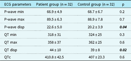
Electrocardiographic assessment revealed that all patients had sinus rhythm and a normal QRS axis. No significant differences were found between the Wilson’s and control groups with respect to P-wave amplitude and duration, QRS duration, P-R interval, and QT interval. Left ventricular hypertrophy was not observed in either group. Incomplete right bundle branch block (n = 1), right ventricular hypertrophy (n = 1), early repolarisation (n = 1), and U wave (n = 1) were observed in the Wilson’s group. No statistically significant difference was found between the two groups for the minimum and maximum values of P wave and QT interval. The QT and P-wave dispersion values had statistically significant differences (p = 0.02 and 0.04, respectively), with higher values in the Wilson’s group. Ventricular and atrial ectopy, as well as ST segment and T-wave abnormalities, were not observed in the Wilson’s group. Table 1 shows the electrocardiographic assessments.
Results of ECG assessment

Disp = dispersion; ECG = electrocardiogram; Max = maximum; Min = minimum.
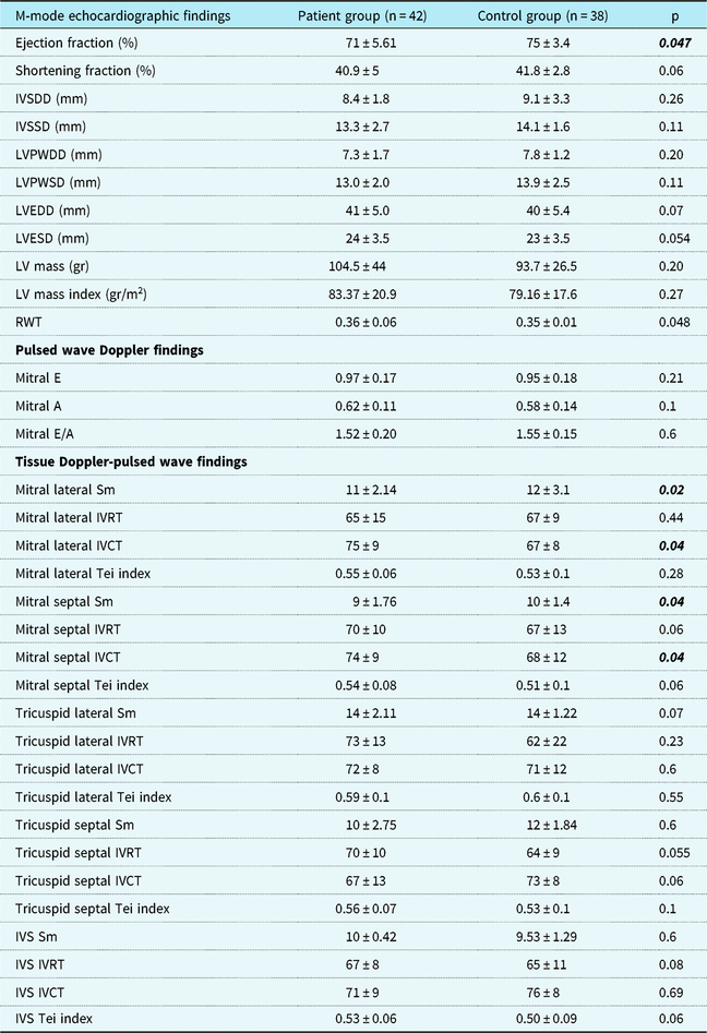
Standard two-dimensional and pulsed-wave Doppler echocardiographic assessment of systolic and diastolic function of the left ventricle was also normal in both groups. The mean left ventricular ejection fraction was 75 ± 3.4% in the control group and was significantly lower in the Wilson’s group (p = 0.047); however, we did not find the difference meaningful, hence the normal values of functional measurements in both groups.
On tissue Doppler echocardiography assessment, mitral early and mitral late velocity values, and mitral early/mitral late ratio were normal, and no statistically significant difference between the two groups was found. Mitral lateral and septal systolic annular velocity values were significantly lower in the Wilson’s group when compared to the control group (p = 0.02 and 0.04, respectively). Also, mitral lateral and septal isovolumetric contraction times were significantly higher in the Wilson’s group when compared to the control group (p = 0.04).
On tissue Doppler echocardiography assessment, mean values for the myocardial performance index, also known as the “Tei index”, for mitral lateral, mitral septal, tricuspid lateral, tricuspid septal, and inter-ventricular septum were higher in the Wilson’s group; however, the difference was not statistically significant. Left ventricular parameters were also compared between the two groups and no statistically significant difference was found. While the left ventricular mass and left ventricular mass index values also did not differ between the two groups, relative left ventricular wall thickness was higher in the Wilson’s group when compared to the control group. This difference was statistically significant (p = 0.04). Concentric remodelling was found in 7 (16%) of 42 patients in the Wilson’s group. Table 2 shows the assessment of the tissue Doppler echocardiography.
Results of Tissue doppler echocardiography assessment

IVCT = isovolumetric contraction time; IVRT = isovolumetric relaxation time; IVS = inter-ventricular septum; IVSDD = inter-ventricular septum diastolic diameter; IVSSD = inter-ventricular septum systolic diameter; LV = left ventricle; LVEDD = left ventricle end diastolic diameter; LVESD = left ventricle end systolic diameter; LVPWDD = left ventricle posterior wall diastolic diameter; LVPWSD = left ventricle posterior wall systolic diameter; Mitral A = atrial contraction; Mitral E = early diastolic flow; Mitral E/A = E/A ratio; RWT = relative wall thickness; Sm = peak systolic velocity; Tei = myocardial performance index.
Discussion
Wilson’s disease is a multisystem genetic disorder that leads to excess copper storage, and high levels of free ionic copper cause damage in affected organs due to oxygen free radicals.Reference Gaffney, Fell and O’Reilly 12 Symptoms of Wilson’s disease are nonspecific and may easily mimic other hepatic diseases; therefore, a combination of biochemical results, liver biopsy, and in some cases genetic testing are required for a precise diagnosis.Reference Hedera 13 , Reference Socha, Janczyk and Dhawan 14 The disease commonly presents with hepatic and neurological manifestations, and cardiac involvement is rare among children. In previous studies that were primarily based on clinical presentation and demographic findings of the disease, no cardiac findings have been reported in children, neither at the time of diagnosis nor at follow up, due to the lack of routine cardiac assessment. Cardiac manifestations such as arrhythmias, autonomic dysfunction, cardiomyopathy, and sudden cardiac death have been found and previously reported in adult patients with Wilson’s disease.Reference Arat, Kacar and Golbasi 3 – Reference Grandis, Nah and Whitman 6
Kuan et al were the first to report electrocardiographic abnormalities, cardiomyopathy, and sudden cardiac death in 18 out of 53 Wilson’s disease patients, and they suggested that cardiac involvement could be related to functional alterations in myocardial tissue due to low or high copper levels.Reference Kuan 15 Cardiovascular autonomic dysfunction due to involvement of both parasympathetic and sympathetic systems was previously reported in adult Wilson’s disease patients with neurological presentation.Reference Soni, Shukla, Singh, Goyal and Behari 16 , Reference Li, Lindauer and Haase 17 In the current study, the standard electrocardiographic assessment was normal in both groups, and QT intervals and minimum and maximum P-wave values were not different. However, QT and P-wave dispersion values were significantly higher in the Wilson’s group. P-wave dispersion is a sign of predisposition to atrial arrhythmias, and a study by Arat et alReference Arat, Kacar and Golbasi 3 showed an increased incidence of P-wave dispersion in adult Wilson’s disease patients with no cardiac symptoms; they suggested that this could be an early sign of cardiac involvement and a high risk of atrial fibrillation.Reference Okutucu, Aytemir and Oto 18 QT dispersion is an indirect measure of repolarisation abnormalities and is used as a prognostic marker to predict tendency to ventricular arrhythmias and sudden cardiac death.Reference Malik and Batchvarov 19 To the best of our knowledge, increased QT dispersion has not been previously reported in adults or in children with Wilson’s disease. It can be a useful non-invasive diagnostic method to predict arrhythmias. Furthermore, as a limitation of the present study, to reveal more precise evidence of tendency to arrhythmia in Wilson’s disease patients, 24-hour Holter monitoring should be performed in all patients at the time of diagnosis, even if the standard electrocardiogram is normal.
Even though it is rare, depending on its degree, cardiomyopathy could become a major problem in terms of mortality and morbidity in patients with Wilson’s disease. At the cellular level, an increased level of copper leads to oxidative stress, free radical formation, and mitochondrial dysfunction, possibly leading to cellular death in the myocardium.Reference Bandmann, Weiss and Kaler 1 Myocardial copper deposition was well described in a cardiac biopsy report in the late 1970s and in a comprehensive post-mortem study.Reference Azevedo, Scaff, Barbosa, Neto and Canelas 20 , Reference Factor, Cho, Sternlieb, Scheinberg and Goldfischer 21 In the literature, no pathognomonic cardiac finding has been attributed to Wilson’s disease, and these findings can be observed in other cardiomyopathies. The reported pathological myocardial changes were interstitial and replacement fibrosis, intra-myocardial small vessel sclerosis, focal inflammatory cell infiltration to a variable degree, myocarditis, cardiac hypertrophy, and occlusive atherosclerosis.Reference Meenakshi-Sundaram, Sinha, Rao and Prashanth 4 , Reference Factor, Cho, Sternlieb, Scheinberg and Goldfischer 21 The study by Arat et al showed that left ventricular wall thickness, left ventricular diameter and left ventricular ejection fraction were similar in both Wilson’s patients and the control group.Reference Arat, Kacar and Golbasi 3 The similar results were published by Elkiran et al and Hlubocka et al.Reference Elkiran, Karakurt and Selimoglu 22 , Reference Hlubocka, Marecek and Linhart 23 We suggested that, even though patients in these studies are mostly well treated and had neither severe neurological impairment nor cardiac symptoms, a more comprehensive investigation is needed to evaluate cardiac involvement in a totally asymptomatic patient group. Tissue Doppler echocardiography is a more useful echocardiographic technique to evaluate global and regional myocardial systolic as well as diastolic function when compared to standard echocardiography, particularly for the assessment of asymptomatic patients. Tissue Doppler echocardiography technique assesses myocardial velocities throughout the cardiac cycle and also gives a chance to provide direct information about myocardial motion.Reference Dragulescu and Mertens 24
Hlubocka et al reported no systolic dysfunction; however, decreases in mitral early wave velocity and mitral early/mitral late ratio were detected, in addition to prolongation in isovolumetric relaxation time values. This indicates a diastolic dysfunction in Wilson’s disease patients by pulsed wave Doppler assessment.Reference Hlubocka, Marecek and Linhart 23 Similarly, Elkiran et al reported prolongation in isovolumetric relaxation time values on tissue Doppler echocardiography in patients with Wilson’s disease, which indicates a diastolic dysfunction; nevertheless, left ventricular systolic function was within the normal range in both groups. Also, no difference was found between the groups regarding left ventricular wall thickness and left ventricular mass values. They concluded that the normal results for systolic function and left ventricular measurements were due to the young age of the patients and the short duration of the disease (there had not been enough time to trigger a left ventricular remodelling process.Reference Azevedo, Scaff, Barbosa, Neto and Canelas 20 The present study results are consistent with the study by Hlubocka et al: while left ventricular mass and left ventricular mass index values did not differ significantly between the two groups, relative left ventricular wall thickness was higher in the Wilson’s group when compared to the control group (p = 0.04). Also, although the incidence is lower, consistent with the study by Hlubocka et al, in the present study concentric remodelling was found in 7 (16%) of 42 patients.Reference Hlubocka, Marecek and Linhart 23 Left ventricular concentric remodelling is a subtle early change in cardiac geometry characterised by increased relative wall thickness with normal overall muscle mass; it is already known as an important and independent predictor of increased cardiovascular risk in hypertensive patients.Reference Verdecchia, Schillaci and Borgioni 25 Concentric remodelling as an indicator of increased left ventricular wall thickness can be related to intra-cardiac copper accumulation, leading to excess of free radicals that alter structural changes in cardiac muscle. In the present study, the difference between the two groups was not remarkable. The measurement in some patients was just above normal limits, thus we did not observe any clinical impact of concentric remodelling in the present study similar to the study by Hlubocka et al.Reference Hlubocka, Marecek and Linhart 23 Therefore, our results suggest that Wilson’s disease could trigger a remodelling process in the left ventricle despite the young age of the patients.
In contrast to previous studies, the most important finding in the current study regarding myocardial systolic function was that the mean values of mitral lateral annular (p = 0.02) and mitral septal (p = 0.04) velocities were lower in the Wilson’s group, and this was statistically significant. Myocardial performance index, also known as the Tei index, incorporates both systolic and diastolic time intervals in expressing global systolic and diastolic ventricular function. Systolic dysfunction prolongs pre-ejection (isovolumetric contraction time), causing a shortening of the ejection time. Both systolic and diastolic dysfunction result in abnormal myocardial relaxation, which prolongs the relaxation period (isovolumetric relaxation time).Reference Tei, Ling and Hodge 26 , Reference Rudski, Lai and Afilalo 27 In the current study, the Tei index for the mitral lateral, mitral septal, tricuspid lateral, tricuspid septal, and inter-ventricular septum was higher in patients with Wilson’s disease; however, the differences were not statistically significant. This indicates a subtle subclinical global dysfunction in the myocardium, despite the young age of the patients and the short duration of the disease. For this reason, patients who are particularly noncompliant with treatment must be followed for the development of cardiac abnormalities.
The present study has potential limitations. First, the current study is a single-centre study; evaluation of a larger number of patients in a longitudinal, multi-centre study with cardiac evaluation of the patients at the time of diagnosis is needed. Second, though tissue Doppler echocardiography is more sensitive than conventional echocardiographic methods, it is angle dependent; and if the acquisition of the angle exceeds 20°, it may lead to an underestimation in tissue velocity. The interpretation of tissue Doppler echocardiography requires more experience than conventional methods, so this can sometimes be an obstacle.Reference Kadappu and Thomas 28 Also, measurements from the echocardiographic assessment were taken from clinical reports, thus the reading physician was not blinded to diagnosis.
To date, only a few studies evaluated the effects of copper accumulation on the myocardium in children. To the best of our knowledge, the current study is the largest series of children having Wilson’s disease with evaluation of cardiac function by means of echocardiography. Although our patients were all adherent to treatment and were asymptomatic, the present study showed that Wilson’s disease patients may have some subclinical systolic dysfunction without any cardiac symptoms, and this subclinical dysfunction can be detected using tissue Doppler echocardiography. The present study also suggested that despite the young age of the patients, the disease could trigger a remodelling process in the left ventricle. This should be assessed with further studies starting from the time of diagnosis and at follow-up. Furthermore, P-wave dispersion and QT dispersion abnormalities were detected, and this can be used to predict a tendency to arrhythmia, even at a young age.
In conclusion, evaluation of cardiac function with tissue Doppler echocardiography in patients with Wilson’s disease is useful during their follow-up. There may be early detection of dysfunction, and this may decrease cardiac morbidity and mortality.