Introduction
A neopharynx is a surgically reconstructed pharynx. It can be primarily reconstructed from the remnant of pharyngeal mucosa left following laryngectomy. However, if less than half the circumference of the pharynx is available, stricture formation may result. The alternatives are to graft a neopharynx totally or partially using myocutaneous, gastro-omental or jejunal free tissue flaps. These techniques have been used to good effect; however, neopharynges created partially or totally from graft material are associated with higher morbidity and mortality rates.Reference Stafford and Mathews1–Reference Carlson, Thourani, Codner and Grist4
Post-operative dysphagia has been reported in 10–16 per cent of cases following total laryngectomy.Reference Sullivan and Hartig5, Reference Gibbons, Halvorsen, Foster, Warner, Roberts and Postlethwait6 Some studies have found no association between neopharynx lumen diameter and functionality, whereas others have suggested that a tumour-free lumen of at least 1.5 cm (2.5 cm stretched) is needed to allow for adequate swallowing. Removal of the larynx results in increased resistance to flow through the neopharynx, due to a loss of superior and anterior laryngeal movement that would normally assist in the opening of the upper oesophageal segment. The above factors, combined with increased tortuosity of the pharynx as a result of tracheostomy, can lead to physiological dysphagia. Therefore, in patients with a neopharynx who present with dysphagia, it may be easy to miss benign pathology (such as strictures, pouches, gastroesophageal reflux and fistulae) as well as malignantpathology (either primary, recurrent or a second primary (either synchronous or metachronous)) involving the neopharynx. Gibbons et al. reported a 50 per cent detection rate for abnormalities of the neopharynx and oesophagus on barium swallow, in their series of 204 patients following total laryngectomy.Reference Gibbons, Halvorsen, Foster, Warner, Roberts and Postlethwait6
Patients with head or neck cancer are at increased risk of synchronous and metachronous lesions, in comparison with the general population. A high degree of suspicion and low threshold for investigation are imperative for such individuals. Sonographic, radiological and endoscopic methods of screening the neopharynx have been proposed to increase the detection rate of neopharyngeal tumours.
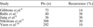
Tumour recurrences have been reported (see Table I) up to 38 per cent.Reference Balfe, Koehler, Setzen, Weymen, Baron and Ogura7, Reference Jung and Adams8 All cases in the neopharynx were that of recurrent squamous cell carcinoma and associated factors were; salvage surgery, degree of tumour extension, lymph node infiltration, poor tumour differentiation and trans-glottic/sub-glottic localisation of the primary tumour.Reference Nikolaou, Markou, Petridis, Vlachtsis, Nalbantian and Daniilidis9 Second primary malignancies are thought to be independent from primary tumour staging and distant and delayed metastases.Reference Spector, Sessions, Haughey, Chao, Simpson and El-Mofty11
Reported neopharynx tumour recurrence rates following total laryngectomy
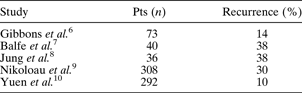
Pts = patients
By definition, a diagnosis of extra-pulmonary small cell carcinoma requires a histological diagnosis at the primary site, a normal chest radiograph and computed tomography (CT) scan, and normal sputum cytology or negative bronchoscopy.19
We present a patient with primary extra-pulmonary small cell carcinoma in the neopharynx, the first reported case of its kind. We also review the literature on the management of neopharyngeal carcinoma, and we discuss the management of extra-pulmonary small cell carcinoma in the neopharynx.
Case report
An 82-year-old, laryngectomised man presented to the head and neck clinic with dysphagia and loss of weight, four years after laryngectomy surgery and adjuvant radiotherapy.
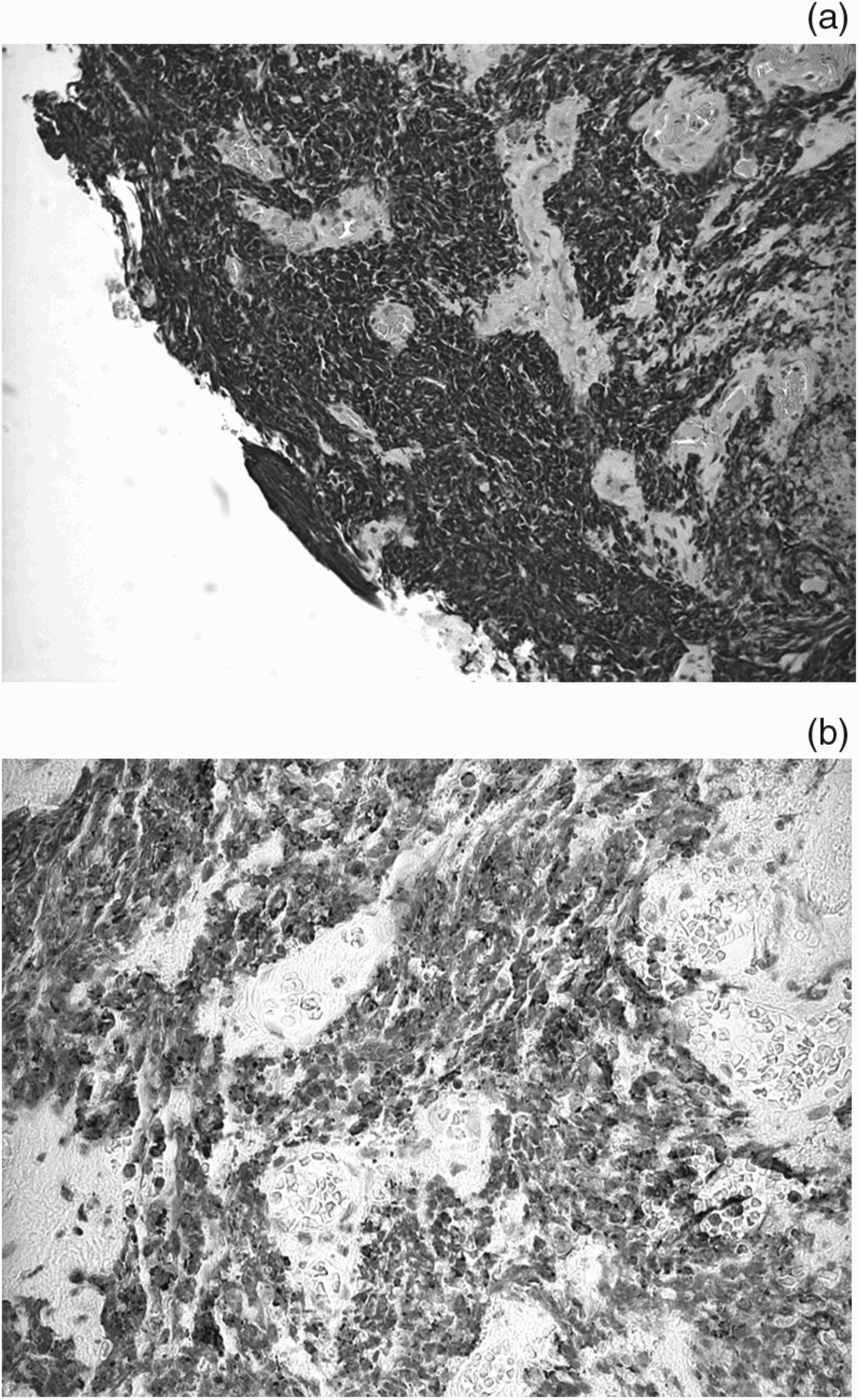
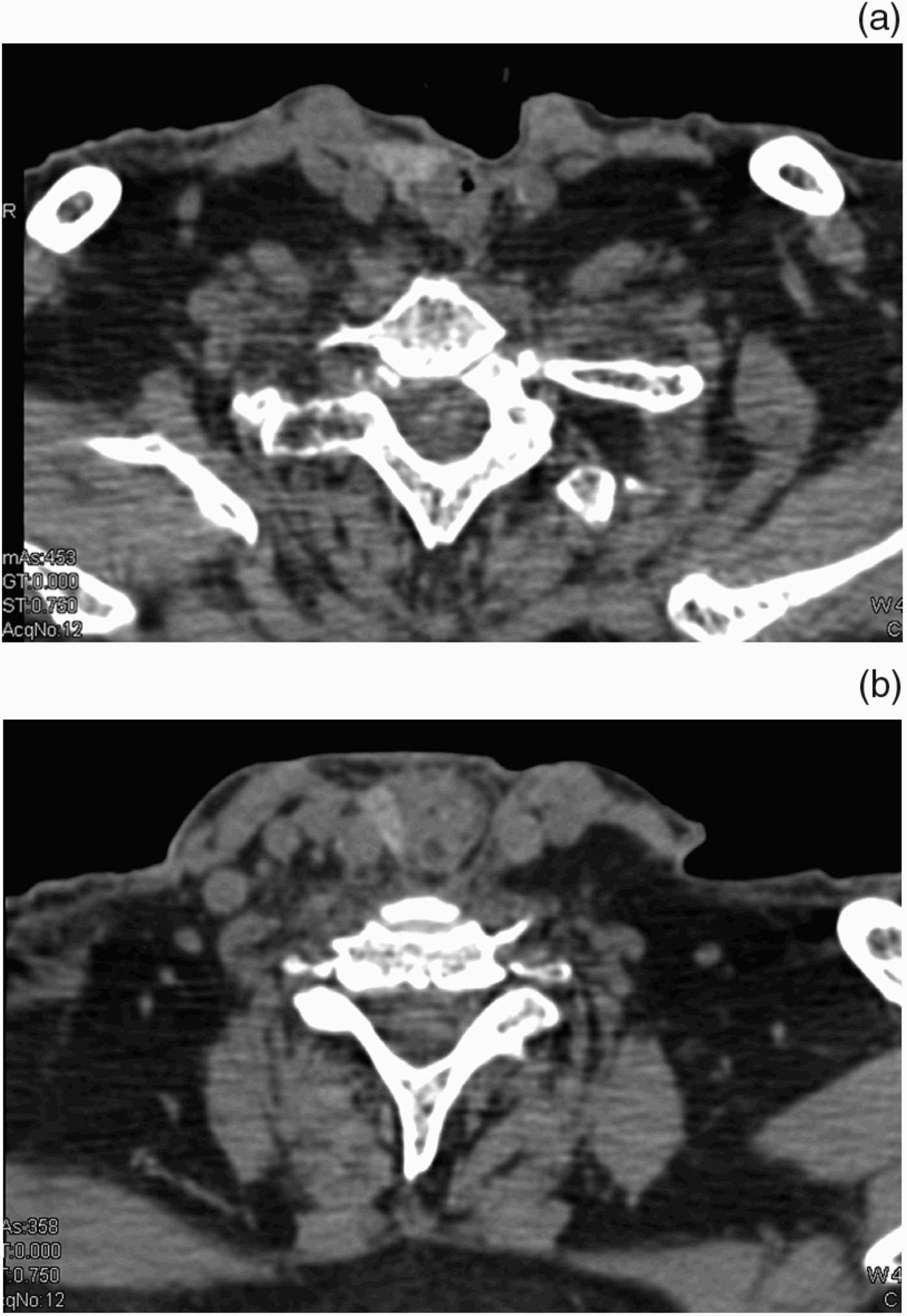
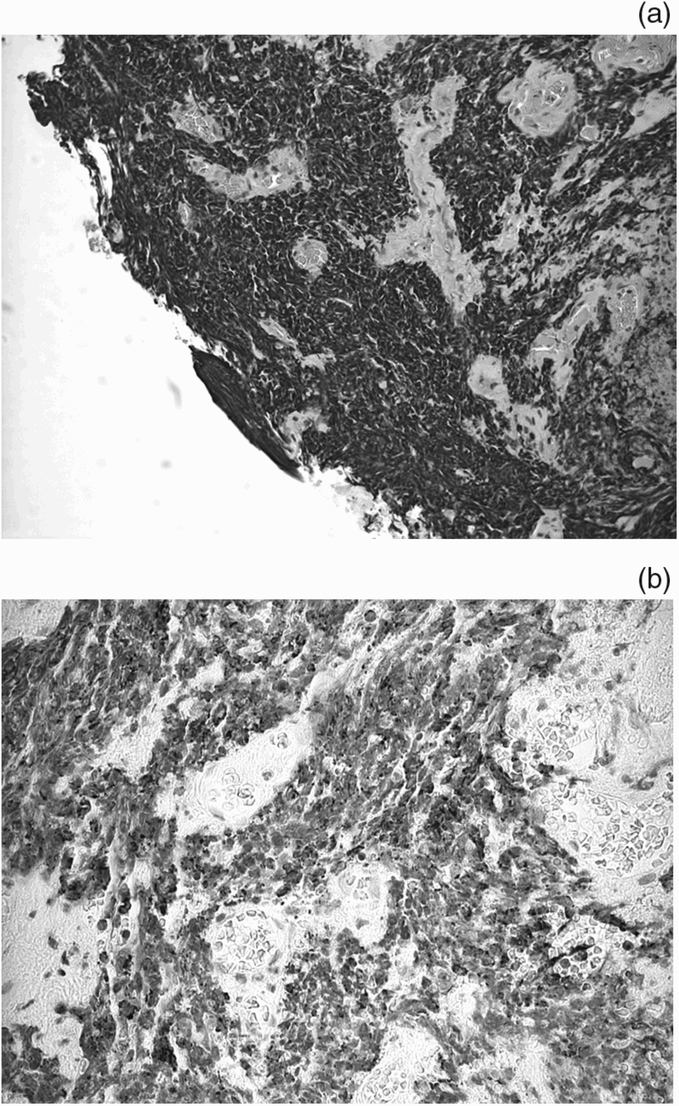
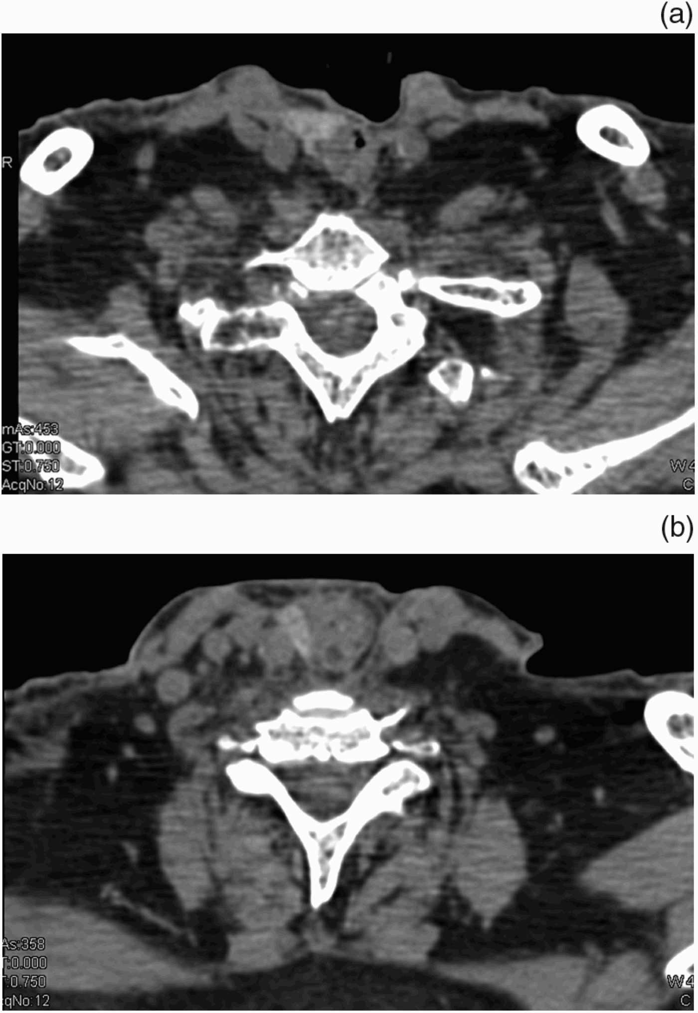
Pharyngoesophagoscopy under general anaesthetic demonstrated a circumferential lesion confined to the neopharynx, causing partial stenosis. A recurrence of the primary laryngeal poorly differentiated squamous cell carcinoma (T4 N0 M0) was suspected. However, histological analysis of biopsy material revealed a neuroendocrine-type small cell carcinoma (Figure 1) (see Appendix 1 for report). Subsequent CT staging to exclude a lung small cell primary showed evidence of scarring from previous tuberculosis but no primary cancer. Staging also detected liver metastases, which were not biopsied. Computed tomography imaging of the tumour clearly demonstrated the tumour's position in the neopharynx (Figure 2).
(a) Photomicrograph of new primary tumour arising in the neopharynx, composed of small, uniform cells with dark, hyperchromatic nuclei and scanty cytoplasm arranged in dense, ‘patternless’ sheets. Note absence of keratinisation as well as characteristic nuclear ‘smudging’ (H&E; 1×20). (b) Photomicrograph showing characteristic perinuclear, dot-like immunopositivity for epithelial cell adhesion marker (CAM5.2) seen in small cell carcinoma.
(a) Axial computed tomography (CT) scan showing tumour at the level of the oropharynx. (b) Axial CT scan demonstrating virtual obstruction of the neopharynx.
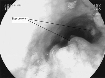
In the two weeks following diagnosis, a per-cutaneous Gastrostomy feeding tube was inserted. Flexible endoscopy showed extension of the lesion into the oropharynx, and the development of skip lesions along the length of the oesophagus (See Figure 3).
Endoscopic image demonstrating the primary lesion and skip lesions within the oesophagus.

Palliative chemo-radiotherapy was commenced.
Treatment was further complicated by the patient requiring continuous ambulatory peritoneal dialysis for chronic renal failure.
Discussion
Small cell carcinoma is an aggressive, locally spreading and distant metastasising tumour most commonly associated with the lung (where it accounts for 25 per cent of pulmonary malignancies).Reference Ferlito and Rinaldo12 It was first documented as a primary lesion outside the lung by Duguid et al. in 1930.Reference Cicin, Karagol, Uzunoglu, Ugun, Usta and Kocak13 Since then, the number of documented cases has been limited, with a reported incidence of 0.1–0.4 per cent.Reference Vrouvas and Ash14 Therefore, no valid conclusions can be drawn on prognosis and management. The commonest site of extra-pulmonary small cell carcinoma is the larynx.Reference Ferlito and Rinaldo12
From a histogenesis point of view, small cell tumours have embryological links with cells derived from the neural crest, namely the amine precursor uptake and decarboxylation (‘APUD’) system.Reference Gould, Memoli, Chejfec and Johannessen15 This may explain the diversity of sites in which extra-pulmonary small cell carcinomas are found. Small cell carcinomas can also be found in association with squamous cell carcinomas and mucin-producing adenocarcinomas, both within and outside the lung. This has prompted an alternative hypothesis that extra-pulmonary small cell carcinomas derive from pluripotent stem cells.Reference Briggs and Ibrahim16 Extra-pulmonary small cell carcinoma sites documented in the literature include: the paranasal sinuses, minor and major salivary glands, hypopharynx, larynx, trachea, thymus, oesophagus, pancreas, stomach, small intestine, colon, rectum, gall bladder, uterine cervix, kidney, urinary bladder, prostate, breast, and skin.Reference Vrouvas and Ash14 Survival rates have been shown to differ depending on the site of disease. Extra-pulmonary small cell carcinoma of the larynx and pharynx has been described as an aggressive disease with a poor prognosis.Reference Weng, Chu, Liu and Chen17 Two- and five-year survival rates of 16 and 5 per cent, respectively, have been quoted.Reference Batsakis, El Naggar and Luna18 Surgery is usually not an option because of the high incidence of metastatic disease, so a combination of chemotherapy and radiotherapy is advocated.
Extra-pulmonary small cell carcinoma has often been misdiagnosed as a secondary metastasis from a pulmonary lesion. In recent times, however, it has become recognised as a clinicopathological entity in its own right.
The paucity of such cases has resulted in many departments managing extra-pulmonary small cell carcinoma in a similar way to pulmonary small cell carcinoma. However, Cicin et al. have demonstrated a significant difference in the outcomes of patients with extra-pulmonary small cell carcinoma, compared with patients with pulmonary small cell cancers.Reference Cicin, Karagol, Uzunoglu, Ugun, Usta and Kocak13 This may influence the choice of therapy.
• In patients with a neopharynx, physiological dysphagia following laryngectomy can mask tumour recurrence
• Neopharyngeal tumour recurrence is commonly due to squamous cell carcinoma
• Primary extra-pulmonary small cell carcinoma of the neopharynx has not previously been reported
• Survival rates for extra-pulmonary small cell carcinoma are dependent on the site of disease
• Combination chemo-radiotherapy, as opposed to surgery, is advocated for the treatment of extra-pulmonary small cell carcinoma of the pharynx
Certainly, the histological classification for both extra-pulmonary and pulmonary small cell carcinoma is the same.19 The diagnosis is based on morphology (i.e. small, uniform cells with hyperchromatic nuclei and sparse cytoplasm arranged in dense, ‘patternless’ sheets with conspicuous nuclear moulding) and immunohistochemistry (i.e. positive staining for at least one neuroendocrine marker, and perinuclear, dot-like positivity for epithelial markers).
Once the diagnosis is clear, chemotherapy (shown to produce a good response in pulmonary small cell carcinoma patients) should be commenced alongside radiotherapy. Radiotherapy has been found to have disappointing results in treating oesophageal small cell carcinoma, and consequently would not be recommended as the sole treatment modality.Reference Medyesy, Wolf, Putnam and Ajani20 Improved outcomes will depend on the development of improved chemotherapy regimens.Reference Weng, Chu, Liu and Chen17
The surgical treatment for neopharyngeal recurrence in patients with total laryngectomy is circumferential pharyngectomy with reconstruction or with gastric pull-up for pharyngoesophagectomy. Yuen et al. studied a series of 292 laryngectomy patients, including 24 with neopharyngeal recurrence.Reference Yuen, Wei, Hui and Ho10 Of these 24 patients, 10 were deemed suitable for salvage surgery with reconstruction. Recurrent tumour was deemed inoperable if there was fixation onto the skull base or prevertebral muscles, or infraclavicular extension, or if the resultant defect would be unreconstructable. Of the 10 operated patients, one died from a carotid blowout, three died of tumour recurrence, two died of a second primary (surviving 122 and 67 months, variously), two died from apparently unrelated causes (at four and 114 months), and the remaining two were disease-free at five and 145 months. Interestingly, averaging these patients' results revealed a mean survival of 76 months after salvage surgery. Although 40 per cent of the patients were tumour-free at three years, no quality of life measures were used to enable demonstration of true benefit from salvage surgery.
Conclusion
Neopharyngeal small cell carcinoma of the neuroendocrine type has not previously been reported. This neoplasm should be managed in the same way as other extra-pulmonary small cell carcinomas occurring within the pharynx or larynx, with a combination of multi-drug chemotherapy and radiotherapy. Surgery has a limited role due to the aggressiveness of the disease and the high chance of local and distant spread at presentation.
Appendix 1. Neopharynx biopsy report
The biopsy comprises four fragments of completely necrotic tissue and two fragments of squamous mucosa infiltrated by a malignant tumour composed of small, uniform cells with small, hyperchromatic nuclei and little cytoplasm, arranged in dense, patternless sheets, which are focally crushed.
There is conspicuous nuclear moulding. Apoptotic bodies as well as mitotic figures are frequent and there is extensive necrosis.
Immunohistochemistry shows strong and diffuse staining for TTF-1 and synaptophysin. CAM5.2 and MNF116 show perinuclear, dot-like positivity. S100, PGP9.5, NSE, CD56, LCA and chromogranin are negative.
Comment: The immunoprofile is that of a small cell carcinoma of the lung. In the absence of dysplasia in the overlying squamous epithelium, this raises the possibility of a metastasis.