





Introduction
Exposure to environmental toxicants prior to and during pregnancy can have significant effects on placental structure and function, and this may have long-term consequences for offspring brain health. Research in mouse models of pregnancy have played an important role in elucidating Developmental Origins of Health and Disease (DOHaD), allowing the study of direct cause-and-effect relationships over the lifetime (e.g., maternal exposure to a toxicant and its effect on offspring brain structure). Reference Heindel1 Per-/polyfluoroalkyl substances (PFAS), sometimes referred to as “forever chemicals,” are a class of organic pollutants used in household products (e.g., textiles, cookware, cosmetics) that have been associated with placental dysfunction in animals and humans and with adverse health outcomes in adulthood. Reference Blake and Fenton2,Reference Aghaei, Steeves, Jobst and Cahill3 Perfluorooctane sulfonic acid (PFOS) and perfluorooctanoic acid (PFOA) are two legacy PFAS contaminants that have been phased out of commercial use because of their link to numerous health issues, Reference Melzer, Rice, Depledge, Henley and Galloway4,Reference Barry, Winquist and Steenland5 including pregnancy complications (e.g., preterm birth Reference Meng, Inoue, Ritz, Olsen and Liew6 and low birth weights. Reference Johnson, Sutton and Atchley7 ) PFOS and PFOA have been shown to bioaccumulate in the placenta and readily cross the placenta in humans, Reference Chen, Yin, Kelly and Liu8 and animals Reference Fenton, Reiner and Nakayama9,Reference Neita, Kiefte, Adams, Mercer, Schneider and Cahill10 accumulating in the offspring. Reference Onishchenko, Fischer and Wan Ibrahim11 This raises concerns about how maternal PFAS exposure impacts offspring health. For example, several groups have studied the impact of maternal exposure to PFAS on offspring bone health, finding negative associations between PFAS concentrations and bone mineral content/density. Reference Højsager, Sigvaldsen and Andersen12,Reference Tao, Zeng and Deng13 Another system that may be vulnerable to PFAS exposure during pregnancy is the developing brain. As described in recent review papers, the neurotoxic effects of PFAS is an active area of investigation with heterogeneous findings, gaps in mechanistic understanding, and variability across different PFAS, exposure windows, and outcomes. Reference Starnes, Rock, Jackson and Belcher14,Reference Bharal, Ruchitha and Kumar15 For example, in mice, while studies are in agreement that early life exposure to PFOS impacts motor activity, the direction of the effect is inconsistent. Reference Fuentes, Vicens, Colomina and Domino16,Reference Johansson, Fredriksson and Eriksson17 Similarly for cognitive function, rats exposed to PFOS during gestation showed impaired spatial memory in one study Reference Wang, Liu, Zhang, Zhao and Quan18 and no significant differences in another. Reference Lau, Thibodeaux and Hanson19 While the evidence base in limited, recent studies in humans have reported sex-specific associations between legacy PFAS levels during pregnancy and offspring brain outcomes, including lower performance IQ and abnormal white matter development measured by magnetic resonance imaging (MRI). Reference Goodman, Till and Green20–Reference Wang, Wen, Huang, Sun, Huang and Wang22
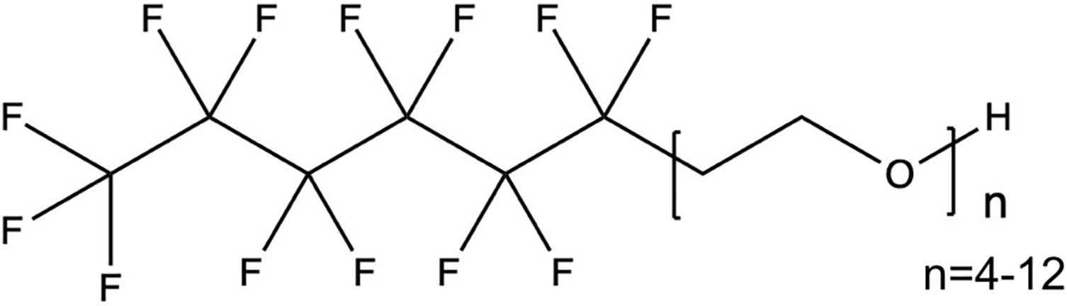
The majority of animal studies examining the developmental effects of PFAS have focused on PFOS and PFOA exposures. Reference Aghaei, Steeves, Jobst and Cahill3 Global restrictions have resulted in the decline of these legacy PFAS Reference Land, de Wit and Bignert23 ; however, new PFAS have been introduced to the market and, while meant to be less bioaccumulative and toxic, may be equally or more harmful. Reference Gomis, Vestergren, Borg and Cousins24 Our group recently identified using mass spectrometry a novel, unregulated class of PFAS, fluorotelomer ethoxylates (FTEOs), in indoor dust and industrial wastewater Reference Steeves, Bissram and Kleywegt25 (Fig. 1). Using high-frequency ultrasound and nuclear magnetic resonance (NMR) metabolomics, we found maternal exposure to FTEOs resulted in developmental toxicity in mice. In the FTEO-exposed group, we observed a decrease in umbilical artery blood flow and evidence of brain sparing, an adaptation to preserve oxygen delivery to the fetal brain and an indicator of fetal distress. Reference Steeves, Hanrahan, Harvey, Jobst and Cahill26 Placental metabolism was also impacted, with decreased relative concentration of asparagine and lysine and increased creatine in the FTEO-exposed placentas compared to controls. Reference Adams, Hanrahan and Kiefte27 The feto-placental weight ratio was also found to decrease with increasing concentration of FTEOs, suggesting a decrease in placental efficiency. Reference Steeves, Hanrahan, Harvey, Jobst and Cahill26,Reference Adams, Hanrahan and Kiefte27 The ability of FTEOs to bioaccumulate and their long-term impact on offspring health, particularly the consequences of exposure on brain development, remain unknown.
Chemical structure of 6:2 fluorotelomer ethoxylates (FTEOs).

In the present study, we use 19F solid-state NMR to determine the bioaccumulation of FTEOs in tissue samples of pregnant mice (maternal liver and the placenta). In addition, we investigated the effect of maternal exposure to FTEOs on early postnatal brain development using three-dimensional high-resolution ex vivo MRI.
Method
Animals
CD-1 female mice (10 ± 2 weeks of age) were purchased from Charles River Laboratories (St. Constant, QC) and bred in-house. Health reports indicated that the mice were free of viral and parasitic pathogens. The mice were singly housed in standard cages and had ad libitum access to food and drinking water. All animal experiments were approved by the Institutional Animal Care Committee at Memorial University of Newfoundland (23-03-LC) and were conducted in accordance with the guidelines established by the Canadian Council on Animal Care.
Experimental design
After evidence of a successful mating (embryonic day 0.5 (E0.5)), the dams were randomly assigned to one of two groups: control (n = 4 for maternal and fetal/placental weights from Steeves et al., Reference Steeves, Hanrahan, Harvey, Jobst and Cahill26 n = 6 for MRI (12 female and 12 male offspring)) or FTEO-exposed (n = 5 for maternal and fetal/placental weights, n = 1 for 19F NMR and n = 5 for MRI (10 female and 10 male offspring)). There were no differences in body weight between the two groups for the CD-1 mice at E0.5. The control group received filtered drinking water (three-phase filtration system at 5, 1 and 0.2 µm) and the exposure group received filtered drinking water containing a commercial mixture of 6:2 FTEOs (Capstone F-30, CAS Number 1640092-35-8, Apollo Scientific, Dredbury, Stockport, UK) (50 mg/L for 19F NMR and 100 ng/L for MRI). The concentration of 50 mg/L was chosen for the 19F NMR experiments to allow comparison to accumulation of PFOA. Reference Neita, Kiefte, Adams, Mercer, Schneider and Cahill10 While similar to other mouse exposure studies, Reference Aghaei, Steeves, Jobst and Cahill3 this concentration does not represent a typical human exposure and, due to the inherent insensitivity of the NMR, was chosen to ensure the resonances are visible in the tissue samples. Drinking water is one of the main routes of human exposure to PFAS and the concentration for the MRI study (100 ng/L) matches the upper end of individual FTEOs in industrial wastewater Reference Steeves, Bissram and Kleywegt25 and is in the range of maximum reported concentrations of other PFAS in drinking water (e.g., fluorotelomer carboxylates = 39 ng/L). Reference Kaboré, Duy and Munoz28 A concentration of 100 ng/L was also chosen based on previous work that showed placental insufficiency and abnormal placental metabolism following exposure and no overt signs of toxicity for the dams (e.g., impacts on weight gain, activity levels, grooming behavior). Reference Steeves, Hanrahan, Harvey, Jobst and Cahill26,Reference Adams, Hanrahan and Kiefte27 For the NMR cohort, the dams were sacrificed at E17.5 (full term is E18.5–19.5) using an intraperitoneal injection of ketamine/xylazine (150 and 10 mg/kg, respectively) and decapitation. Maternal liver and placentas were excised and flash-frozen in liquid nitrogen. Fetal skin samples were collected for fetal sex genotyping. For the MRI cohort, the offspring were weighed on postnatal days (PND) 2, 9, 16, and 23. At weaning (PND23), two females and two males were randomly selected from each litter and sacrificed using a transcardiac perfusion to remove the blood from the brain and infuse an MRI contrast agent (ProHance).
19F magic angle spinning solid-state nuclear magnetic resonance
19F magic angle spinning (MAS) NMR experiments with 1H decoupling were performed using a Bruker NEO 600 MHz spectrometer (1H and 19F Larmor frequencies are 600.29 and 564.80 MHz, respectively) and a 3.2 mm triple-tuned MAS solid-state NMR probe. The sample preparation and scan optimization is described in detail in Neita et al. Reference Neita, Kiefte, Adams, Mercer, Schneider and Cahill10 The tissue types were chosen because of our detection of PFOA in all murine liver samples (n = 9/9) and in 66.7% (n = 8/12) of male placentas. Reference Neita, Kiefte, Adams, Mercer, Schneider and Cahill10 Briefly, frozen samples were thawed and a small piece (∼0.5 cm3) was sectioned and packed into a 3.2 mm zirconium NMR rotor. Spectra were acquired using a single pulse experiment with a 90° pulse length of 4 µs, a recycle delay of 3 s, a spinning speed of 4 kHz and bearing gas temperature of 298 K. The number of scans was 89,753 for the liver sample (time = 75 hours) and 102,400 for the placenta (time = 85 hours). The spectra were processed using MestReNova (version 15.1.0, Mestrelab, Research S.L., Norwich, CT). They are referenced to polytetrafluoroethylene (−126 ppm) and processed with exponential line broadening (100 Hz) and automatic phase correction. No quantification of the FTEOs concentration was performed.
Ex vivo high-resolution magnetic resonance imaging
The transcardiac brain perfusion involved perfusion of 30 mL phosphate buffered saline (PBS), 1 µL/mL heparin and 2 mM ProHance, followed by 30 mL of 4% paraformaldehyde (PFA) and 2 mM ProHance in PBS through the left ventricle. Reference Cahill, Laliberté and Ellegood29 The brain samples were incubated in 4% PFA and 2 mM ProHance overnight at 4°C and then stored in a solution of PBS, 2 mM ProHance, and sodium azide and shipped to the Mouse Imaging Centre at the Hospital for Sick Children (Toronto, Ontario, Canada). Three-dimensional high-resolution ex vivo T2-weighed MRI scans of the whole brain were acquired on a multi-channel 7.0 T Bruker magnet. Using a custom-built solenoid array, multiple brain samples were imaged in parallel using the following scan parameters optimized for grey-white matter contrast: T2-weighted fast spin-echo sequence with a cylindrical k-space acquisition (TR = 30 ms, TE = 12 ms, TEeff = 30 ms, echo train length = 6, four averages, field-of-view = 20.2 mm x 20.2 mm x 25.2 mm, matrix size = 504 × 504 × 630, isotropic resolution = 40 µm3). Images from two female FTEO-exposed, one male FTEO-exposed and two female control mice had to be excluded because of damage during sample preparation.
MRI data analysis
To assess anatomical differences related to maternal FTEO exposure, an automated image registration approach was used. Reference Avants, Tustison and Song30–Reference Nieman, van Eede, Spring, Dazai, Henkelman and Lerch32 An average MR image was created through a series of linear (6 parameters followed by 12 parameters) and nonlinear steps. The changes in brain volume were evaluated for both individual voxels (three-dimensional pixel) and brain structures, providing complementary information about changes in morphometry on the microscopic and macro-/mesoscopic level. Reference Lerch, van der Kouwe and Raznahan33 The image registration provided deformation fields for each brain and the Jacobian determinants that describe the volumetric differences at each voxel. The multiple automatically generated templates (MAGeT) segmentation was used to determine the absolute volumes of 182 brain structures for each image. Reference Chakravarty, Steadman and van Eede34 The method uses the output of the six-parameter linear alignment and multiple labeled templates to accurately segment each structure. As the total brain volume was not different between groups, relative brain volumes were calculated by normalizing to total brain volumes to eliminate the variance from overall brain size.
Statistical analysis
Statistical analysis was performed using the R programming software (www.r-project.org). Data are presented as means and 95% confidence intervals. Maternal body weights were compared using a one-way ANOVA and the fetal and placental weights were compared using a linear mixed effects model with group (control, FTEOs) as the fixed effect and litter as the random effect. The body weight growth curves were fit with a natural spine with three degrees of freedom. A linear mixed effects model was used to determine whether there was an effect of group (control, FTEOs) on the spline coefficients after accounting for the longitudinal study design. For analysis of the neuroanatomical differences from the ex vivo MRI data, we used a linear model at every voxel and for every brain structure with the main effects of group (control, FTEOs) and sex (female, male) and a group-by-sex interaction. Multiple comparisons were controlled for using a 10% threshold for the false discovery rate (FDR). Reference Genovese, Lazar and Nicols35
Results
FTEOs bioaccumulate in the dam and the placenta
There were no differences in maternal, fetal or placental weights between FTEO-exposed and control groups at E17.5 (N = 4−5 litters/group, n = 34−64 fetuses/placentas/group, Supplementary Figure 1). 19F MAS solid-state NMR was used to investigate the bioaccumulation of FTEOs in pregnant mice and the placenta. Fig. 2 shows the 19F MAS NMR spectra for a maternal liver and a male placental tissue sample. The increased 19F signal intensity in the maternal liver demonstrates higher bioaccumulation compared to the placenta.
19F NMR spectra of a (a) maternal liver and (b) placenta tissue sample from a mouse exposed to FTEOs for 17 days.

Maternal exposure to FTEOs does not impact postnatal offspring weights
Both groups consumed the same amount of drinking water throughout gestation and lactation (15 ± 3 mL/day, Supplementary Figure 2). The control and FTEO-exposed dams had a similar body weight at E17.5 and there were no visible impacts of FTEO exposure on the dams (e.g., lethargy, unkempt fur, hunched posture). Analysis of variance of the postnatal growth curves showed no significant differences between the control and FTEO-exposed offspring (N = 5–6 litters/group, n = 50–57 offspring/group, Supplementary Figure 3).
Maternal exposure to FTEOs induces sex-specific focal volume changes in the offspring brain
Analysis of three-dimensional high-resolution ex vivo MR images allowed determination of differences in offspring neuroanatomy that results from indirect exposure to FTEOs throughout gestation and lactation. There were no significant differences between control and FTEO-exposed brains in total brain volume or at the structure-level (N = 5–6 litters/group, n = 8–12 brains/group/sex). Using voxelwise analysis of relative brain volumes, significant focal differences resulting from FTEO exposure were observed across the whole brain (t-statistic = 2.82, 10% FDR, Fig. 3a). Areas of the brain that demonstrated a significant decrease in volume in the FTEO-exposed mice were subregions of the cortex (secondary auditory, secondary motor, temporal association, ectorhinal, entorhinal, secondary visual), cerebral aqueduct, corpus callosum, fimbria, subregions of the hippocampus (CA3 oriens layer and CA3 radialis layer), inferior colliculus, medulla, periaqueductal grey, stria terminalis, and in the grey matter of the cerebellum. Other areas of the brain were significantly larger in the FTEO-exposed mice including the bed nucleus of stria terminalis, cerebellar peduncle, cingulate cortex, dentate gyrus, globus pallidus, internal capsule, lateral septum, midbrain, primary somatosensory cortex, striatum, superior colliculus, and thalamus.
In the control group, several of the brain areas showed the expected sex differences in volume for mice including the thalamus (larger in males Reference Spring, Lerch and Henkelman36 ), cingulate cortex (larger in females Reference Guma, Beauchamp and Liu37 ), dentate gyrus (larger in males Reference Maderia and Lieberman38 ), and secondary auditory cortex (larger in females Reference Guma, Beauchamp and Liu37 ). A significant sex-by-group interaction was observed, demonstrating that many of the neuroanatomical differences between the FTEO-exposed and control offspring were dependent on sex (F-statistic = 8.16, 10% FDR, Fig. 3b). For example, compared to controls, the striatum (Fig. 3c), thalamus (Fig. 3d) and dentate gyrus (Fig. 3f) showed a volume increase in the female FTEO-exposed offspring and not the males, while the cingulate cortex (Fig. 3e), secondary auditory cortex (Fig. 3g), and medulla (Fig. 3j) were decreased in only the female FTEO-exposed offspring. In contrast, only the male FTEO-exposed offspring showed an increase in the volume of the inferior colliculus compared to controls and there was no difference in the females (Fig. 3i). The volume of the superior colliculus was impacted in both sexes following FTEO exposure, with a decrease in the female FTEO-exposed and an increase in the male FTEO-exposed compared to controls (Fig. 3h).
(a) Representative coronal MR images showing anatomical differences in the brains of control and FTEO-exposed offspring. The images are overlaid with color maps that indicate regions of relative volume that are significantly larger (hot colors) and significantly smaller (cool colors) in FTEO-exposed mice compared to controls (t-statistic = 2.82, 10% FDR). (b) Representative images showing a significant sex-by-group interaction (F-statistic = 8.16, 10% FDR). (c-j) Relative volume of voxels (indicated by asterisk in B) to demonstrate sex differences. The vertical axis shows the value of the relative Jacobian determinant. Data shown as means and 95% confidence intervals. N = 5–6 litters, n = 8–12 mice/group/sex. A2, secondary auditory cortex; BNST, bed nucleus of stria terminalis; CA3Or, CA3 oriens layer; CA3Rad, CA3 radialis layer; ca, cerebral aqueduct, CbGM, cerebellar grey matter, CC, cingulate cortex; cc, corpus callosum; cp, cerebellar peduncle; DG, dentate gyrus; EctC, ectorhinal cortex; EntC, entorhinal cortex; fi, fimbria; GP, globus pallidus; IC, inferior colliculus; ic, internal capsule; ls, lateral septum; Med, medulla; MB, midbrain; M2, secondary motor cortex; NAc, nucleus accumbens; PAG, periaqueductal grey; S1, primary somatosensory cortex; st, stria terminalis; Str, striatum; SC, superior colliculus; TA, temporal association cortex; Th, thalamus; V2, secondary visual cortex.

Figure 3. Long description
The brain areas that were impacted included the secondary auditory cortex, bed nucleus of stria terminalis, CA3 oriens layer in the hippocampus, CA3 radialis layer in the hippocampus, cerebral aqueduct, cerebellar grey matter, cingulate cortex, corpus callosum, cerebellar peduncle, dentate gyrus, ectorhinal cortex, entorhinal cortex, fimbria, globus pallidus, inferior colliculus, internal capsule, lateral septum, midbrain, secondary motor cortex, nucleus accumbens, periaqueductal grey, primary somatosensory cortex, stria terminalis, striatum, superior colliculus, temporal association cortex, thalamus, secondary visual cortex
Discussion
19F solid-state NMR and 1H high-resolution ex vivo MRI revealed bioaccumulation of FTEOs in maternal and placental tissue and developmental neurotoxicity following maternal exposure to FTEOs. This bioaccumulation potential and toxicity, combined with the previously documented persistence in the environment, Reference Steeves, Bissram and Kleywegt25 provides evidence that FTEOs can be harmful to human health and should be considered for government regulation. Consistent with the DOHaD hypothesis, the results of this study show that indirect exposure to FTEOs in early life can impact brain structure in adolescence. This may be partially explained by FTEO-associated placental dysfunction, Reference Steeves, Hanrahan, Harvey, Jobst and Cahill26 a known cause of adverse long-term neurodevelopmental outcomes. Reference Lodefalk, Chelslín, Patriksson Karlsson and Hansson39 While the fetal brain sparing phenomenon observed following maternal FTEO exposure is neuroprotective, Reference Steeves, Hanrahan, Harvey, Jobst and Cahill26 it has been associated with neurodevelopmental injury in some cases Reference Benítez-Martín, Marín-Clavijo, Blanco-Elena, Jiménez-López and González-Mesa40 and therefore may not fully protect the FTEO-exposed offspring.
In the 19F NMR experiments, the higher detection of FTEOs in the maternal liver compared to the placenta is consistent with previous biodistribution studies of PFOA in humans and animals using mass spectrometry Reference Kim, Heo, Lee, Hwang, Lee and Cho41,Reference Nielsen, Fischer, Leth and Grandjean42 and our recent work using 19F solid-state NMR. Reference Neita, Kiefte, Adams, Mercer, Schneider and Cahill10 The liver serving as a target organ is not surprising since PFAS are known to bind to fatty acid-binding proteins in the liver. Reference Golden-Mason, Salomon and Matsuba43 Similarly, legacy PFAS (PFOS, PFOA, perfluorohexane sulfonic acid (PFHxS)) have been shown to bioaccumulate in the placenta. Reference Chen, Yin, Kelly and Liu8,Reference Neita, Kiefte, Adams, Mercer, Schneider and Cahill10,Reference Mamsen, Björvang and Mucs44 However, the FTEO concentrations in both the liver and placenta are much lower than PFOA concentrations measured by 19F solid-state NMR in Neita et al., Reference Neita, Kiefte, Adams, Mercer, Schneider and Cahill10 suggesting FTEOs are not as bioaccumulative as PFOA. This may be due to the differences in chemical structure (e.g., alkyl chain length, end group) and adds to our understanding of how the chemical structure of PFAS impacts bioaccumulation potential.
As a measure of systemic toxicity, we measured offspring body weights. The lack of an impact of FTEO exposure on prenatal and postnatal body weights is consistent with previous work, where there was no difference in fetal weights between the FTEO-exposed and control groups at E17.5. Reference Steeves, Hanrahan, Harvey, Jobst and Cahill26,Reference Adams, Hanrahan and Kiefte27 While prenatal PFAS has been associated with abnormal postnatal growth trajectories, Reference Kadawathagedara, de Lauzon-Guillain and Botton45 the exposure concentration used in this study (100 ng/L) may be too low to observe an effect on body weight.
Next, we used structural MRI to determine the neurotoxicity of maternal FTEO exposure. Despite the normal body growth trajectory, we found focal changes in the FTEO-exposed offspring brain in grey and white matter regions that are involved in motor function, hearing, learning and memory, motivation, waste removal, sensory processing, and physiological functions. These changes are consistent with a recent review article observing that impairments to motor function, and learning and memory are the most common functional deficits resulting from PFAS exposure. Reference Brown-Leung and Cannon46 In humans, exposure to PFAS has been also associated with hearing impairment. Reference Li47 In a human birth cohort study, prenatal PFAS exposure (perfluorononanoic acid (PFNA), PFOA, PFHxS) was associated with specific changes in the brain of the offspring (age 5 years) including in the corpus callosum and hypothalamus. Reference Barron, Dickens and Tuulari48 Two recent human studies reported associations between maternal PFAS concentrations (PFOS, PFHxS, perfluorodecanoic acid (PFDeA), perfluoroundecanoic acid (PFUA), perfluorododecanoic acid (PFDoA)) and white matter microstructure. Reference England-Mason, Reardon and Reynolds21,Reference Wang, Wen, Huang, Sun, Huang and Wang22 A recent study of replacement short chain PFAS (GenX and perfluorobutanoic acid (PFBA)) reported that maternal exposure in rats resulted in neuroinflammation and impaired cognitive function in the offspring. Reference Lorenzini, Moretti and Zanardello49 These neurological effects may be the result of abnormal placental and fetal development or of direct crossing of the blood-brain barrier, allowing PFAS to interfere with neurons and synapses or cause oxidative stress and inflammation. Reference Starnes, Rock, Jackson and Belcher14,Reference Iqubal, Ahmed, Ahmad, Ranjan Sahoo, Iqubal and Haque50,Reference Lagostena, Magnelli, Rotondo and Dondero51 More work needs to be done to replicate these findings and determine the severity of the neurotoxicity of different classes of PFAS.
Our study revealed that the impact of maternal exposure to FTEOs on neuroanatomy was dependent on sex. PFAS are known to be endocrine disruptors, interfering with thyroid and steroid hormones. While it is unclear what is causing the sex differences observed in this study, hormones may play a role. The majority of the brain areas with significant sex-dependent effects showed changes in female FTEO-exposed offspring compared to control offspring and not in males, suggesting females may be more vulnerable to the neurotoxic effects of FTEOs. Sex differences in the study of prenatal exposure to PFAS have been reported, including increased vulnerability of female offspring to neurodevelopmental effects. Reference Goudarzi, Nakajima and Ikeno52,Reference Pang, Wang and Gao53 However, it is important to note that several studies have reported increased susceptibility in males or no sex differences following PFAS exposure and this remains an ongoing field of study.
The present study has several limitations. We only focused on exposure to a single class of PFAS (FTEOs), while biomonitoring data shows that humans are typically exposed to multiple PFAS. Future studies should address this by assessing the effects of real-world exposure to a mixture of PFAS. The use of a single exposure concentration in the current study limits the interpretation of dose–response relationships and prevents the assessment of threshold effects. Another limitation is the delivery of the FTEOs via drinking water. FTEOs have not yet been detected in treated drinking water (only in wastewater); inhalation of indoor dust may be the more common route to exposure. While our previous work demonstrated the utility of 19F solid-state NMR to detect the known accumulation of PFOA in tissue, Reference Neita, Kiefte, Adams, Mercer, Schneider and Cahill10 we do not have a positive control for comparing the degree of neurotoxicity using MRI. Finally, the use of ex vivo structural MRI does not allow monitoring of the developmental trajectory within each mouse, with the present study limited to a single adolescent timepoint, and also lacks the spatial resolution to detect changes on the cellular level. Future studies should investigate the impact of the neuroanatomical differences on behavior (e.g., tests of motor and cognitive function), determine the brain outcomes into adulthood, and explore intergenerational effects.
In summary, maternal murine exposure to FTEOs, a new class of replacement PFAS, resulted in bioaccumulation in a maternal liver and placenta tissue sample and resulted in focal changes in neuroanatomy that differed between females and males. Currently, there is limited information available about the bioaccumulation potential of replacement PFAS. This is important for predicting organ-specific toxicity. The persistence, bioaccumulation potential, and toxicity of FTEOs raises significant concerns about its impact on brain health. As shown with legacy PFAS, Reference Mariussen54 the neurotoxic effects of FTEOs may be more pronounced when they occur during early life, representing a critical window for protection. This work revealed sex- and region-specific areas of the developing brain that are susceptible to the toxicological effects of FTEOs. This animal neurotoxicology work motivates future studies to understand the long-term consequences of maternal FTEO exposure on other organ systems (e.g., skeletal system) in the mouse and on offspring health in humans.
Supplementary material
The supplementary material for this article can be found at https://doi.org/10.1017/S2040174426100804.
Acknowledgements
None.
Financial support
This work was supported by the Canadian Institutes of Health Research (PJT-206067) and a Natural Science and Engineering Council of Canada Discovery Grant.
Competing interests
None.
Ethical standard
The authors assert that all procedures contributing to this work comply with the ethical standards of the relevant national guidelines on the care and use of laboratory animals (The Canadian Council on Animal Care) and has been approved by the institutional committee (Animal Care Committee at Memorial University of Newfoundland).


 Open access

