INTRODUCTION
Blastocystis is one of the most frequently encountered protozoan parasites reported in humans (Amin, 2002; Windsor et al. 2002). Although Blastocystis has previously been detected in various hosts from Australia, including humans, cats, dogs and chickens (Duda et al. 1998; Lee and Stenzel, 1999; Hellard et al. 2000), they were not genetically characterized. Moreover, Australian native fauna had not previously been screened for Blastocystis.
There are several methods used to detect Blastocystis infections, which include wet smears, concentration methods and in vitro amplification which was recently shown to be the most sensitive method to detect Blastocystis from faecal samples (Zaman and Khan, 1994; Leelayoova et al. 2002; Suresh and Smith, 2004; Termmathurapoj et al. 2004). However, due to the variable morphological forms that Blastocystis exhibits, it is impossible to distinguish among different species and subspecies of Blastocystis solely based on morphology without the use of molecular techniques (Yoshikawa et al. 2004b). It has been reported that the PCR detection of Blastocystis directly from faecal specimens is rather insensitive (Termmathurapoj et al. 2004). Hence, in vitro propagation is still used widely to facilitate molecular characterization.
Recent molecular studies have shown that Blastocystis displays considerable genetic heterogeneity in conserved genes, such as the small subunit of nuclear ribosomal RNA gene (SSU) and the elongation factor-1α gene (Ho et al. 2000; Arisue et al. 2003; Yoshikawa et al. 2003, 2004a,c; Abe et al. 2003a,b; Noel et al. 2003, 2005; Abe, 2004). Studies based on the SSU rRNA gene have revealed the presence of 7 groups or subtypes of Blastocystis displaying low host specificity. It is possible that these groups or subtypes correspond to different species of Blastocystis (Noel et al. 2005).
The aims of the present study were to develop a reliable PCR-based method for the detection and characterization of Blastocystis directly from faeces and to compare this method to in vitro culture for the diagnosis of Blastocystis infections. Moreover, we aimed to genetically characterize isolates of Blastocystis from a variety of animal hosts in Australia and Thailand, including isolates from animals and humans living in a localized endemic focus of parasitic infection, in order to explore the zoonotic potential of this parasite.
MATERIALS AND METHODS
Sampling strategy
Fresh faecal samples were obtained from these species of Australian native small marsupials in Julimar (100 km from Perth, Western Australia): chuditch (Dasyurus geoffroii), woylie (Bettongia penicillata), quenda (Isoodon obesulus) and brushtailed possums (Trichosurus vulpecula). Samples were also collected from the following captive primates and their human handlers (zoo-keepers) from the Perth Zoo, Western Australia: Hamadryas baboon (Papio hamadryas hamadryas), black and white ruffed lemur (Varecia variegatus), crested macaque (Macaca nigra nigra), colobus (Colobus guereza), orang utan (Pongo pygmaeus abelii), ring tailed lemur (Lemur catta), spider monkey (Ateles geoffroyi), vervet (Cercopithecus aethiops) and white cheeked gibbon (Nomascus leucogenys). The samples were screened for Blastocystis using both PCR directly from faecal samples, as well as PCR following in vitro propagation (see Table 1) and formed the basis for the comparative study.

In addition, faecal samples were collected from a tiger (Panthera tigris sumatrae), rhinoceros (Ceratotherium simum simum), otter and fishing cat (Prionailurus viverrinus) from the Perth Zoo. Faecal samples from domestic cats and dogs were collected from veterinary clinics and animal refuges and from Aboriginal communities in the central desert in Western Australia. Blastocystis-positive human faecal samples were obtained from a medical diagnostic laboratory in Perth, Western Australia. Two human and 3 dog faecal samples for Blastocystis were also collected from a localized parasite endemic community 150 km east of Bangkok, Thailand. The aforementioned samples were fixed in 20% dimethylsulfoxide (DMSO) and subjected to PCR only.
In vitro cultivation
Fifty mg of fresh faecal material was inoculated into a sterile screw-top tube containing 3 ml of Jones' medium supplemented with 10% horse serum (Jones, 1946; Suresh and Smith, 2004). Each sample was cultured in duplicate and incubated at 37 °C for 24–48 h. The cultures were examined for the presence of various morphological forms of Blastocystis using light microscopy at 400× magnification.
Prior to DNA extraction, 1 of the duplicate cultures was pelleted via centrifugation at 400 g for 10 min and the supernatant discarded. The resultant pellet was stored at −20 °C until required.
Blastocystis purification
The remaining duplicate culture was purified using the Blastocystis purification method as previously described (Hoevers et al. 2000; Snowden et al. 2000) in order to reduce bacterial contamination and to concentrate Blastocystis organisms. The other duplicate culture was pelleted via centrifugation at 400 g for 10 min at 22 °C. The supernatant was discarded and the pellet was resuspended in 3 ml of phosphate-buffered saline (PBS, pH 7·4). This suspension was overlaid on to a Ficoll-Paque column and centrifuged at 2000 g for 20 min at 22 °C (Suresh and Smith, 2004). Blastocystis separated into a band approximately 1 cm from the surface. This layer was collected and resuspended in 8 ml of PBS and centrifuged at 500 g for 5 min, which was repeated 6 times. The resultant pellet was resuspended in 1 ml of PBS and centrifuged at 500 g for 5 min. The resultant pellet was stored at −20 °C until required.
DNA extraction
DNA was extracted from faeces fixed in 20% DMSO, as well as from pelleted and purified cultures of Blastocystis using QIAamp DNA Stool Mini Kit (Qiagen, Germany). The manufacturer's protocol was used, with the following exceptions. (i) Faecal material fixed in 20% DMSO was washed 3 times with distilled water prior to extraction. (ii) The faecal material was suspended in 1·4 ml of ATL lysis buffer as specified by the manufacturer's protocol (Qiagen, Germany) and ruptured by 3–5 freeze-thaw cycles in liquid nitrogen and 95 °C water bath, respectively. (iii) The DNA was eluted from the matrix with 50 or 100 μl of AE elution buffer (Qiagen, Germany).
PCR amplification
A nested PCR reaction was used to amplify an approximately 1100 bp region of the SSU rDNA. The primary PCR utilized previously published forward and reverse primers (RD3, 5′-GGG ATC CTG ATC CTT CCG CAG GTT CAC CTA C-3′; RD5, 5′-GGA AGC TTA TCT GGT TGA TCC TGC CAG TA-3′) for PCR amplification under the conditions described by Clark (1997). The secondary PCR utilized previously published forward and reverse primers (forward, 5′-GGA GGT AGT GAC AAT AAA TC-3′; reverse, 5′-CGT TCA TGA TGA ACA ATT AC-3′) under the conditions described by Bohm-Gloning et al. (1997). The PCR was carried out in 0·2 ml tubes in a Perkin Elmer GeneAmp 2400 thermocycler.
Sequencing and phylogenetic analysis
Bands representing amplified PCR products were excised from a gel and purified using the UltraClean™ GelSpin DNA Purification Kit (MO BIO Laboratories, Inc.). Manufacturer's kit protocols were followed, except that DNA was eluted using 30 μl of ultrapure PCR water and incubated at room temperature for 10 min prior to centrifugation at 10000 g for 30 s. The PCR products were sequenced in both directions using an ABI 3730 capillary sequencer. Sequences were analysed using SeqEd v 1.03 (Applied Biosystems) and compared with previously published sequences from GenBank™ using the BLAST 2.2.9 program (http://www.ncbi.nlm.nih.gov/blast).
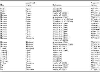
Phylogenetic analysis was conducted using sequence data for a region of the SSU rDNA (~850 bp). Sequences were aligned using the program CLUSTAL W (Thompson et al. 1994) and then manually adjusted. Phylogenetic analysis of sequence data from Blastocystis isolates from this study as well as 26 other Blastocystis isolates retrieved from GenBank™ (Table 2) was carried out using MEGA v3.1 (Kumar et al. 2004). (http://www.megasoftware.net). Distance-based analysis was undertaken using Kimura-2-parameter and the tree was conducted using the UPGMA algorithm. Bootstrap values were calculated by the analysis of 1050 replicates from the UPGMA tree. Proteromonas lacertae was used as the outgroup.

RESULTS
Detection of Blastocystis using in vitro amplification
From a total number of 64 samples subjected to in vitro propagation, 12 (19%) were positive for Blastocystis (Table 1). The dominant morphological form in the cultures was the vacuolar form, with some cultures containing a few granular forms.
The use of the Blastocystis purification method
The Blastocystis purification method did not affect or improve the PCR amplification from DNA from in vitro cultures. The amplicons produced from both purified and unpurified cultures were of a similar abundance, yielding more than the required amount of DNA for sequencing purposes.
Detection of Blastocystis directly from the faeces using PCR
From a total number of 107 samples screened for Blastocystis using PCR directly from the faeces, 37 (35%) were positive for the organism. From the subset of 64 samples, which were also subjected to in vitro propagation, 27 (42%) samples were shown to be positive for Blastocystis (Table 1).
Phylogenetic analysis
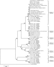
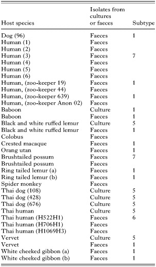
DNA sequences obtained from 22 Blastocystis isolates (Table 3) were included in the phylogenetic analysis. The rooted UPGMA tree identified 7 clades, which corresponded to the 7 known groups/subtypes identified in previous molecular studies (Arisue et al. 2003; Abe et al. 2003a,b; Noel et al. 2003, 2005). There was strong bootstrap support for the clustering of subtypes 2 and 4 to the exclusion of subtypes 1, 3, 5, 6 and 7, but only moderate support for the delineation of subtypes 1, 5 and 6 from subtypes 3 and 7, as well as subtype 2 from 4 (see Fig. 1). With the exception of subtypes 1 and 5, which clustered together, the other subtypes were clearly separated from each other with strong bootstrap support.

Fig. 1. UPGMA tree displaying the relationships among Blastocystis isolates, inferred by distance based analysis of SSU rDNA sequence data using Kimura's-2-parameter distance estimates. Some sequences used for comparison were from GenBank™ Scale bar shows 0·2 substitutions (corrected) per base pair.

All isolates of Blastocystis isolated from primates and their human handlers at the Perth Zoo were placed within subtypes 1 or 5, as were 3 dog and 1 human isolate from Thailand, and 1 dog isolate from Australia. The sequences of most primate isolates of Blastocystis were identical to each other and placed in subtype 1. However, isolates from the in vitro cultures (from baboon, vervet and black and white ruffed lemur) were not identical to those amplified directly from the faeces of the baboon, black and white ruffed lemur and vervet. Analyses of the sequence data representing the isolates amplified directly from faecal samples revealed the presence of mixed peaks in the sequencing chromatograms, whereas no mixed peaks were evident in the sequencing chromatograms from isolates obtained by in vitro cultivation. A Blastocystis isolate from 1 of the zoo-keepers (zoo-keeper (19) faeces) was 99·6% similar in sequence compared with those from the primates (baboon culture and white cheeked gibbon faeces (a)) at the Perth Zoo. A single Blastocystis isolate from a Thai human was shown to be 100% similar to an isolate from a dog living in the same community. Another human isolate from Thailand was placed within subtype 6. Blastocystis sequences from a possum and human in Australia were also shown to be 100% similar to each other and were placed in subtype 7.
DISCUSSION
Comparing PCR detection directly from the faeces to in vitro propagation for the detection of Blastocystis
Prior to this study, in vitro propagation was considered to be the most sensitive method for the detection of Blastocystis infections (Leelayoova et al. 2002; Suresh and Smith, 2004). An advantage of the in vitro culture method is that low numbers of Blastocystis can be cultured for use in other (e.g., genotypic and/or phenotypic) tests (Suresh and Smith, 2004). A previous study comparing detection methods for the diagnosis of Blastocystis infections revealed that out of 23 Blastocystis positive samples, 98% and 52% were positive using in vitro culture and PCR detection directly from the faeces, respectively (Termmathurapoj et al. 2004). In the present study, 19% and 42% of samples were shown to be positive for Blastocystis using in vitro amplification and PCR detection directly from the faeces, respectively. This result suggests that the DNA extraction method used in the present study was more effective compared with the method used by Termmathurapoj et al. (2004), the PCR-based method used in this study for the detection of Blastocystis infections is more sensitive compared to in vitro amplification and that in vitro cultivation does not need to be performed to achieve increased sensitivity of molecular tests.
This is the first study to detect Blastocystis in Australian native marsupials. It should be noted that none of these samples were positive by culture, and some were positive by PCR. It may be possible that some isolates require longer than 48 h for the establishment of growth in vitro, as growth in some cultures may take up to 72 h of incubation before parasite stages can be detected, with subculturing into fresh medium every 48–72 h. In some studies, subculturing is performed up to 5 times before a sample is considered to be negative for the presence of Blastocystis (Yoshikawa et al. 2004c).
In previous studies using Jones' medium supplemented with 10% horse serum (Salim et al. 1999; Leelayoova et al. 2002; Suresh and Smith, 2004; Termmathurapoj et al. 2004; Yakoob et al. 2004; Yoshikawa et al. 2004c), only human faecal samples were screened for Blastocystis. However, several different types of media to cultivate Blastocystis have been reported, and it has been suggested that the usefulness of cultivation may depend on the reagents and protocols employed (Zaman and Khan, 1994; Leelayoova et al. 2002). Perhaps an alternative to Jones' medium should be evaluated for the detection of Blastocystis in non-human hosts, since Blastocystis is susceptible to changes in environmental conditions (Stenzel and Boreham, 1996; Leelayoova et al. 2002; Tan, 2004). Other types of medium, such as diphasic-agar slant medium have been effective for propagating Blastocystis from cattle, pigs and chickens (Yoshikawa et al. 1996, 1998; Abe et al. 2003a,b,c), and should be evaluated (along with other types of medium) in comparative diagnostic studies.
This study has shown that it is possible to detect Blastocystis directly from faeces solely using molecular tests with a greater sensitivity compared with in vitro propagation. A clear advantage of using molecular techniques to detect Blastocystis directly from the faeces for the diagnosis of Blastocystis infections is that the method is not dependent upon factors such as animal host species, viability of Blastocystis organisms, and the various reagents and protocols used in in vitro propagation. Another advantage of using molecular techniques compared with conventional methods is that it is less time consuming, and can provide more information in terms of genetic variability and determining zoonotic relationships (Traub et al. 2005).
Preferential growth of Blastocystis in vitro
Previous molecular characterization studies of Blastocystis have only analysed Blastocystis isolates amplified in vitro (Ho et al. 2000; Arisue et al. 2003; Noel et al. 2003, 2005; Yoshikawa et al. 2004c). The present study was the first to compare isolates obtained from in vitro cultures and those amplified directly from the faeces by the PCR. According to the phylogenetic tree constructed, in vitro isolates of vervet and black and white ruffed lemur origin belonged to subtype 5, whereas the isolates amplified directly from the faeces from these hosts belonged to subtype 1 (black and white ruffed lemur faeces and vervet faeces). The other in vitro isolate (from baboon) was identical to an isolate from a white cheeked gibbon (white cheeked gibbon faeces (a)) but was not identical to the baboon isolate amplified directly from the faeces (baboon faeces). These findings indicate that not only did these primates have mixed infections, which were evident due to the mixed peaks in the sequencing chromatograms for the isolates amplified directly from the faeces (there were no mixed peaks in the sequencing chromatograms from in vitro isolates), but also that preferential growth of a particular isolate can occur in vitro if the host is infected with more than one isolate. In the present study, Blastocystis subtype 5 overgrew subtype 1 in vitro.
Although in vitro propagation is considered one of the most sensitive methods currently available for the detection of Blastocystis infections, the organisms grown in culture may not accurately represent the parasite population sampled. In the early studies on genetic variability in Giardia, Assemblage A was considered to be the most commonly isolated genotype. However, it was later shown that the in vitro culturing methods used were preferentially selecting for Assemblage A genotypes (Thompson and Monis, 2004). As with more recent studies on Giardia, the ability to genetically characterize isolates of Blastocystis directly from faeces or environmental samples will provide a way of avoiding the potential selectivity imposed by laboratory amplification in vitro.
Zoonotic potential
In the present study, all 4 faecal samples collected from the zoo-keepers were positive for Blastocystis. Two of these samples were sequenced and characterized successfully. Blastocystis isolated from the zoo-keepers at the Perth Zoo were found to be similar to some isolates from the primates, which clustered into the same subtype (subtype 1). It has been reported that the close contact between animals in a zoo environment can facilitate the transmission of Blastocystis, and intimate association between humans and animals in such environments (Salim et al. 1999; Abe et al. 2002).
Similarly, a dog isolate (dog 676 culture) and a human isolate (human culture) from the same village in Thailand were identical genetically and belonged to subtype 5. The present study is the first to report a human isolate (human (Th522H1) faeces) belonging to subtype 6, which has been considered to be specific to pig and cattle isolates (Noel et al. 2005). These findings may be reflective of the interactions between humans and animals in rural Thailand, and provide evidence suggesting zoonotic transmission.
In addition, a human isolate (human (3) faeces) and a brushtailed possum isolate (brushtailed possum faeces) from the present study were genetically identical to each other, and to rat and human isolates from Singapore and Japan, respectively. The findings from the present study agree with previous studies, indicating the presence of 7 distinct subtypes which are not host specific, comprising isolates from humans and various other animals (Yoshikawa et al. 1996, 1998; Clark, 1997; Snowden et al. 2000; Arisue et al. 2003; Noel et al. 2005).
In conclusion, the present study demonstrated that PCR-based methods were more sensitive than in vitro propagation for the detection of Blastocystis. Comparisons between Blastocystis isolates from in vitro cultures and those amplified directly from faeces revealed that preferential amplification can occur in vitro. The present study also identified a number of isolates which may be of zoonotic significance.