Heterotaxy syndromes may be associated with various venous anomalies, including Abernethy malformation, which is an anomalous porto-systemic communication. The presence of this anomaly in a post-Kawashima patient can have detrimental effects on the univentricular circuit by causing pulmonary artery hypertension, or by acting as a veno-venous collateral causing desaturation.
Case report
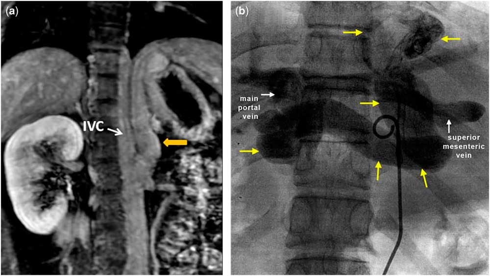
A 17-year-old girl, diagnosed to have situs ambiguous, mesocardia, hypoplastic right ventricle, ventricular septal defect, and severe pulmonary stenosis, had undergone Kawashima operation at 7 years of age. Her oxygen saturation after the surgery was 86%. She presented now with deepening cyanosis and effort intolerance. A catheterisation study showed mean superior caval vein and pulmonary artery pressures of 16 mmHg. There were no significant pulmonary arterio-venous malformations. A trans-thoracic echocardiogram showed a large anomalous venous channel (measuring 18 mm at the narrowest part), from the portal system joining the inferior caval vein, which was confirmed by Contrast CT angiogram. A diagnosis of type II Abernethy malformation was made and she was taken up for occlusion of the malformation. Her serum ammonia and other liver function tests were normal. Inferior caval vein angiogram showed the anomalous vein arising at the level of the left renal vein and coursing up to the left of the midline liver, before taking a tortuous turn to join the main portal vein (Fig 1a, Supplementary video 1). The vein was joined by the right and left renal veins at the lower end and by a branch of the superior mesenteric vein at the upper end (Fig 1b, Supplementary video 2). Portal venous pressure was 16 mmHg, which on balloon occlusion of the anomalous vein decreased to 13 mmHg, and the oxygen saturation also improved from 74 to 91%. The vein was closed with a 22-mm Amplatzer vascular plug II (St. Jude Medical, now Abbott, Golden Valley, Minnesota, United States of America) (Supplementary video 3) midway between the entry of renal veins and superior mesenteric veins into the channel. Post-occlusion angiogram showed no flow into the portal venous system from inferior caval vein.
( a ) Abdomen contrast CT showing the origin of the anomalous vein from the inferior caval vein (IVC) and its upward and leftward course towards the midline liver. ( b ) Selective venogram of the Abernethy malformation showing the tortuous course of the vein (yellow arrows) and its termination by joining the main portal vein (white arrow). A branch of the superior mesenteric vein (white arrow) is seen joining the anomalous vein.

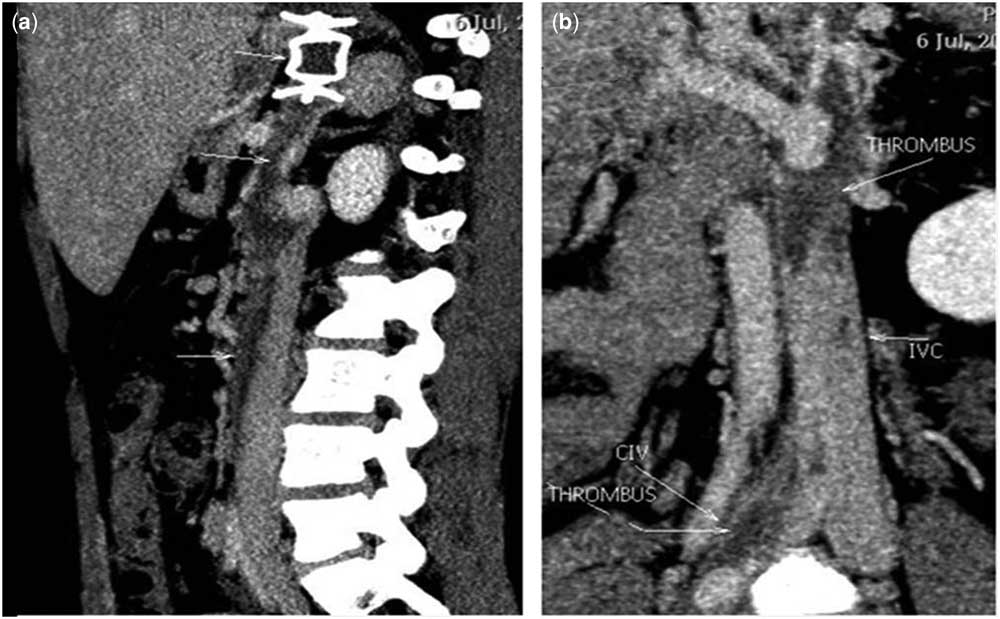
She presented a week later with severe pain in the right thigh, and venous Doppler ultrasonogram showed extensive thrombus extending from the anomalous channel distal to the plug into the inferior caval vein and to both external iliac and common femoral veins. Thrombus was also extending from the vascular plug into the portal system. The renal veins were free of thrombus. She was managed conservatively with anti-coagulation – heparin infusion – and started on a single anti-platelet drug and oral anti-coagulant. Contrast CT venogram taken 1 week later showed significant resolution of thrombus (Fig 2a and b). At a duration of 2 months after the procedure, venous Doppler ultrasound of the lower limb showed good resolution of the thrombus.
( a ) CT contrast venogram showing thrombosis in the anomalous venous channel extending to the inferior caval vein (IVC). The vascular plug is seen in the anomalous channel. ( b ) Thrombus is seen in the inferior caval vein and common iliac vein (CIV).

Discussion
Abernethy malformation is defined as any congenital communication between the portal and systemic venous system.Reference Morgan and Superina 1 In all, 8–9% of Abernethy malformations may be associated with other congenital cardiac anomalies such as heterotaxy syndromes.Reference Kothari 2 , Reference Ratnasamy, Kubegov and Swaminathan 3 The manifestations of this malformation are due to the effects of unmetabolised intestinal nitrogenous substances including ammonia on the systemic circulation causing symptoms of raised ammonia and hepatic encephalopathy. On the pulmonary vasculature, this can lead to pulmonary arterio-venous malformations or pulmonary artery hypertension.
In Abernethy malformation, typically the blood flow is from the portal system to the systemic veins. When Abernethy malformation is coexistent in a patient who has undergone Kawashima operation, the inferior caval vein reflects the pulmonary artery pressure and when the pulmonary artery pressure is elevated for any reason, flow in the malformation can be predominantly from the inferior caval vein to the portal vein. This can act as a veno-venous collateral and contribute to significant desaturation.Reference Doraiswamy and Sivakumar 4 The shunt can transiently reverse and flow from the portal vein to the inferior caval vein, and can cause the unmetabolised intestinal nitrogenous substances including ammonia to reach the lung, leading to pulmonary artery hypertension, which in turn can worsen the inferior caval vein to portal vein shunt. Thus, the patient can be symptomatic owing to the desaturation caused by the reversed Abernethy flow from inferior caval vein to the portal vein, as well as owing to the raised pulmonary artery pressure induced by the transient flow from the portal vein to inferior caval vein. Our patient had elevated pulmonary artery pressures (mean 16 mmHg) and significant desaturation (SaO2 74% in room air).
Catheter intervention of Abernethy malformation is safe and effective.Reference Knirsch, Benz and Bühr 5 Pre-procedure planning of closure of Abernethy malformation in heterotaxy syndrome requires detailed study of venous anatomy, as these patients may have other venous anomalies as well. The vascular plug has to be meticulously placed to avoid occluding any systemic vein. The anomalous Abernethy collateral in our patient communicated above to a tributary of the superior mesenteric vein and below to both renal veins. Therefore, the malformation was closed with a plug positioned midway between the entry of the renal veins and the superior mesenteric veins, and thus the renal veins drained to the inferior caval vein end of the channel, and the superior mesenteric vein to the portal end. Post procedure, her oxygen saturation improved from 74 to 91%. Liver function tests remained normal before the procedure and at 2 months’ follow-up.
We do not routinely prescribe anti-coagulants following vascular plug closure of Abernethy malformation. However, a patient with heterotaxy syndrome, particularly one who has undergone Kawashima procedure, is prone for venous thrombosis,Reference Kothari 2 and it is advisable to start them on oral anti-coagulation and an anti-platelet medication after the procedure. Our patient developed significant inferior caval vein and portal vein thrombosis and had to be anti-coagulated with heparin. She is presently asymptomatic (at 2 months of follow-up) with an oxygen saturation of 90%, with near complete resolution of the venous thrombus, and she is on optimal anti-coagulation.
Conclusion
Closure of Abernethy malformation in a failing Kawashima patient results in improved oxygen saturation and reduced pulmonary artery pressure. It can be safely and effectively done by catheter intervention. Meticulous planning is needed for accurate positioning of the occluding device to avoid systemic venous occlusion, particularly in heterotaxy syndrome. In view of the high risk of venous thrombosis in a Kawashima patient, oral anti-coagulation and an anti-platelet agent is mandatory after the procedure and may have to be continued indefinitely.
Financial Support
This research received no specific grant from any funding agency or commercial or not-for-profit sectors.
Acknowledgements
None.
Conflicts of Interest
None.
Ethical Standards
Informed consent has been obtained from the patient.
Supplementary materials
To view supplementary material for this article, please visit https://doi.org/10.1017/S1047951118000185