Introduction
Cysticercosis is caused by Cysticercus cellulosae, the larval stage of the tapeworm Taenia solium (Nsengiyumva et al., Reference Nsengiyumva, Druet-Cabanac, Ramanankandrasana, Bouteille, Nsizabira and Preux2003). Besides the large economic losses that cysticercosis causes to pig production (Infante et al., Reference Infante, Reyna and Farias1997), this domestic mammal plays an important role in the epidemiology of the parasitic zoonosis (Gonzalez et al., Reference Gonzalez, Cama, Gilman, Tsang, Pilcher, Chavera, Castro, Montenegro, Verastegui, Miranda and Bazalar1990; Kaur et al., Reference Kaur, Joshi, Ganguly, Mahajan and Malla1995). In addition, human cysticercosis due to C. cellullosae constitutes an important public health problem for both emerging and industrialized countries (Pammenter & Rossouw, Reference Pammenter and Rossouw1987; Vega et al., Reference Vega, Pinero, Ramanankandrasana, Dumas, Bouteille, Fleury, Sciutto, Larralde and Fragoso2003; Bucardo et al., Reference Bucardo, Meza-Lucas, Espinoza, Garcia-Jeronimo, Garcia-Rodea and Correa2005).
Several methods have been used to diagnose cysticercosis in humans and animals. The most common are computed tomography, nuclear magnetic resonance, immunoelectrophoresis, complement fixation, agglutination and enzyme-linked immunosorbent assay (ELISA) (Ferreira et al., Reference Ferreira, Vaz, Nakamura, Sasaki, Ferreira and Livramento1997; Fleury et al., Reference Fleury, Bouteille, Garcia, Marquez, Preux, Escobedo, Sotelo and Dumas2001; Husain et al., Reference Husain, Jyotsna, Bagchi, Husain, Mishra and Gupta2001). The immunological tests represent a valuable tool to diagnose cysticercosis (Fleury et al., Reference Fleury, Bouteille, Garcia, Marquez, Preux, Escobedo, Sotelo and Dumas2001). ELISA and Western blotting (Rodriguez-Canul et al., Reference Rodriguez-Canul, Fraser, Allan, Dominguez-Alpizar, Argaez-Rodriguez and Craig1999) are the tests mostly used for this purpose, and have been described in other studies (Arambulo et al., Reference Arambulo, Walls, Bullock and Kagan1978; Rosas et al., Reference Rosas, Sotelo and Nieto1986; Kaur et al., Reference Kaur, Joshi, Ganguly, Mahajan and Malla1995; Fleury et al., Reference Fleury, Bouteille, Garcia, Marquez, Preux, Escobedo, Sotelo and Dumas2001). However, both tests are sophisticated and require expensive equipment and, consequently, are not available in emerging countries where cysticercosis is an endemic disease (Infante et al., Reference Infante, Infante and Flores-Gutiérrez2002).
A variant of the immunoenzymatic tests, the immunobinding dot-blot assay, which does not require electronic equipment for reading the results (Infante et al., Reference Infante, Infante and Flores-Gutiérrez2002; Santellano-Estrada et al., Reference Santellano-Estrada, Infante, Díaz-Aparicio and Flores-Gutiérrez2004), has been used successfully for diagnosing cysticercosis in both humans and pigs (Correa et al., Reference Correa, Sandoval, Harrison, Parkhouse, Plancarte, Meza-Lucas and Flisser1989; Vaz et al., Reference Vaz, Ferreira, Camargo, Nakamura and Camargo1990, Reference Vaz, Nakamura, Camargo, Camargo and Ferreira1996; Infante et al., Reference Infante, Reyna and Farias1997) and also for other infectious diseases (Infante et al., Reference Infante, Infante and Flores-Gutiérrez2002; Flores-Gutiérrez et al., Reference Flores-Gutiérrez, Infante, Salinas-Meléndez, Thomas, Estrada-Bellmann and Briones-Encinia2004; Santellano-Estrada et al., Reference Santellano-Estrada, Infante, Díaz-Aparicio and Flores-Gutiérrez2004). Therefore, the objective of this study was to modify and evaluate an immunobinding dot-blot assay (DBA) on nitrocellulose paper for the serological diagnosis of human cysticercosis.
Materials and methods
Antigen production
The antigen was prepared from a sample of crude and whole C. cellulosae of swine origin, which was diluted in Tris buffer saline (TBS) (Trizma hydrochloride+Trizma base (Sigma Chemical Company, St. Louis, Missouri, USA) in distilled water, pH 7.5), sonicated for 20 min and dialysed for 12 h in TBS. Protein concentration of the antigen was determined by the Bradford method as described previously (Bradford, Reference Bradford1976); this protein concentration was used to determine the antigen working dilution and the immunological sensitivity of the DBA.
Serum samples
The DBA was standardized using 15 positive and 15 negative serum samples as controls; which were obtained from patients with, respectively, positive and negative neurocysticercosis confirmed by ELISA and magnetic resonance. These control serum samples were diluted in TBS in five tenfold dilutions, from 1:5 to 1:10,000 in order to obtain the accurate serum dilution. Once the DBA was standardized, its evaluation was carried out with 110 serum samples of unknown status regarding cysticercosis or other parasitic diseases.
Immunobinding dot-blot assay procedures
The DBA was standardized by modifying procedures described elsewhere (Infante et al., Reference Infante, Reyna and Farias1997; Flores-Gutiérrez et al., Reference Flores-Gutiérrez, Infante, Salinas-Meléndez, Thomas, Estrada-Bellmann and Briones-Encinia2004) and using only the positive and negative aforementioned control serum samples.
The nitrocellulose paper (NCP) (Applied Scientific Company, Richmond, California, USA) with a capacity to concentrate 80 μg protein/cm2, was cut into strips of 10 × 5 mm, on which 5 μl of the antigen was spotted. The NCP strip was incubated with blocking solution (TBS+0.5% v/v Tween 20+1.0% v/v bovine gelatin+0.5 mol/l NaCl) (Sigma), in order to avoid unspecific reactions.
Serum samples, diluted 1:1000 in blocking solution, were incubated on the NCP followed by incubation with biotinylated goat anti-human IgG (H+L) diluted 1:1000 in blocking solution; the second step consisted of incubation with strepavidin–peroxidase (Zymed Laboratories, San Francisco, California, USA) diluted 1:500 in blocking solution. After this, horseradish peroxidase (Zymed Laboratories) was diluted 1:500 in blocking solution and incubated with the NCP.
The substrate consisted of a mix of Solution A (10 ml cold methanol+30 mg 4-chloro-naphthol (Sigma)) and Solution B (50 ml TBS+30 μl H2O2 30% (Sigma)), which was prepared a few minutes before use. Utilizing this substrate, a positive reaction was revealed as a dark-blue spot on the NCP; the presence of this colour reaction was considered as the cut-off point to decide whether there were positive or negative results.
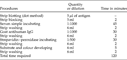
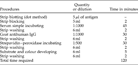
Between each incubation period, the antibodies and conjugates that had not bound during the reaction were removed with washing solution (TBS+0.05% v/v Tween 20 (Sigma)). All the steps of the DBA were carried out at room temperature (table 1).
Procedures and time required for the immunobinding dot-blot assay on nitrocellulose paper for the serological diagnosis of human cysticercosis.
Immunobinding dot-blot assay evaluation
The reproducibility of the DBA was evaluated by performing this test four times on each of the 15 positive and 15 negative control serum samples.
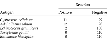
The immunological sensitivity (the smallest amount of antigen that DBA could detect with very low or no noticeable background staining of the NCP strip) was determined with the protein concentration in each of the five tenfold dilutions from 1:10 to 1:104, and observing the reaction produced by the DBA. The immunological specificity of the DBA was determined carrying out the test with antigens of adult Taenia solium, Echinococcus granulosus, Toxoplasma gondii and Entamoeba histolytica, which were prepared in an analogous way to the C. cellulosae antigen.
For the statistical evaluation of the DBA, the assumed gold standard was ELISA. Both tests, the developed DBA and ELISA, were performed on the 110 serum samples of unknown status, and diagnostic validity parameters were obtained. The epidemiological sensitivity (the absence of false-negative results) and epidemiological specificity (the absence of false-positive results) and predictive values (the probability of a true diagnosis, no matter whether positive or negative) were calculated using Episcope 2.0 software (Veterinary School, University of Zaragoza, Spain) with a 95% confidence level. Also, the results of both tests were analysed statistically using the kappa test with the same confidence level.
Results
The prepared antigen had a protein concentration of 8 mg/ml. The antigen working dilution was 1:10 and the antibody working dilution was 1:1000. These dilutions gave a positive colour reaction that did not fade after a positive result was obtained. The developed DBA could be carried out in a total time of 2 h (table 1).
Immunobinding dot-blot assay evaluation
The reproducibility of the DBA was adequate, because the test reproduced the same results four times on each of the 15 positive and 15 negative control serum samples, using the antigen of crude and whole C. cellulosae of swine origin.
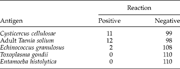
The immunological sensitivity of the DBA was 0.08 mg of antigen protein/ml. However, cross-reactions of the DBA were observed with adult T. solium and E. granulosus antigens (table 2).
Determination of immunological specificity of the immunobinding dot-blot assay on nitrocellulose paper for the serological diagnosis of human cysticercosis.
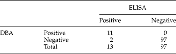
After analysing the 110 serum samples of unknown health status regarding cysticercosis with ELISA as the gold standard and the DBA (table 3), the developed assay obtained the following parameters of diagnostic validity: an epidemiological sensitivity of 84.615% and epidemiological specificity of 100.00%, and 100.00% and 97.98% for positive and negative predictive values, respectively.
Results obtained by ELISA and the immunobinding dot-blot assay (DBA) on nitrocellulose paper for the serological diagnosis of human cysticercosis.
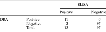
In addition, there was a disagreement between ELISA and the DBA in two samples; in contrast, both tests showed a concordance in 108 samples or an observed proportion of agreement of 0.982; in spite of this, a kappa value of 0.907 suggested that there were no significant statistical differences between the tests (P < 0.05).
Discussion
Successful treatment and control of cysticercosis depends on accuracy, specificity and sensitivity. Recently, the dot-blot assays or Dot-ELISA, which are considered simple, rapid and cheaper methods (Flores-Gutiérrez et al., Reference Flores-Gutiérrez, Infante, Salinas-Meléndez, Thomas, Estrada-Bellmann and Briones-Encinia2004), have been developed and evaluated for the diagnosis of cysticercosis (Vaz et al., Reference Vaz, Ferreira, Camargo, Nakamura and Camargo1990, Reference Vaz, Nakamura, Camargo, Camargo and Ferreira1996; Agudelo et al., Reference Agudelo, Botero and Palacio2005).
In the present study, the immunological sensitivity of the developed DBA was similar to that of other immunoenzymatic tests that utilize antigens from C. cellulosae (Kaur et al., Reference Kaur, Joshi, Ganguly, Mahajan and Malla1995; Infante et al., Reference Infante, Reyna and Farias1997; Biswas et al., Reference Biswas, Parija and Narayan2004). However, the DBA showed cross-reactions with the adult T. solium antigen; this was not unexpected, because some researchers have recommended the use of T. solium instead of C. cellulosae as antigen (Fleury et al., Reference Fleury, Bouteille, Garcia, Marquez, Preux, Escobedo, Sotelo and Dumas2001). In addition, since the 110 serum samples of unknown status regarding cysticercosis were not diagnosed for E. granulosus, the two serum samples determined as positive for the DBA for this parasite, could be considered as false-positive results or a mixed infection.
The whole antigens obtained from larval or adult stages produce low values of sensitivity and specificity when they are used for the immunological diagnosis of cysticercosis (Bruckner, Reference Bruckner1985). In spite of this, the detection of IgG against C. cellulosae, as in the case of the DBA developed in this study, gives the highest sensitivity and specificity, when compared with the detection of other immunoglobulins (Odashima et al., Reference Odashima, Takayanagui and Figueiredo2002).
The developed DBA obtained similar results of sensitivity and specificity as other studies, which utilized antigens of C. fasciolaris (Husain et al., Reference Husain, Jyotsna, Bagchi, Husain, Mishra and Gupta2001), C. cellulosae (Costa, Reference Costa1986; Morakote et al., Reference Morakote, Nawacharoen, Sukonthasun, Thammasonthi and Khamboonruang1992) and cysticercus of Taenia crassiceps (Peralta et al., Reference Peralta, Vaz, Pardini, Macedo, Machado, De Simone and Peralta2002). In addition, the sensitivity and specificity of the DBA were higher than those reported for the immunoenzymatic diagnosis of cyticercosis in serum and cerebrospinal fluid samples (Rosas et al., Reference Rosas, Sotelo and Nieto1986; Baveja et al., Reference Baveja, Arya, Goyle, Sharma and Gupta1990; Gonzalez et al., Reference Gonzalez, Cama, Gilman, Tsang, Pilcher, Chavera, Castro, Montenegro, Verastegui, Miranda and Bazalar1990; Vianna et al., Reference Vianna, Costa-Cruz, Macedo, De Souza and Moreira1992).
Similar studies have been reported for the diagnosis of cysticercosis. Utilizing a matrix of polyester fabric treated with N-methylol-acrylamide resin and different parts of C. cellulosae and C. longicollis as antigens, Vaz et al. (Reference Vaz, Nakamura, Camargo, Camargo and Ferreira1996) obtained values that ranged from 95.1% to 100.0% for epidemiological sensitivity and epidemiological specificity, respectively, which are considered acceptable for the serodiagnosis of cysticercosis. Although the diagnostic validity of the assay developed by Vaz et al. (Reference Vaz, Ferreira, Camargo, Nakamura and Camargo1990, Reference Vaz, Nakamura, Camargo, Camargo and Ferreira1996) had slightly higher epidemiological sensitivity than that obtained for the DBA developed in this study, the latter is less complicated and time consuming.
Other Dot-ELISA assays for diagnosing cysticercosis (Biswas et al., Reference Biswas, Parija and Narayan2004) have reported values of 56.25%, 92%, 87.09% and 68.66% for sensitivity, specificity, positive predictive value and negative predictive value, respectively. These values were less than those obtained by DBA in the present study. In addition, the assay reported by Biswas et al. (Reference Biswas, Parija and Narayan2004) was more time consuming than that reported here.
However, although the present study indicates that DBA is not as sensitive as ELISA, we conclude that this assay constitutes an easier and faster alternative for the diagnosis of human cysticercosis, which could be used as a screening test or in epidemiological studies, in laboratories that not are equipped to conduct other sophisticated immunoenzymatic assays.
Acknowledgements
This paper is part of the Masters degree thesis of E. Hernández-Cruz. The study was supported by Universidad Autónoma de Tamaulipas. The publication of this paper was supported in full by the Fondo Mixto de Fomento a la Investigación Científica y Tecnológica CONACYT – Gobierno del Estado de Tamaulipas (México). We thank Dr Carlos Larralde, Instituto de Ciencias Biomédicas, Universidad Nacional Autónoma de México, for providing positive and negative control serum samples.