INTRODUCTION
Fasciolosis is a major parasitic disease of livestock and humans (Keiser and Utzinger, Reference Keiser and Utzinger2004, Reference Keiser and Utzinger2005; Fairweather, Reference Fairweather2005; Mas-Coma et al. Reference Mas-Coma, Bargues and Valero2005). In the UK, there has been a sharp increase in the incidence and spread of fasciolosis in recent years, a phenomenon attributed to climate change, with the disease appearing in areas previously believed to be free from it (Mitchell, Reference Mitchell2002; Pritchard et al. Reference Pritchard, Forbes, Williams, Salimi-Bejestani and Daniel2005). Control of fasciolosis is heavily dependent on the use of a single drug, triclabendazole. Continued use of the drug is under threat, due to the development of resistance in countries throughout Europe and elsewhere (Fairweather, Reference Fairweather2005; Alvarez-Sanchez et al. Reference Alvarez-Sanchez, Mainar-Jaime, Perez-Garcia and Rojo-Vasquez2006; Brennan et al. Reference Brennan, Fairweather, Trudgett, Hoey, McCoy, McConville, Meaney, Robinson, McFerran, Ryan, Lanusse, Mottier, Alvarez, Solana, Virkel and Brophy2007). So, for fasciolosis, there is a two-pronged dilemma of increasing prevalence of the disease and declining efficacy of the principal drug used for its control. A number of general strategies have been proposed to deal with anthelmintic resistance: they involve either novel ways of utilizing existing drugs or seeking new compounds. With regard to novel compounds for use as anthelmintics, there has been a growing interest in making use of natural plant products that have been used as traditional medicines in developing countries (Hammond et al. Reference Hammond, Fielding and Bishop1997; Kayser et al. Reference Kayser, Kiderlen and Croft2003; Iqbal et al. Reference Iqbal, Akhtar, Sindhu, Khan and Jabbar2003, Reference Iqbal, Lateef, Ashraf and Jabbar2004; Buenz et al. Reference Buenz, Schnepple, Bauer, Elkin, Riddle and Motley2004, Reference Buenz, Bauer, Johnson, Tavana, Beekman, Frank and Howe2006; Anthony et al. Reference Anthony, Fyfe and Smith2005; Balunas and Kinghorn, Reference Balunas and Kinghorn2005; Chin et al. Reference Chin, Balunas, Chai and Kinghorn2006; Crump, Reference Crump2006; Newman and Cragg, Reference Newman and Cragg2007; Stepek et al. Reference Stepek, Lowe, Buttle, Duce and Behnke2007).
Genistein is the compound under investigation in the present study. It is one of the active ingredients of Flemingia vestita, a plant indigenous to Northeast India and known locally as Soh-Phlang; genistein is also present in the soybean and soy products. Roots and peel of the root tuber of F. vestita are consumed raw by the local people to cure helminth infections (Rao, Reference Rao1981). Genistein and other isoflavone components of the plant have been shown to possess activity against a range of native nematode, cestode and trematode parasites (Yadav et al. Reference Yadav, Tandon and Rao1992; Tandon et al. Reference Tandon, Pal, Roy, Rao and Reddy1997); also, against hydatid organisms (Naguleswaran et al. Reference Naguleswaran, Spicher, Vonlaufen, Ortega-Mora, Torgerson, Gottstein and Hempill2006). Isoflavonoid extracts of the West African Turburku plant, Millettia thonningii have activity against different life-cycle stages of Schistosoma mansoni (Lyddiard et al. Reference Lyddiard, Whitfield and Bartlett2002) and isoflavones from the bark of the leguminous plant, Dalbergia frutescens have potency against the protozoan parasite, Giardia intestinalis (Khan et al. Reference Khan, Avery, Burandt, Goins, Mikell, Nash, Azadegan and Walker2000). As well as their anti-parasitic activity, isoflavones have a variety of uses in human medicine, including cancer and cardiovascular disease (Dixon and Ferreira, Reference Dixon and Ferreira2002; Watanabe et al. Reference Watanabe, Uesugi and Kikuchi2002; Sarkar et al. Reference Sarkar, Adsule, Padhye, Kulkarni and Li2006; Siow et al. Reference Siow, Li, Rowlands, Winter and Mann2007). In this study, the activity of genistein against the liver fluke, Fasciola hepatica has been evaluated in vitro. Morphological changes were assessed by means of scanning and transmission electron microscopy and its effect on neuromuscular activity determined using muscle strip preparations of the fluke.
MATERIALS AND METHODS
F. hepatica isolate
The Cullompton isolate of Fasciola hepatica was used for this study. It was originally obtained as a field isolate from an abattoir in Cullompton, Devon, UK, in 1998 and has been shown to be susceptible to triclabendazole (Robinson et al. Reference Robinson, Trudgett, Hoey and Fairweather2002; McCoy et al. Reference McCoy, Fairweather, Brennan, Kenny, Ellison and Forbes2005), albendazole (Buchanan et al. Reference Buchanan, Fairweather, Brennan, Trudgett and Hoey2003), clorsulon (Meaney et al. Reference Meaney, Fairweather, Brennan, McDowell and Forbes2003, Reference Meaney, Fairweather, Brennan and Forbes2004, Reference Meaney, Haughey, Brennan and Fairweather2005a, Reference Meaney, Haughey, Brennan and Fairweatherb, Reference Meaney, Allister, McKinstry, McLaughlin, Brennan, Forbes and Fairweather2006, Reference Meaney, Allister, McKinstry, McLaughlin, Brennan, Forbes and Fairweather2007) and nitroxynil (McKinstry et al. Reference McKinstry, Fairweather, Brennan and Forbes2003, Reference McKinstry, Brennan, Halferty, Forbes and Fairweather2007). Adult male Sprague-Dawley rats were each infected orally with 20 metacercarial cysts under light ether anaesthesia by means of a stomach tube.
In vitro drug treatment
Adult flukes (at least 12 weeks old) were recovered from the bile ducts of infected rats at autopsy, under sterile conditions in a laminar flow cabinet, and placed in warm (37°C) sterile NCTC 135 culture medium containing antibiotics (penicillin, 50 IU/ml; streptomycin, 50 μg/ml; NCTC was obtained from MP Biomedicals, London, UK; antibiotics were obtained from Sigma, Poole, Dorset, UK). Whole flukes were transferred to fresh culture medium containing genistein at a concentration of 0·27 mg/ml (=1 mm). This concentration was less than the 0·5 mg/ml concentration used in previous studies on genistein with the trematodes, Fasciolopsis buski (Roy and Tandon, Reference Roy and Tandon1996; Kar and Tandon, Reference Kar and Tandon2000, Reference Kar and Tandon2004; Kar et al. Reference Kar, Tandon and Saha2002, Reference Kar, Tandon and Saha2004) and Paramphistomum sp. (Tandon et al. Reference Tandon, Pal, Roy, Rao and Reddy1997) and with the cestode, Raillietina echinobothrida (Tandon et al. Reference Tandon, Pal, Roy, Rao and Reddy1997; Pal and Tandon, Reference Pal and Tandon1998a, Reference Pal and Tandonb; Das et al. Reference Das, Tandon and Saha2006). Genistein was initially prepared as a stock solution in dimethyl sulphoxide (DMSO) and added to the culture medium to give a final solvent concentration of 0·1% (v/v). Controls were prepared by incubating flukes in sterile NCTC culture medium containing 0·1% (v/v) DMSO.
Fluke motility was observed at regular intervals and the time taken for flukes to become totally inactive recorded. A gradual decline in movement was observed after 30 min and cessation of activity was observed after 3 h of drug exposure. The experiment was terminated at this point and the flukes (both treated and control) were processed for scanning and transmission electron microscopy, as described below. Controls at 0 h were also prepared. A total of 11 treated flukes were prepared for scanning electron microscopy (SEM) and 5 treated flukes were prepared for transmission electron microscopy (TEM). Eight control flukes were prepared for SEM: four 0 h controls and four 3 h controls; and 4 flukes were prepared for TEM: two 0 h controls and two 3 h controls.
Tissue preparation for scanning electron microscopy
Initially, the flukes were lightly flat-fixed for 1 h at room temperature in 4% (w/v) aqueous glutaraldehyde and subsequently free-fixed at 4°C in a 3:1 mixture of 4% (w/v) aqueous glutaraldehyde and 1% osmium tetroxide overnight. Flukes were dehydrated in an ascending series of ethanols, dried in hexamethyldisilazane, mounted on aluminium stubs and sputter-coated with gold-palladium. The flukes were viewed using an FEI Quanta 200 scanning electron microscope operating at an accelerating voltage of 10 keV.
Tissue preparation for transmission electron microscopy
Flukes were flat-fixed for 1 h at room temperature in 4% (w/v) glutaraldehyde and subsequently dissected into 4 body regions, namely, oral cone (including ventral sucker), anterior midbody, posterior midbody and tail. Tissues were free-fixed for a further 3 h at room temperature in 4% (w/v) glutaraldehyde in 0·1 m sodium cacodylate buffer, pH 7·6, then rinsed briefly in 0·1 m sodium cacodylate buffer (pH 7·6) and placed in fresh buffer at 4°C overnight. Following further division of each of the body regions into thin strips, the tissues were post-fixed for 1 h at room temperature in 1% osmium tetroxide, rinsed in 0·1 m sodium cacodylate buffer (pH 7·6), dehydrated in an ascending series of ethanols, infiltrated and embedded in Agar 100 resin and polymerized for 48 h at 60°C. Ultrathin sections (60–70 nm thick) were cut with glass knives using a Reichert Ultracut E ultramicrotome, mounted on uncoated copper grids, double-stained with uranyl acetate (10 min) and lead citrate (6 min) and viewed in an FEI CM 100 transmission electron microscope operating at an accelerating voltage of 100 keV.
Tissue preparation for muscle strip physiology
Spontaneously active muscle strips were obtained from adult flukes recovered from the bile ducts of experimentally infected laboratory rats. The strips were obtained by making a horizontal cut below the ventral sucker and by removing the posterior end and lateral margins of the fluke. The remaining midbody tissue section was cut into two longitudinally, resulting in 2 muscle strips approximately 3 mm wide and 10 mm long. Five muscle strips were used for each experiment. The muscle strips were tied with cotton thread at both ends so that they could be suspended vertically in 4-ml organ baths. The end of one of the cotton threads was attached to an isometric force transducer (Harvard Instruments, 50g model) and output of muscle activity, that is, frequency and amplitude of muscle contractions, was recorded on a chart recorder. For full details of the method, see Graham et al. (Reference Graham, McGeown and Fairweather1999, Reference Graham, Fairweather and McGeown2000). The organ baths were perfused continually with Hédon-Fleig saline buffered with 10 mm HEPES and adjusted to pH 7·4 using 1 m NaOH and maintained at 37°C. Regularization of spontaneous activity of muscle strips was observed after around 60 min. At this point, test solutions of genistein at concentrations of 1 μM, 10 μM, 100 μM were allowed to perfuse the organ bath and any changes in the spontaneous muscle activity of the strips were recorded via the chart recorder. Following drug exposure, a 15-min washout was carried out where Hédon-Fleig saline was allowed to perfuse the organ bath and any change in muscle activity recorded. Due to considerations of cost, an alternative method of drug delivery was carried out for the higher concentration of 1 mm; for this, the perfusion pump was switched off for 15 min prior to drug addition. Test solutions of 1 mm were added directly to the 3·5 ml organ bath via a pipette. Addition of a little air to the organ bath via the pipette ensured even mixing. A control experiment was carried out in which 3·5 μl of Hédon-Fleig saline was added directly to the organ bath to rule out the effect of the syringe on muscle strips. After 15 min drug exposure, Hédon-Fleig saline was allowed to perfuse the system for the washout period. Genistein was prepared as a stock solution in DMSO and the maximum solvent concentration used was 0·5% (v/v). Control muscle strips were exposed to this concentration of solvent without drug. Each experiment was repeated a minimum of 5 times, using muscle strips derived from separate flukes. Contraction amplitude and frequency were analysed separately and only contractions of an amplitude >/=0·5 mN were counted. The statistical significance of any changes to the mean frequency and amplitude of muscle contractions in each of the 5-min periods of drug exposure and washout stages was compared to the last 5 min of the control stage. A repeated measures design analysis of variance (ANOVA) with post-hoc testing using Tukey-Kramer multiple comparisons tests was carried out to assess any significant changes to the frequency and amplitude of muscle contractions, with significance set to the 95% level.
RESULTS
Scanning electron microscopy
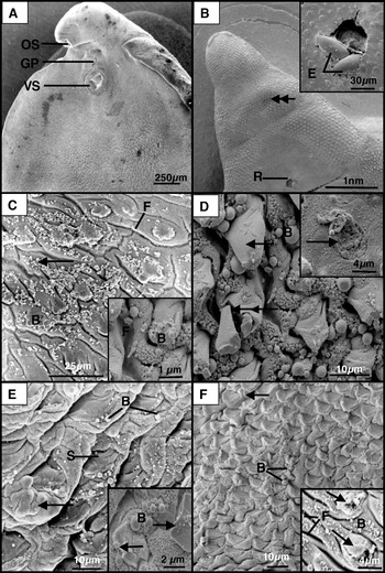
After 3 h incubation of whole flukes in genistein, there was severe swelling of the tegument in the oral cone region, on both surfaces, and especially along the lateral margins (Fig. 1A–D). In one specimen, the tegument had ruptured, along with the lining of the uterus, and eggs could be seen projecting from the surface (Fig. 1B inset). The tegumental surface was covered with blebs in the inter-spine areas (Fig. 1C and D); blebs were also present on the tips of the spines (Fig. 1C and D). On the dorsal surface, some spines appeared to be ‘cracked’ and some spine loss was evident (Fig. 1D).
Scanning electron micrographs (SEMs) of adult Fasciola hepatica treated in vitro with genistein for 3 h. (A) Low-power micrograph of the ventral oral cone region, showing genital pore (GP), oral sucker (OS), ventral sucker (VS). (B) SEM of dorsal oral cone region showing swelling of the tegument (double arrow). A rupture (R) of the tegument is visible, with eggs protruding from the surface. Inset shows the eggs (E) projecting from the rupture. (C) High-power SEM of the ventral oral cone region, showing the general swelling and furrowing (F) of the tegument. Blebbing (B) is present on the tips of the spines and in the inter-spine areas (arrow). Inset shows the blebbing (B) on and between the spines and furrowing (F) of the tegument at higher magnification. (D) High-power SEM of dorsal oral cone. A carpet of blebs (B) covers the inter-spine tegumental surface. Some of the spines appear to have lost their tegumental covering (arrow) and one spine has split (double arrow). Inset shows spine loss (arrow). (E) Ventral anterior midbody region. The tegument is extremely swollen (arrow), so that the spines (S) appear sunken and the surface is carpeted with blebs (B). Inset shows blebbing (B) of the tegument and disruption of the spines (arrows). (F) SEM of dorsal anterior midbody region, showing the swelling (arrow) and blebbing (B) of the tegument in the inter-spinal areas. Inset shows disruption to the spines in that the tegumental covering has split (arrows). Furrowing (F) of the tegument is visible and blebs (B) cover the interspine surface.

In the anterior midbody region, the tegument on the ventral surface was swollen and thrown into a number of furrows: the swelling was such that the spines appeared to be embedded in the swollen tegument surrounding them (Fig. 1E). Swelling was less severe on the dorsal surface, but the disruption to the spines was greater, in that their tegumental covering had split (Fig. 1F).
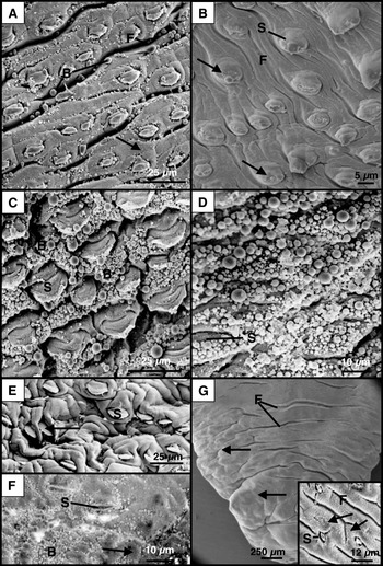
The posterior midbody region was the worst affected region of all. There was severe swelling and furrowing of the tegument (Fig. 2A and B). The spines appeared to be submerged by the tegument surrounding them (Fig. 2A). Blebbing of the tegumental surface was extensive and was so severe at times that the spines were barely visible (Fig. 2C and D).
Scanning electron micrographs (SEMs) of adult Fasciola hepatica treated in vitro with genistein for 3 h. (A) Ventral posterior midbody region, showing swelling (arrow), furrowing (F) and blebbing (B) of the tegument. (B) Dorsal posterior midbody region, showing swelling and furrowing (F) of the tegument. The tegumental covering of the spines (S) is swollen and blebbing is evident on the tips (arrows). (C) High-power SEM of ventral posterior midbody region, showing extensive blebbing (B) on the tips of the spines (S) and in the inter-spine areas. (D) High-power SEM of dorsal midbody region, showing the tegument and spines (S) covered with a carpet of blebs. (E) Ventral tail region. The tegument is severely swollen and many of the spines (S) appear sunken (arrow). (F) Ventral tail region, showing widespread blebbing (B). The tegument is swollen (arrow) and the spines (S) appear sunken. (G) SEM of dorsal tail region, showing swelling (arrows) and furrowing (F) of the tegument. Inset of the surface at higher magnification, showing furrowing (F). The surface has a roughened appearance (arrows), with microvillus-like projections. The spines (S) appear sunken.

In the tail region, swelling and furrowing of the tegument was evident and the spines appeared to be sunken (Fig. 2E–G). Patches of blebs occurred on the ventral surface (Fig. 2F), but were less conspicuous on the dorsal surface. The latter appeared roughened, due to microvillus-like projections (Fig. 2G).
The 0 h and 3 h control flukes showed a normal surface morphology. The morphology matched the images presented by Bennett (Reference Bennett1975) and Fairweather et al. (Reference Fairweather, Threadgold, Hanna and Dalton1999).
Summary
Disruption to the flukes mainly took the form of blebbing, although swelling and furrowing of the tegument was also observed. The ventral surface of the fluke was slightly more severely affected than the dorsal side. The severity of the changes increased posteriorly and was most pronounced in the posterior midbody region. Disruption was also more severe along the lateral margins than in the central region of the fluke's body.
Transmission electron microscopy
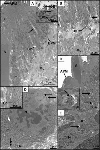
On the basis of the scanning electron microscope results, sections from the posterior midbody region of the fluke were examined. In the tegument, there was slight swelling of the mucopolysaccharide masses associated with the basal infolds (Fig. 3A and B). ‘Open’ bodies were observed at the apex of the syncytium, but there were relatively few secretory bodies in the apical cytoplasm (Fig. 3A). The mitochondria retained their normal distribution within the syncytium (Fig. 3A). One notable abnormal feature of the syncytium was the presence of ‘packets’ of cytoplasm that contained concentrations of T1 and T2 secretory bodies (Fig. 3C). The muscle blocks beneath the basal lamina appeared normal (Fig. 3A and B). The tegumental cell bodies contained few secretory bodies (Fig. 3D). The Golgi complexes appeared reduced, with few discernible cisternae, and were totally absent in some cells (Fig. 3D and E). There was evidence of autophagic activity in the cells (Fig. 3D and E).
Transmission electron micrographs (TEMs) of adult Fasciola hepatica treated in vitro with genistein for 3 h. (A) TEM of the tegumental syncytium, showing the apical plasma membrane (APM) and basal plasma membrane (BPM). The basal infolds (BI) are slightly swollen and chains of mitochondria (arrow) lie between them. Blocks of muscle (Mu) lie below the basal plasma membrane. S, spine. Inset of the apical region of the syncytium showing T1 secretory bodies (T1), a number of ‘open’ bodies (OB) and mitochondria (M). (B) Basal region of the syncytium and underlying blocks of muscle (Mu). The mucopolysaccharide masses associated with the basal infolds are slightly swollen (arrow). BPM, basal plasma membrane. (C) ‘Packets’ of T1 and T2 secretory bodies (arrows) occur throughout the syncytium. The apical plasma membrane (APM) is convoluted. S, spine. Inset shows one of the ‘packets’ of secretory bodies at higher magnification (arrow). (D) TEM of a tegumental cell containing a small number of T1 secretory bodies (arrows). The Golgi complex (Gc) is abnormal and there is some evidence of autophagy (double arrow). m, mitochondria; N, nucleus. Inset shows the reduced and abnormal organization of the Golgi complex (Gc). (E) TEM of a tegumental cell showing the presence of autophagic vacuoles (arrows). No Golgi complexes are present and few secretory bodies. N, nucleus.

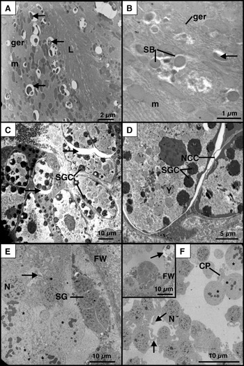
The gastrodermal cells contained a large number of autophagic vacuoles (Fig. 4A). The surface lamellae and cisternae of granular endoplasmic reticulum appeared normal and a number of secretory bodies were present in the cells (Fig. 4A and B).
Transmission electron micrographs (TEMs) of adult Fasciola hepatica treated in vitro with genistein for 3 h. (A) Gastrodermal cells containing large numbers of autophagic vacuoles (arrows). ger, cisternae of granular endoplasmic reticulum; L, gut lamellae; m, mitochondria. (B) High-power TEM of a gastrodermal cell containing autophagic vacuoles (arrow). Also present are secretory bodies (SB), cisternae of ger (ger) and mitochondria (m). (C) TEM of a vitelline follicle, showing a predominance of mature vitelline cells (asterix). Within the cells, the shell globule clusters (SGC) have lost their normal organization and distribution. The cells are pulling away from the nurse cell cytoplasm, leaving spaces (arrow) between the cells. (D) Mature vitelline cell. The shell globule clusters (SGC) are abnormal. Spaces are evident between the cells and the nurse cell cytoplasm (NCC). Y, yolk globule. (E) TEM of a testis follicle. Spermatogonia (SG) lie at the periphery of the follicle. Later stages of development are abnormal and the cells are connected by thin strands of cytoplasm (arrow). The nuclear material in some nuclei (asterix) is clumped. FW, follicle wall; N, nucleus. (F) Testis follicle. Cells are connected by thin strands of cytoplasm (arrows), creating syncytial groups of cells. A cytophore (CP) is present, but is not connected to the cells. Nuclear material appears clumped in some of the nuclei (asterix). Inset shows a large autophagic vacuole (arrow) adjacent to the follicle wall (FW). N, nucleus.

Within the vitelline follicle there appeared to be an abnormal preponderance of mature cells, with fewer-than-normal numbers of earlier developmental stages (Fig. 4C). Spaces were present between the cells and the extensions of nurse cell cytoplasm that permeate throughout the follicle (Fig. 4C and D). In the mature cells, the shell globule clusters were fewer in number and smaller than normal: they appeared to be loosely-packed and some occupied a more central position in the cell, rather than being distributed around the periphery of the cell (Fig. 4C and D).
The testis follicles were relatively empty and loosely filled with spermatogenic cells (Fig. 4E and F). The spermatogonia at the periphery of the follicle appeared relatively normal: but the later spermatocyte stages were disrupted. Cells were joined by thin strands of cytoplasm and appeared more syncytial than being arranged in typical rosettes (Fig. 4E and F). In one instance, the cytophore connecting a group of cells had broken down (Fig. 4F). The nuclear material in a number of cells appeared clumped (Fig. 4E and F) and there was evidence of autophagic activity in the follicle (Fig. 4F). Spermatids and spermatozoa were absent.
The fine structure of the tegumental syncytium, tegumental cells, gastrodermal cells, vitelline cells and spermatogenic cells in the control specimens remained normal. For images of normal structure, the reader is referred to the papers by Threadgold (Reference Threadgold1963, Reference Threadgold1967) (for tegument); by Robinson and Threadgold (Reference Robinson and Threadgold1975) (for gut); by Irwin and Threadgold (Reference Irwin and Threadgold1970) and Colhoun et al. (Reference Colhoun, Fairweather, Brennan and Woods1998) (for vitelline cells); and by Stitt and Fairweather (Reference Stitt and Fairweather1990) (for spermatogenic cells).
Muscle strip physiology
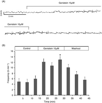
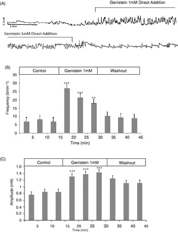
There was no observable change in muscle activity on perfusion of muscle strips with genistein at a concentration of 1 μm. On perfusion of strips with genistein at a concentration of 10 μm, there was a significant increase (P<0·05) in the frequency of muscle contractions between 10–15 min of the control period and 25–30 min of the drug exposure time-period (Fig. 5). Perfusion of 100 μm genistein solution resulted in a significant (P<0·01) increase between the 10–15 min control period and first 5 min of the drug exposure; and significant (P<0·001) increases in the frequency of muscle contractions between the same control period and the 20–25 and 25–30 min drug exposure periods. On addition of genistein solution directly to the organ bath via a syringe, giving a bath concentration of 1 mm, there was a significant increase (P<0·001) in the frequency of muscle contractions between the control period (10–15 min) and the first 10 min of drug exposure. Significant increases (P<0·01) in the frequency of muscle contractions were observed between the same control period and the remaining 25–30 min block of drug exposure. Addition of 1 mm genistein solution also resulted in a significant increase (P<0·001) in the amplitude of muscle contractions between the last 5 min of the control period and all of the drug exposure periods. There were significant increases (P<0·01) in the amplitude of contractions between the control period and 30–35 min and 40–45 min of the washout period (Fig. 6). On perfusion with Hédon-Fleig saline during the washout stage, normal contraction amplitude and frequency returned after some time.
(A) A typical trace showing isometric tension as recorded from a Fasciola hepatica midbody muscle strip, showing muscle activity during control, genistein (10 μm) exposure and washout periods. (B) Bar chart showing mean frequency of muscle contractions during each 5-min period of control, genistein (1 µm) exposure and washout periods. *P<0·05.

(A) A typical trace showing isometric tension as recorded from a Fasciola hepatica midbody muscle strip showing muscle activity during control, genistein (1 mm) exposure and washout periods. (B) Bar chart showing mean frequency of muscle contractions during each 5-min period of control, genistein (1 mm) exposure and washout periods. (C) Bar chart showing mean amplitude of muscle contractions during each 5-min period of control, genistein (1 mm) exposure and washout periods. **P<0·01; ***P<0·001.

Controls
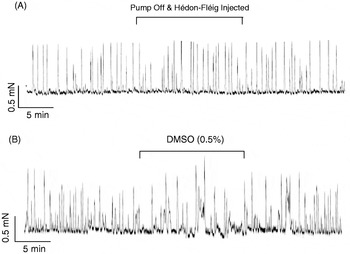
The first control was conducted to determine whether the spontaneous contractions were affected by the switching on and off of the perfusion pump and if the injection of a solution into the chamber and its subsequent mixing had any effect on spontaneous contractions. Neither procedure had any effect on the baseline tension and there were no significant changes in the mean values of amplitude or frequency of contractions during the last 5-min control period and any of the 5-min periods of either the pump being switched off, a solution being injected, or the pump being switched back on (Fig. 7A). Control muscle strips were also exposed to Hédon-Fleig saline containing DMSO at a maximum concentration of 0·5% (v/v). There was no effect on the mean baseline tension and there was no significant change in the mean value of amplitude or frequency during the last 5-min control period and any of the 5-min periods of DMSO or washout (Fig. 7B). Any changes in muscle activity, therefore, could be attributed to the action of genistein, and the addition of solutions via the syringe had no effect on muscle activity. (For further details on the analysis of control data, see Wells, Reference Wells2008.)
(A) A typical trace showing isometric tension recorded from a muscle strip when the pump was switched off, Hédon-Fleig saline was injected into the chamber and mixed, and the pump was switched on again. (B) A typical trace showing isometric tension recorded from a muscle strip before, during and after superfusion with DMSO (0·5%).

DISCUSSION
The results of the present study have demonstrated the in vitro activity of genistein against F. hepatica. The disruption induced by genistein is comparable to its effect on a range of helminth parasites (Yadav et al. Reference Yadav, Tandon and Rao1992; Pal and Tandon, Reference Pal and Tandon1998a, Reference Pal and Tandonb; Kar and Tandon, Reference Kar and Tandon2000; Das et al. Reference Das, Tandon and Saha2006). There was extensive surface disruption following in vitro incubation, indicating that the tegument of F. hepatica is extremely sensitive to this plant extract. The gut, and more especially the reproductive structures, of the fluke were also highly susceptible to the action of this compound. A pronounced neuromuscular effect was also observed, bringing about a gradual loss of spontaneous movement from after incubation for 20 min, leading to complete immobilization at 3 h. Disruption of the morphology and changes in the spontaneous movements of parasites have been used as two of the parameters of the anthelmintic efficacy of various botanical extracts (Robinson et al. Reference Robinson, Williams, Lindo, Terry and Mansingh1990; Tandon et al. Reference Tandon, Pal, Roy, Rao and Reddy1997).
Scanning electron microscope observations revealed extensive surface disruption following a 3-h incubation with genistein. Blebbing was observed over the whole of the fluke on both surfaces, but the extent of blebbing appeared to be more severe in certain areas. The most-affected region was the posterior midbody, especially along the lateral margins of the fluke. Blebbing is a stress response by the fluke to drug treatment that allows the fluke to replace damaged tegument. This reaction has been noted in previous studies on the liver fluke following treatment with the fasciolicides triclabendazole (Stitt and Fairweather, Reference Stitt and Fairweather1994), nitroxynil (McKinstry et al. Reference McKinstry, Fairweather, Brennan and Forbes2003, Reference McKinstry, Brennan, Halferty, Forbes and Fairweather2007) and clorsulon (Meaney et al. Reference Meaney, Fairweather, Brennan, McDowell and Forbes2003, Reference Meaney, Fairweather, Brennan and Forbes2004, Reference Meaney, Haughey, Brennan and Fairweather2005a, Reference Meaney, Haughey, Brennan and Fairweatherb, Reference Meaney, Allister, McKinstry, McLaughlin, Brennan, Forbes and Fairweather2006, Reference Meaney, Allister, McKinstry, McLaughlin, Brennan, Forbes and Fairweather2007). Perhaps the regional severity of damage may be due to weak spots on the tegumental surface, which make them particularly sensitive to the effects of genistein. Alternatively, regional variation in severity of damage may be as a result of differences in the activity of ion pumps in the apical plasma membrane in different regions of the fluke, as suggested by Anderson and Fairweather (Reference Anderson and Fairweather1988).
Widespread swelling of the tegument was observed and was most severe along the lateral margins of the midbody and in the tail region of the fluke. Pronounced surface disruption by genistein of a range of helminths has been noted previously. For example, Flemingia vestita crude root extract caused swelling and cracking of the tegument of Raillietina echinobothrida (Tandon et al. Reference Tandon, Pal, Roy, Rao and Reddy1997), and genistein brought about structural distortion of the tegument of R. echinobothrida, manifested primarily as severe blebbing (Pal and Tandon, Reference Pal and Tandon1998a, Reference Pal and Tandonb). Wrinkles, tegumental sloughing, loss of spines and deformity of suckers were observed in the digenetic flukes Artyfechinostomum sufrartyfex and Fasciolopsis buski treated with F. vestita extract (Roy and Tandon, Reference Roy and Tandon1996). Comparable drug effects, such as blebbing, swelling of the tegument, loss of spines and distortion to suckers have been observed in F. gigantica treated with the natural compound, propolis (10–30 μg/ml). Propolis (bee glue) is a resinous bee-hive product, and has been shown to have potent anti-viral, anti-bacterial and anti-protozoal properties; it is considered to be a viable natural fasciolicidal drug (Hegazagi et al. Reference Hegazagi, Abd El Hady and Shalaby2007).
At the ultrastructural level, open bodies at the apex of the syncytium indicate the release of secretory bodies to maintain tegumental integrity (Rogan and Threadgold, Reference Rogan and Threadgold1984), and the presence of ‘packets’ of secretory bodies throughout the syncytium as seen in the present study is a unique phenomenon. The fluke is able to replace damaged tegument via the continual turnover of Type 1 (T1) and Type 2 (T2) secretory bodies supplied from the tegumental cells via the cytoplasmic connections (Fairweather and Boray, Reference Fairweather and Boray1999; McConville et al. Reference McConville, Brennan, McCoy, Castillo, Hernandez-Campos, Ibarra and Fairweather2006). However, secretory bodies were sparsely distributed within the tegumental cells following genistein treatment. Their production appeared to be inhibited, with Golgi complexes either exhibiting an abnormal morphology or being absent from the cells. More general disruption and possible degeneration of the cells are indicated by the presence of autophagic vacuoles within the cells. Comparable changes, for example, a decline in the number of secretory bodies and a gradual disappearance of Golgi complexes from the cells, was noted in a previous study involving the sulphoxide metabolite of triclabendazole (TCBZ.SO) (Stitt and Fairweather, Reference Stitt and Fairweather1994). Such degenerative changes in the tegumental cells will have an adverse effect on tegumental functioning. Autophagic vacuoles were also common in the gut, again suggesting degeneration and reduced level of activity due to a lack of secretory bodies.
Disruption to the tegument and gastrodermis could be as a result of, or be exacerbated by, interference with the levels and functioning of tegumental and gut enzymes by genistein, as has been demonstrated in F. buski (Kar and Tandon, Reference Kar and Tandon2004). Levels of AcPase and ATPase activity in F. hepatica have been shown to alter following treatment with the anthelmintic luxabendazole (Gorchilova et al. Reference Gorchilova, Poljakova, Spaldonova and Vinarova1990). Interference with levels of such enzymes may have a knock-on effect on membrane transport systems, absorption and digestion in the fluke. Genistein has also been shown to affect levels of the enzymes involved in energy and glycogen metabolism in the cestode, R. echinobothrida, the latter response facilitating the mobilization of energy reserves to deal with anthelmintic stress (Tandon et al. 2003; Das et al. Reference Das, Tandon and Saha2004; Tandon and Das, Reference Tandon and Das2007).
A major observation by Stitt and Fairweather (Reference Stitt and Fairweather1996) – which is similar to that seen in the present study – was a shift in the cell population within the vitelline follicle. In both experiments, existing vitelline cells appear to continue with development, but they are not replaced by stem cells and this leads to a preponderance of mature cells within the follicle. Other effects comparable to those caused by genistein exposure included formation of gaps between the nurse cell cytoplasm and the vitelline cells, loosely-packed and abnormally-distributed shell globule clusters and areas of autophagy within the cell cytoplasm (Stitt and Fairweather, Reference Stitt and Fairweather1996). Disrupted contact between the nurse cells and the vitelline cells may exacerbate drug pressure by compromising the nutrient supply reaching the latter.
Within the testes, too, cell development and differentiation are disrupted during spermatogenesis. It has been shown that genistein can block cell-cycle progression at the G2/M checkpoint (Matsukawa et al. Reference Matsukawa, Marui, Sakai, Satomi, Yoshida, Matsumoto, Nishino and Aoike1993; Singletary et al. Reference Singletary, Frey and Li2002). More specifically, genistein can disrupt spermatogenesis, inhibiting DNA synthesis during meiotic division and differentiation (i.e. during the spermatocyte stages) and leading to degeneration of spermatocytes and reduced numbers of spermatids and spermatozoa in the vertebrate model organism, Oryzias latipes (medaka) and in the rat (Delclos et al. Reference Delclos, Bucci, Lomax, Latendresse, Warbritton, Weis and Newbold2001; Song and Gutzeit, Reference Song and Gutzeit2003; Svechnikov et al. Reference Svechnikov, Supornsilchai, Strand, Wahlgren, Seidlova-Wuttke and Söder2005). In the fluke, cell division at the periphery of the testis follicle appears normal but, as development continues toward the centre of the follicle, few typical rosettes of spermatocytes were present; they were replaced by syncytial masses of cells which appeared to be unable to divide and contained clumped nuclear material.
It has been suggested that genistein can protect against various cancers by inhibiting cell proliferation, inducing apoptosis and interfering with signalling pathways (Agarwal, Reference Agarwal2000; Dixon and Ferreira, Reference Dixon and Ferreira2002; Sarkar and Li, Reference Sarkar and Li2004; Sarkar et al. Reference Sarkar, Adsule, Padhye, Kulkarni and Li2006). Genistein is a recognized inhibitor of tyrosine-specific protein kinases (Akiyama et al. Reference Akiyama, Ishida, Nakagawa, Ogawara, Watanabe, Itoh, Shibuya and Fukami1987), enzymes involved in epidermal growth factor (EGF), transforming growth factor (TGF) and other signalling pathways that are concerned with cell division and development. Elements of these pathways have been identified in parasites such as Schistosoma and Echinococcus (reviewed by Dissous et al. Reference Dissous, Khayath, Vicogne and Capron2006; Knobloch et al. Reference Knobloch, Beckmann, Burmeister, Quack and Grevelding2007). The components have been localized in different tissues of Schistosoma, e.g. tegument, muscle, gonads and vitelline cells. So, the effects observed in the present study and studies on other parasites could be linked to these mechanisms. The degeneration of cells observed in the present study could be linked to the apoptotic action of genistein.
On addition of genistein to muscle strips of F. hepatica, statistically significant increases in contraction frequency and amplitude were observed. In a recent paper on R. echinobothrida, genistein was shown to cause rapid muscular contractions followed by flaccid paralysis (Das et al. Reference Das, Tandon and Saha2006). As calcium (Ca2+) is known to have a role in rapid and sustained muscular contraction, the effects of genistein on Ca2+ homeostasis was investigated. Tissue levels of Ca2+ in R. echinobothrida significantly decreased on exposure to genistein, whereas efflux of Ca2+ into the surrounding test medium was significantly increased. It is believed that ion pumps in the apical plasma membrane control movement of calcium ions into the muscle, bringing about contraction, and control the transport of calcium ions out of the cell to cause relaxation (Fairweather and Boray, Reference Fairweather and Boray1999). It may be useful to carry out analysis on calcium homeostasis in F. hepatica on exposure to genistein, in an effort to ascertain whether a similar phenomenon occurs in this parasite.
The paralysis of F. buski induced by genistein has been attributed to the neurotoxic effects of raised levels of nitric oxide (NO) and nitric oxide synthase (NOS) (Kar et al. Reference Kar, Tandon and Saha2002). A state of ‘oxidative stress’ (Kar et al. Reference Kar, Tandon and Saha2002) is brought about in the fluke, which interferes with various cellular processes, such as disruption of energy metabolism, calcium homeostasis and the functioning of mitochondria. Since paralysis of F. hepatica was observed, as well as some disruption to the mitochondria, alterations in the levels of NO and NOS cannot be ruled out. This could be investigated further via NAPDH-diaphorase histochemical staining to mark NOS activity in the fluke. In a recent paper on R. echinobothrida, it has been shown that genistein causes increases in NOS activity, in NO efflux from the parasite and in cGMP levels; these increases coincided with the onset of a flaccid paralysis (Das et al. Reference Das, Tandon and Saha2007). One function of cGMP is to mediate the cellular actions of NO by affecting Ca2+ channels and Ca2+ homeostasis and this, in turn, could be responsible for the flaccid paralysis and other changes observed in this tapeworm following genistein treatment (Das et al. Reference Das, Tandon and Saha2007). A similar mechanism may operate in F. hepatica.
Texts detailing natural pharmaceuticals date back to the ancient Egyptians in 3100 BC, with many more herbal texts and receptaria (recipe texts) written through the ages. These historical texts provide a starting point in novel plant-based drug discovery (Buenz et al. Reference Buenz, Schnepple, Bauer, Elkin, Riddle and Motley2004), although they remain under-utilized.
Genistein shows broad-spectrum activity against a wide range of helminth parasites, but its fasciolicidal potential has been demonstrated in this study. The tegument, reproductive tissues and gut appear very sensitive to the action of genistein, and the very rapid cessation of activity in the fluke suggests a possible interference in the motility of F. hepatica on exposure to this natural compound. Further work needs to be undertaken to assess the effect of this isoflavone against the damaging migratory juvenile stages of F. hepatica and, in light of the current situation with triclabendazole resistance, its effect upon triclabendazole-resistant flukes should be evaluated.
This work was supported by a grant from the European Union (DELIVER grant, no. FOOD-CT-200X-023025).