Introduction
Molecular data have revolutionized our understanding of the phylogeny of lichen-forming Ascomycota, resulting in a dramatically different current classification of these fungi compared to classifications in the pre-molecular era (Hibbett et al. Reference Hibbett, Binder, Bischoff, Blackwell, Cannon, Eriksson, Huhndorf, James, Kirk and Lücking2007; Schoch et al. Reference Schoch, Crous, Groenewald, Boehm, Burgess, de Gruyter, de Hoog, Dixon, Grube and Gueidan2009; Jaklitsch et al. Reference Jaklitsch, Baral, Lücking, Lumbsch and Frey2016). This encouraged a re-interpretation and more thorough study of phenotypic characters, which previously often focused on ascomatal characters or secondary chemistry (Frisvad et al. Reference Frisvad, Andersen and Thrane2008; Crespo et al. Reference Crespo, Kauff, Divakar, del Prado, Pérez-Ortega, Amo de Paz, Ferencova, Blanco, Roca-Valiente and Núñez-Zapata2010; Divakar et al. Reference Divakar, Crespo, Wedin, Leavitt, Hawksworth, Myllys, McCune, Randlane, Bjerke and Ohmura2015; Maharachchikumbura et al. Reference Maharachchikumbura, Hyde, Jones, McKenzie, Huang, Abdel-Wahab, Daranagama, Dayarathne, D’Souza and Goonasekara2015; Upreti et al. Reference Upreti, Divakar, Shukla and Bajpai2015). However, most Ascomycota also reproduce asexually through conidia, mitotically formed cells produced superficially or in specific asexual conidiomata. Most commonly, lichen-forming fungi form these in pycnidia, but also campylidia, hyphophores, or sporodochia are found in specific clades (Jaklitsch et al. Reference Jaklitsch, Baral, Lücking, Lumbsch and Frey2016). The presence, locality and structure of the pycnidia are characteristic of some clades, and also the branching of conidiophores is taxonomically informative in some groups. For example, the genus Parmeliopsis forms a clade on its own within the parmelioid lichens, characterized by having branched conidiophores (Vobis Reference Vobis1980; Crespo et al. Reference Crespo, Kauff, Divakar, del Prado, Pérez-Ortega, Amo de Paz, Ferencova, Blanco, Roca-Valiente and Núñez-Zapata2010). The conidia form has been shown to characterize clades at generic or subgeneric levels (e.g. Kärnefelt Reference Kärnefelt1998; Crespo et al. Reference Crespo, Kauff, Divakar, del Prado, Pérez-Ortega, Amo de Paz, Ferencova, Blanco, Roca-Valiente and Núñez-Zapata2010; Kirika et al. Reference Kirika, Divakar, Leavitt, Buaruang, Crespo, Mugambi, Gatheri and Lumbsch2017). Hence, the detailed study of pycnidia, conidia, and their development is important in identifying potentially overlooked characteristics of clades.
With this in mind, we have revisited data from the first author on the genus Peltigera Willd. based on material that became available only after his Ph.D. thesis was submitted (Vobis Reference Vobis1980). Pycnidial conidiomata are rarely observed in Peltigera species. Furthermore, these small coelomycete anamorphic structures, usually called pycnidia, have a very special morphological position concerning the dorsiventral organized thallus of Peltigera. They are strictly located at the margin of the thallus lobes. Tulasne (Reference Tulasne1852) was the first to describe the anatomical structures of P. canina (L.) Willd. and P. polydactylon (Neck.) Hoffm. Nylander (Reference Nylander1858–1860) and Lindsay (Reference Lindsay1861) confirmed his observations in P. rufescens and P. canina, respectively. Subsequently, Glück (Reference Glück1899) expanded the general knowledge of the pycnidial conidiomata in lichen-forming fungi and distinguished eight different anatomical types of conidiophore. Detailed descriptions of the conidiophores in P. rufescens are presented and integrated as ‘Peltigera-type’, the simplest type within a morphological ladder of conidiophore complexity. The conidiophores of Peltigera are characterized by a particularly simple construction, which was later confirmed (Lindahl Reference Lindahl1959).
Material and Methods
Material studied
Peltigera degenii Gyeln. Austria: Vorarlberg: Kleinwalsertal, Künenwald near Hirschegg, alt. 1100 m, on dead wood between mosses, 20 vii 1983, M. Schmidt 5 (F).
Peltigera didactyla (With.) J.R. Laundon. Argentina: Prov. Neuquén: Río Limay, Valle Encantado, Cerro Las Chivas, S slope, alt. 720 m, on soil in Austrocedrus chilensis forest, 18 xii 1988, M. Havrylenko (BCRU 01487).
Peltigera laciniata (G. Merr.) Gyeln. Costa Rica: Prov. Cartago: Cordillera de Talamanca, 3.5 km S of El Empalme on Inter-American Highway, 9°41′N, 83°55′W, alt. 2500 m, montane rainforest zone, roadside fields in cloud forest area, 28 xii 1978, A. Henssen 26175 a (H).
Morphology and anatomy
For morphological observations, a Bausch & Lomb Stereozoom 7 dissecting microscope (SM) was used. Scanning electron micrographs (SEM) were made with a Hitachi S-530 microscope with air-dried, gold-coated material. For anatomical studies, sections were made with a freezing microtome, and mounted and stained in lactophenol cotton blue. Squash preparations were made to study the conidiophores and conidiogenous cells (Vobis Reference Vobis1980). The micrographs were taken with a Wild M20 or Olympus BH-2 light microscope (LM).
Ultrastructure
Thallus lobes were kept for 3 d in a moistened chamber at 15 °C before fixation. Small pieces containing pycnidia were cut out and fixed for 2 h with 4% (v/v) glutaraldehyde in sodium cacodylate buffer (0.05 mol l−1, pH 7.0) (Sabatini et al. Reference Sabatini, Bensch and Barrnett1963). After washing for 2 h in buffer, the specimens were postfixed overnight in 1% osmium tetroxide in the same buffer, washed, dehydrated in ethanol, and embedded in epoxy resin (Spurr Reference Spurr1969). Ultrathin sections were made with a Reichert Ultracut 42 microtome with a diamond knife and stained with uranyl acetate (Watson Reference Watson1958) and lead citrate (Reynolds Reference Reynolds1963). Micrographs were taken using a Siemens IA transmission electron microscope (TEM).
Results
Morphology and anatomy of pycnidia
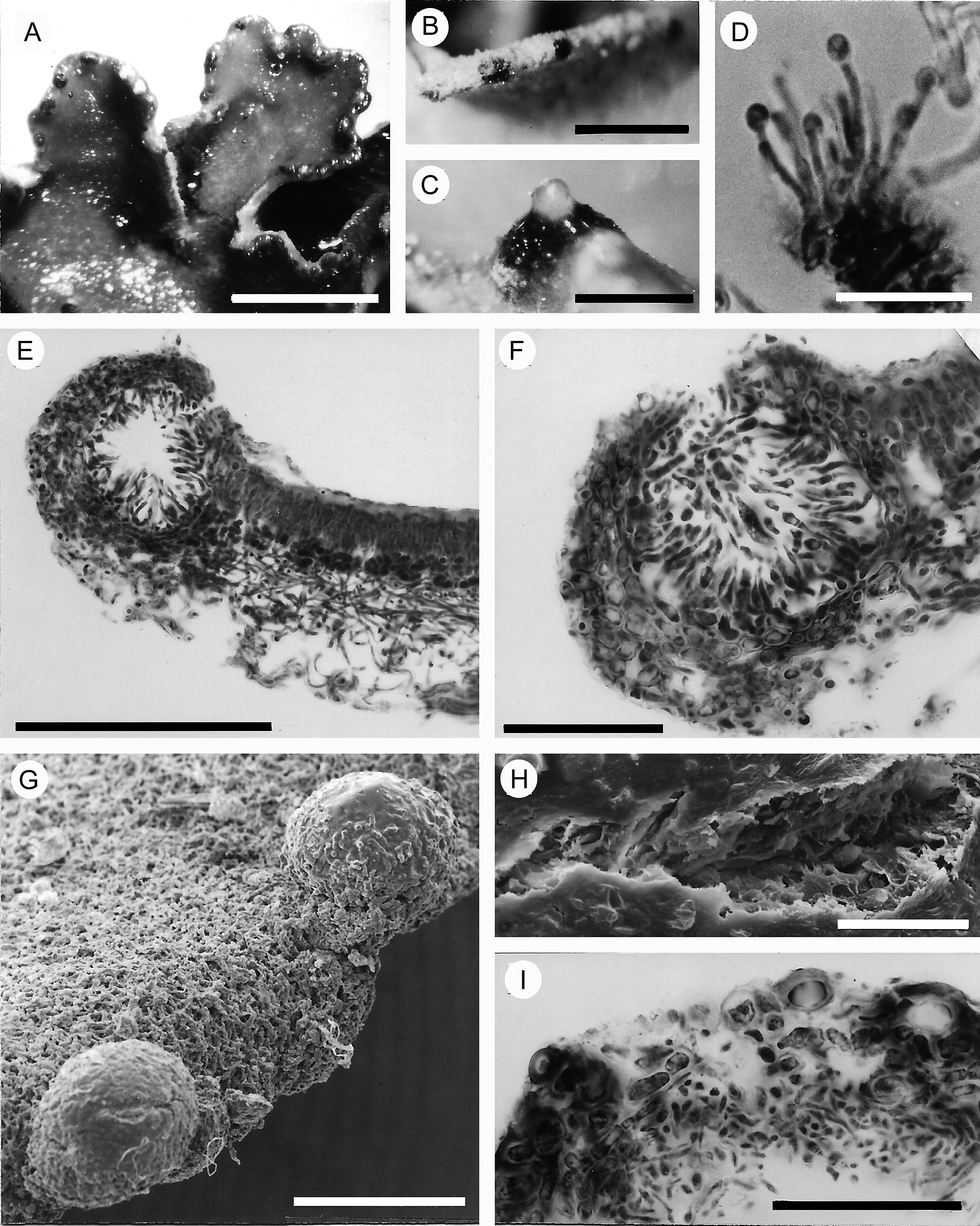
The pycnidia of the Peltigera species studied are restricted to and emerging from the margin of the thallus, and are black when dry (Fig. 1B) and lighter coloured when wet (Fig. 1A), as shown in P. didactyla. In wet material, the mucilaginous substance inside the pycnidium swells and exudes with the conidia through the aperture (Figs 1C & 3H). Vertical sections of the thallus margin of P. degenii (Fig. 1E & F) demonstrate the typical two- to multilayered, parenchymatical upper cortex of Peltigera. In P. laciniata, the cortex is covered with a tomentum, and the upper part of the pycnidium emerges from the surface of the thallus, but free of tomental hyphae (Fig. 1G). The shape of the pycnidium is almost spherical (Fig. 1E & F), expanding to a flattened ellipsoidal shape (Fig. 3G). The pycnidia are unilocular, characterized by a single central cavity, which is lined by the conidia-forming elements. They are oriented centripetally with their conidiophores and conidiogenous cells into the non-chambered pycnidial conidiomata (Figs 1E, 3G & 4D).
Pycnidia in marginal positions in Peltigera didactyla (A–D), P. degenii (E & F) and P. laciniata (G–I). A, moistened thallus lobes with numerous dark pycnidia. B, margin of a dry lobe with two pycnidia. C, moistened pycnidium extruding mass of conidia. D, conidiophores producing conidia at apices. E, transversal section of a lobe with marginal pycnidium. F, young pycnidium. G, margin of a lobe with two pycnidia. H, ostiole aperture in the form of an oblong split on the upper surface of the pycnidium. I, transversal section of a forming ostiole. A–C & I = stereo microscope; D–F = light microscope; G & H = scanning electron microscope. Scales: A = 3.5 mm; B = 0.5 mm; C = 0.25 mm; D & H = 25 μm; E = 150 μm; F = 50 μm; G = 0.3 mm; I = 70 μm.

Figure 1. Long description
Top row, from left to right: Panel A shows moistened thallus lobes of Peltigera didactyla with numerous dark pycnidia along the margins, scale bar 3.5 millimeters. Panel B displays the margin of a dry lobe with two visible pycnidia, scale bar 0.5 millimeters. Panel C presents a moistened pycnidium extruding a mass of conidia, scale bar 0.25 millimeters. Panel D is a close-up under light microscopy of conidiophores producing conidia at their apices, scale bar 25 micrometers. Middle row: Panel E shows a transversal section of a lobe of Peltigera degenii with a marginal pycnidium, scale bar 150 micrometers. Panel F displays a young pycnidium in section, scale bar 50 micrometers. Bottom row: Panel G is a S E M view of a lobe margin of Peltigera laciniata with two pycnidia, scale bar 0.3 millimeters. Panel H is a S E M close-up of an ostiole aperture as an oblong split on the upper pycnidium surface, scale bar 25 micrometers. Panel I shows a transversal section of a forming ostiole, scale bar 70 micrometers. Each panel highlights different developmental stages and anatomical features of marginal pycnidia in three Peltigera species.
The wall of the pycnidium is two-layered. The inner layer is the proper pycnidial wall, composed of pseudoparenchymatous cells with an intimate connection to the base of the conidiophores (Figs 1F, 2A, 3D & 4D). The cells have a cytoplasmic connection by simple septal pores with related Woronin bodies (Fig. 2E). The concentric bodies are located in restricted areas. Large portions of the cytoplasm are taken up by irregularly shaped, intermeshing translucent regions. Their interior forms a net of fine granules. They also include conspicuous, dark, large electron-opaque bodies. In addition, small lipid globules are distributed in the cytoplasm. At the bottom of the pycnidium, the inner wall has direct contact with the hyphae of the medulla (Figs 1E & 3G). Outwardly of the inner wall layer, which is involved in the production of conidia, there occurs the second wall layer. This outer wall of the pycnidium is the direct continuation of the upper cortex of the thallus, with some modifications: the cells are slightly larger and more irregularly shaped (Figs 3G & 4A–D), and their cell walls are thicker and show frequent incrustations of brownish dark pigments (Fig. 3D–G).
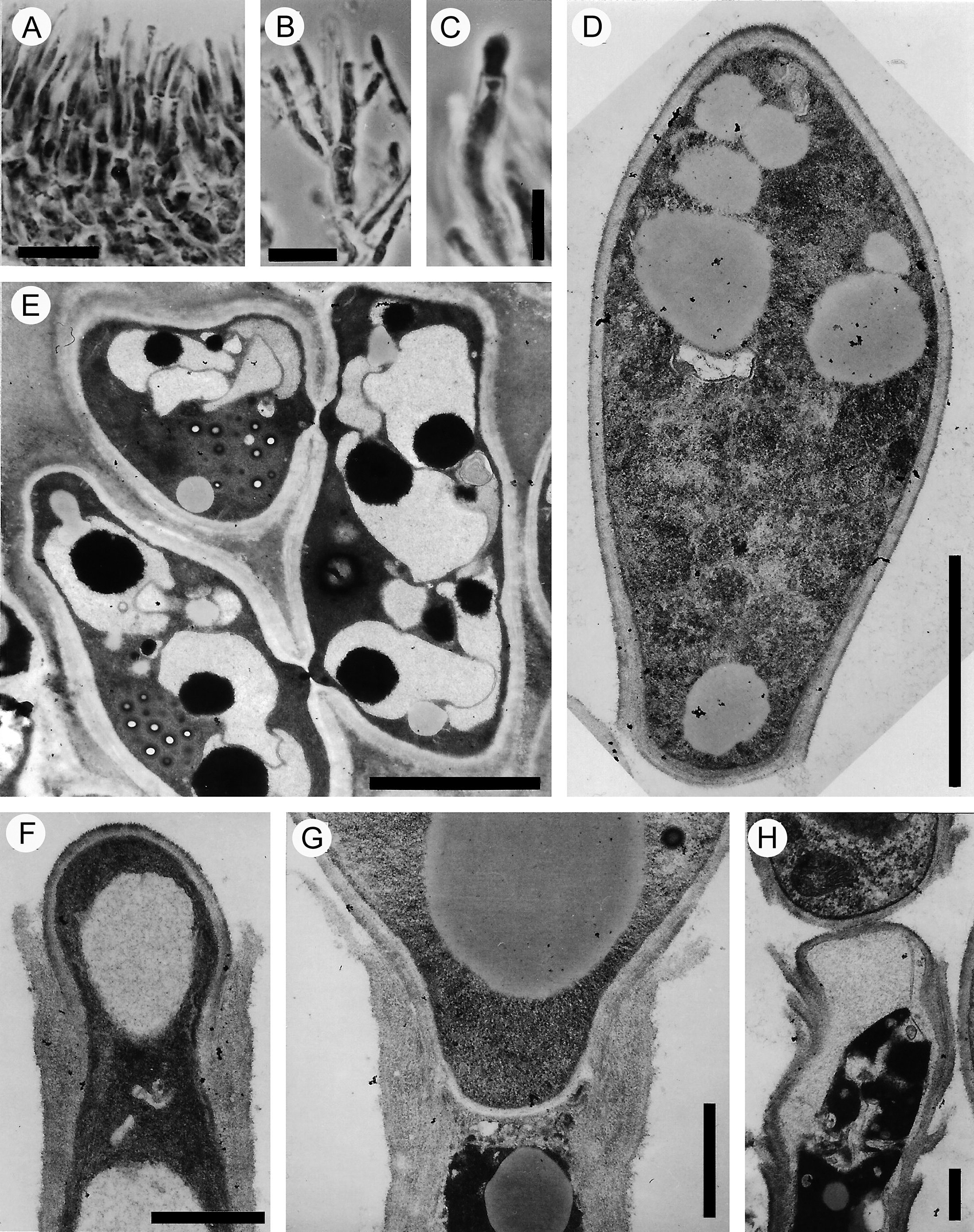
Conidiophores, conidiogenesis and conidia in Peltigera laciniata. A, layer of conidiophores in vertical section. B, articulated conidiophore with elongated conidiogenous cells. C, tip of a conidiogenous cell producing a phialoconidium in acrogenous position. D, unicellular conidium with typical obovate shape. E, anastomosing hyphal cells at the base of conidiophores. F, tip of a conidiogenous cell, starting with enteroblastic formation of a conidium. G, conidium separated transversely from the conidiogenous cell by a conidial septum below the phialidic mouth of the collarette. H, apex of a proliferating conidiogenous cell, phialidic collarettes in varying distances. A–C = light microscope; D–H = transmission electron microscope. Scales: A & B = 10 μm; C = 5 μm; D = 2 μm; E = 3 μm; F & G = 1 μm; H = 0.5 μm.

Figure 2. Long description
Panel A at the top left shows a vertical section of densely packed conidiophores forming a layer, scale bar 10 micrometers. Panel B to the right displays an articulated conidiophore with elongated, branching conidiogenous cells, scale bar 10 micrometers. Panel C further right shows the tip of a conidiogenous cell with a phialoconidium forming at the apex, scale bar 5 micrometers. Panel D at the top right presents a unicellular, obovate conidium with internal granules, scale bar 2 micrometers. Panel E at the bottom left depicts anastomosing hyphal cells at the base of conidiophores, with multiple interconnected cells and dark inclusions, scale bar 3 micrometers. Panel F to the right shows the tip of a conidiogenous cell initiating enteroblastic conidium formation, scale bar 1 micrometer. Panel G further right illustrates a conidium transversely separated from the conidiogenous cell by a septum below the phialidic mouth of the collarette, scale bar 1 micrometer. Panel H at the bottom right displays the apex of a proliferating conidiogenous cell with multiple phialidic collarettes at varying distances, scale bar 0.5 micrometers. All scale bars are black and located at the bottom of each panel.
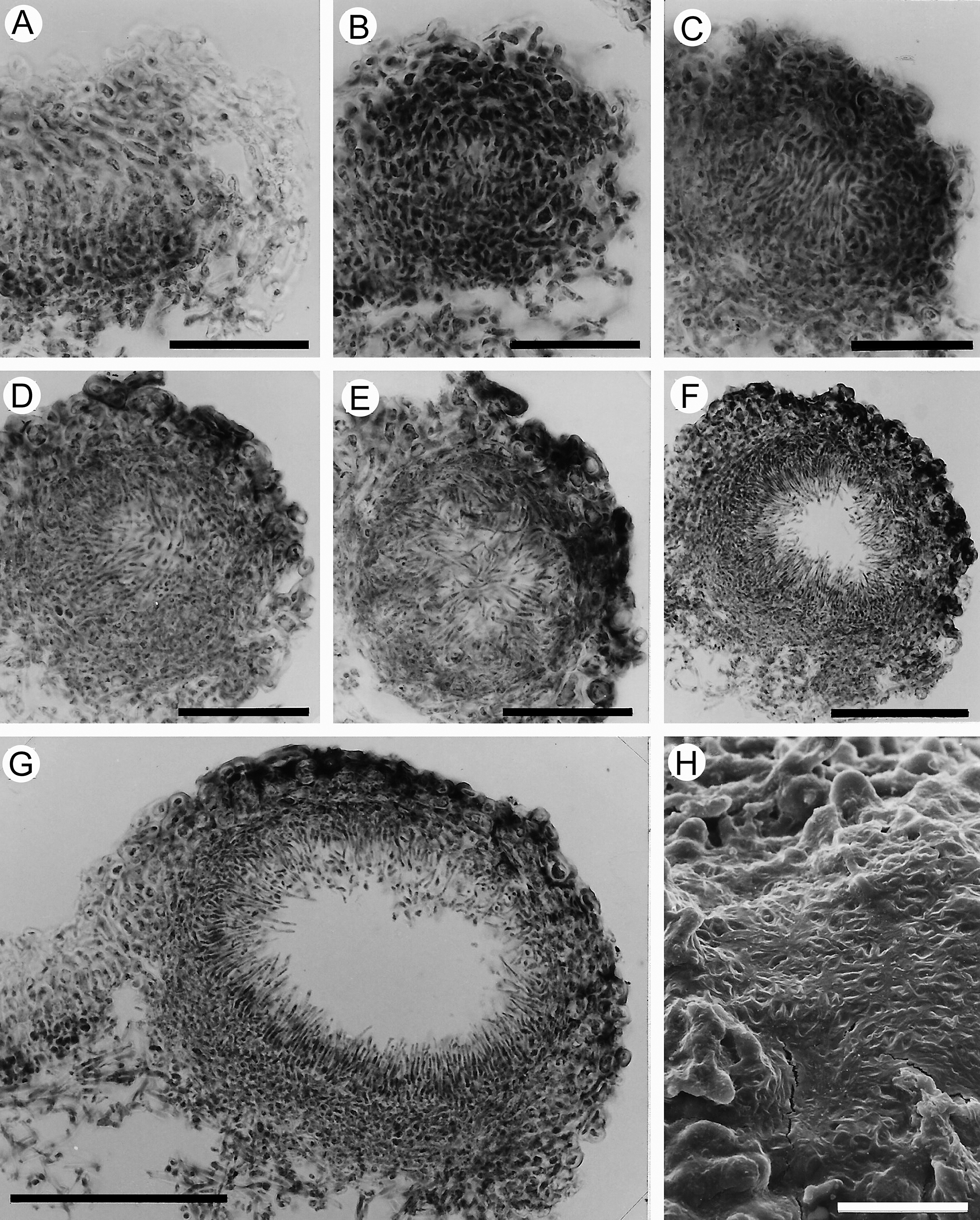
Ontogenetic development of pycnidial conidioma in Peltigera laciniata in a series of microtome sections (A–G) and extruded conidia (H). A, margin of a lobe with the initiation of a primordium consisting of a flattened dense hyphal web. B, the primordium forming a globose shape with a compact cluster of plasma-rich hyphae. C, the inner hyphae are parallel and vertically orientated, surrounded by irregular, isodiametric cells. D, loosening of the hyphal web at the central part. E, beginning of the differentiation of the inner hyphae into future conidiophores. F, expansion of the immature pycnidium with radially orientated young conidiophores, forming a central cavity. G, mature pycnidium with a single cavity, lined by conidia-forming conidiophores, which develop from the inner wall layer. The external wall with its large and darkened cells completely encloses the pycnidium; a typical ostiole is still not formed. H, upper surface of a pycnidium, partly covered with a dried, slimy substance, in which the shapes of the conidia are visible. A–G = light microscope; H = scanning electron microscope. Scales: A–E = 50 μm; F & G = 100 μm; H = 25 μm.

Figure 3. Long description
There are eight panels labeled A through H. Panels A to G are light micrographs arranged in two rows, each showing a transverse section of pycnidial conidioma at different ontogenetic stages. Panel A, top left, shows the margin of a lobe with a flattened, dense hyphal web as the primordium initiates. Panel B, to the right, displays a globose primordium with a compact cluster of plasma-rich hyphae. Panel C, next, reveals inner hyphae that are parallel and vertically oriented, surrounded by irregular, isodiametric cells. Panel D, bottom left, shows loosening of the hyphal web at the center. Panel E, to the right, depicts the beginning of inner hyphae differentiation into future conidiophores. Panel F, next, presents an expanded immature pycnidium with radially oriented young conidiophores forming a central cavity. Panel G, bottom right of the sequence, shows a mature pycnidium with a single central cavity lined by conidiophores, the external wall is composed of large, darkened cells, and no ostiole is visible. Panel H, at the bottom right, is a scanning electron micrograph of the upper surface of a pycnidium, partially covered with a dried, slimy substance, where conidia shapes are visible. Scale bars: A to E equal 50 micrometers, F and G equal 100 micrometers, H equals 25 micrometers.
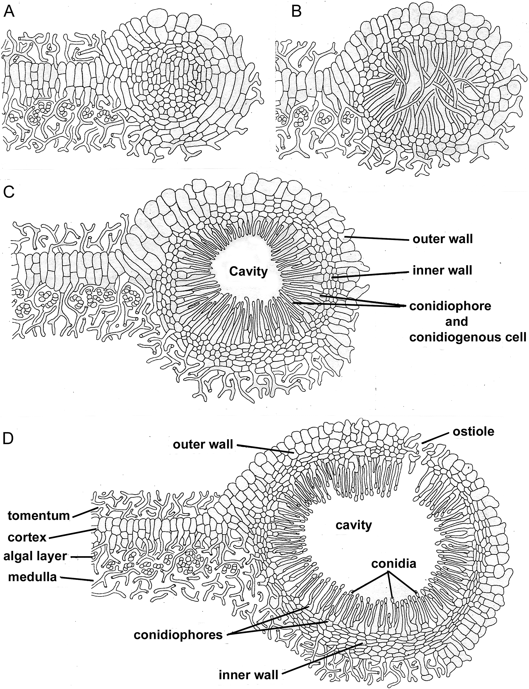
Diagram of pycnidial ontogeny according to the suggested Peltigera-type. A, primordium placed at the margin of a lobe. B, the inner cells of the primordium form a hyphal web. C, immature pycnidium with immature conidiophores, centripetally orientated into the cavity. D, mature pycnidium with production of conidia, and formation of ostiole by rupture of the pycnidial wall on the upper side.

Figure 4. Long description
Panel A, top-left, shows an early primordium at the lobe margin, with densely packed cells forming a rounded structure. Panel B, top-right, depicts the primordium’s inner cells forming a hyphal web. Panel C, bottom-left, presents an immature pycnidium with a central cavity, surrounded by an outer wall and inner wall, and conidiophores with conidiogenous cells oriented toward the cavity. Panel D, bottom-right, illustrates a mature pycnidium: the outer wall is ruptured at the upper side forming an ostiole, the cavity is filled with conidia, and conidiophores line the inner wall. Additional labels in panel D identify the tomentum, cortex, algal layer, and medulla in the surrounding tissue.
Conidiophores
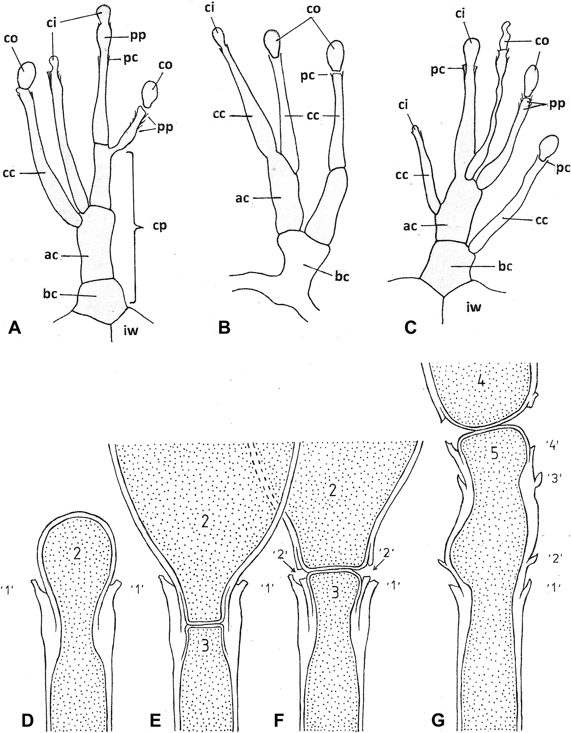
The conidiophores are composed of one to three cells, successive as a very short main axis and the elongated conidiogenous cells, which emerge from this axis (Figs 2B & 5A–C). The conidiophores are integrated with their basic cells into the pseudoparenchymatous tissue of the inner pycnidial wall (Figs 1F, 2A & 4D). The short main axis of the conidiophore can be ramified (Fig. 5B). In general, the conidiogenous cells are born terminally on the axial cells, but also subterminally, even on the basal cell of the conidiophore (Figs 2B & 5C). The conidiogenous cells are thin-walled, slender, non-septate, and strictly directed towards the centre of the pycnidium (Fig. 4D). They form the conidia at their apex (Figs 1D, 2C & 5A–C). The cytoplasm of the conidiogenous cells is similar to that of the base of the conidiophores and the cells of the inner wall layer of the pycnidium (Fig. 2E), with large lipid globules. After the formation of several conidia, the cytoplasmic content of the conidiogenous cells disappears successively, and the cell walls collapse (Fig. 5C).
Peltigera laciniata; diagrams of anatomical variations of conidiophores and conidiogenous cells (A–C), and conidiogenesis (D–G). A, conidiophore with conidiogenous cells in terminal and subterminal positions. B, ramification of the basal cell of the conidiophore. C, formation of conidia in different stages. (Abbreviations: co = conidium; ci = conidium initial; cc = conidiogenous cell; cp = conidiophore; bc = basal cell of conidiophore; ac = axial cell of conidiophore; iw = cell of inner wall; pc = phialidic collarette; pp = proliferating phialide). D, initial stage of enteroblastic development of the second conidium (2) inside a phialidic collarette (‘1’). E, formation of an interspace septum inside the phialidic neck after terminating the growth of the second conidium (2). F, the conidium (2) is pushed out of the neck-channel by the initial of the third conidium (3); a new ‘cuff’ (‘2’) is left behind inside the phialidic collarette (‘1’), provoked by the breaking of the peripheral wall of the longitudinal axis of the conidium (2). G, extended apex of the conidiogenous cell with four collarettes (‘1’–‘4’), originating from proliferating phialidic development of conidia; the fourth conidium (4) is liberated by the force from the initial of the fifth conidium (5).

Figure 5. Long description
The top row contains three panels labeled A, B, and C, each depicting branching conidiophores with labeled parts. In A, the conidiophore (cp) has conidiogenous cells (cc) at terminal and subterminal positions, with conidium initials (ci), conidia (co), phialidic collarettes (pc), proliferating phialides (pp), axial cell (ac), basal cell (bc), and inner wall cell (iw) labeled. B shows ramification of the basal cell (bc) with axial cell (ac), conidiogenous cells (cc), conidium initials (ci), and conidia (co) at the tips. C illustrates multiple stages of conidia formation, with similar labels as A, highlighting proliferating phialides (pp) and phialidic collarettes (pc). The bottom row contains panels D to G, showing conidiogenesis. D depicts the initial stage of enteroblastic development of the second conidium (2) inside a phialidic collarette (‘1’). E shows formation of an interspace septum inside the phialidic neck after the second conidium (2) is formed. F illustrates the second conidium (2) being pushed out by the initial of the third conidium (3), leaving a new ‘cuff’ (‘2’) inside the collarette (‘1’). G shows an extended conidiogenous cell apex with four collarettes (‘1’ to ‘4’), each marking a proliferative event, and the fourth conidium (4) being released by the fifth conidium (5). All abbreviations are labeled as in the caption.
Conidiogenesis and conidia
Conidia are exclusively formed at the tip of the conidiogenous cells (Figs 1D, 4D & 5A–C). In many cases, it is possible to observe a collarette at this apical point (Fig. 2C), which indicates phialidic conidiogenesis (Hawksworth Reference Hawksworth and Galun1988). This is corroborated by electron microscopic studies (Fig. 5D–F).
The position of protrusion of the second conidium is recognizable at a typical collarette, indicated here as ‘1’, because it represents the remains of the production of the first conidium at the tip of the conidiogenous cell. In the middle of the phialidic neck, the cell wall is thickened inwardly, so the conidial primordium appears constrained at this position. The primordial conidium has to pass through this bottleneck and subsequently appears as a swelling head at collarette ‘1’. The cytoplasm of the initial is surrounded from the beginning with its cell wall material (Figs 2F & 5D). When the conidium has reached its complete size, its cytoplasmic connection with the conidiogenous cell is separated transversely by cross-wall layers at the narrowest position of the channel (Fig. 2G). Septal pores were not observed. Simultaneously with the separation of the conidium, new wall material is added to its neighbouring narrow base, forming a cup-like sealing of the conidium (Fig. 5E). This construction is pushed out of the phialidic neck by the upward moving cytoplasm of the conidiogenous cell. The cell wall at the tip of the conidiogenous cell is identical to the septum layer previously formed (Fig. 5F). Subsequently, the conidium is separated when the outer peripheral cell wall layer at the base of the conidium is broken off at the level of the already existing collarette ‘1’. It remains as a collarette, indicated here as ‘2’, between collarette ‘1’ and the upward moving new, third conidium (3) (Fig. 5F). The initial of the conidium (3) is again surrounded by a new cell wall layer. Repetitions of the conidium production lead to an accumulation of layers at the same level and are visible in the light microscope as a collarette of a phialide (Fig. 2C).
Frequently, the production of conidia in repetition shows another variation at the phialidic apex. When the lower cell wall of the conidium does not break off directly at the level of the last-formed collarette, the cuffs left behind do not remain at the same level, but continue their growth simultaneously together with the pushing initial of the following conidium. Subsequent repetitions of this process result in the apex of the conidiogenous cell being lengthened and carrying successive, variably spaced out collarettes (Figs 2H, 5A & G), a pattern previously referred to as proliferation by Vobis (Reference Vobis1980). The conidia are hyaline, unicellular, and their basic shape is obovate. In P. didactyla, they are more spherical (Fig. 1D). The conidium of P. laciniata has a slightly pointed top, and the base is typically edged (Fig. 2D & H). The size range of P. laciniata, calculated from TEM micrographs, is 5.1–6.6 × 2.4–3.5 μm.
Ontogeny of the pycnidia
The development of the pycnidia was studied in Peltigera laciniata (Fig. 3). The first external indication of the early stages is a local brownness at the margin of a lobe. Anatomical studies show an accumulation of cytoplasm-rich hyphae forming a concave structure (Fig. 3A). This concentration of densely interwoven short-celled hyphae expands up to the primordium (Fig. 3B). The inner hyphae of the primordia are parallel in orientation (Figs 3C & 4A). The primordium enlarges and differentiates distinctly into an inner spherical body with tall and loose hyphae, surrounded by small isodiametric hyphae which are outwardly enveloped by irregularly shaped, larger cells. This external cellular layer is pseudoparenchymatous and a continuation of the upper cortex of the thallus (Fig. 4B). Its cell walls become encrusted with dark pigments to form the mature multilayered outer wall of the pycnidium (Fig. 3D & E).
In a further stage, the loosely woven, long hyphae of the inner primordium deviate from each other (Fig. 3E) and change their orientation from parallel to interwoven, finishing in a centripetal formation. These are the first future conidiophores. They attach basally to small, anastomosing cells, which form a thin pseudoparenchymatous layer (Fig. 2E, 4B). With further growth of this pseudoparenchyma, the inner wall of the pycnidium is formed (Fig. 3F). The ultrastructure of the cells of this inner wall is described above under ‘Morphology and anatomy of pycnidia’ (Fig. 2E).
As the volume of the pycnidium doubles (Fig. 3F), the future conidiophores are drawn apart in centrifugal direction by the expanding pycnidial wall and lose their net-like structure. Young conidiophores are produced by the inner wall and pushed between pre-existing ones. Simultaneously, thin and elongated cells are developed from the short basic cells of the conidiophores, which are strongly oriented towards the centre of the pycnidium. These narrow, elongated outgrowths are the first conidiogenous cells (Fig. 4C).
The mature pycnidium is a dark-coloured, spherical to slightly flattened body, located at the margin of the thallus. It can still remain closed at this stage of development (Fig. 3G), although conidia are produced in large numbers (Fig. 4D). The conidia are collected inside the large, central and unilocular cavity, surrounded by conidiogenous cells. The conidiophores occur at the base and along the sides up to the upper part of the unilocular pycnidium (Figs 3G & 4D). The conidia are released by bursting at pre-determined positions of the wall layers (Figs 1I & 4D), either as a circular pore (Fig. 1F) or an oblong split (Fig. 1H). In intensely moistened thalli, the swelling mucous mass of the interior generates sufficient pressure so that the conidia are pressed out and discharged over the surface of the pycnidium (Figs 1C & G, 3H). These types of bursting ostioles do not open with any special ostiolar apparatus.
The inner wall layer of the pycnidium forms the base for the conidiophores, connected to the outer wall layer on the external area of the pycnidium (Figs 1F, 3G & 4D). The outer wall layer is dark, consisting of large luminous, isodiametrical cells (Figs 1E & F, 3G, 4A–D). A special hyphal arrangement suitable for constructing an ostiole could not be observed during the ontogenetic development of the conidiomata.
Discussion
The pycnidial conidiomata, as well as the initials of the hemiangiocarpous ascomata of the genus Peltigera, have the same morphological position on the thallus. Both are located at the margins of the thallus lobes (Henssen et al. Reference Henssen, Keuck, Renner, Vobis and Reynolds1981a). The mature pycnidia remain half-immersed at the same position as in their initial stages (Glück Reference Glück1899; Lindahl Reference Lindahl1959). In spite of the closeness of generative structures, fertilization by transfer of conidial nuclei via trichogynes to ascogones has never been observed, nor has any cellular contact between conidium and trichogyne been demonstrated, as occurs in Cladonia furcata (Huds.) Baumg. (Honegger Reference Honegger1984). Whether somatogamy plays a role in Peltigera, as suggested by Moreau (Reference Moreau1928), is unclear.
Condiomatal ontogeny
The conidiomatal ontogeny of Peltigera laciniata follows the general patterns of other lichenized fungi (Vobis & Hawksworth Reference Vobis, Hawksworth, Cole and Kendrick1981) but also shows some specific characteristics that are discussed below.
The primordia arise in the algal layer and, due to their marginal position, the contact with the photobiont layer is only one-sided. This remains until the pycnidium matures. The initial state with short-celled or isodiametric hyphae, forming a globose complex, is known in many species of distantly related lichenized clades within Lecanoromycetes, Arthoniomycetes and Eurotiomycetes (Glück Reference Glück1899; Letrouit-Galinou Reference Letrouit-Galinou1972, Reference Letrouit-Galinou1984, Reference Letrouit-Galinou1986; Vobis Reference Vobis1980; Janex-Favre & Wagner Reference Janex-Favre and Wagner1986). From the early stages, the primordium is surrounded by a two-layered cell cover formed by the cortical layer. Their initial palisade-like arrangement is modified to irregular isodiametric hyphal cells with slightly thickened cell walls, which turn slightly brownish. The hyphae of the tomentum covering the cortex are not involved in this process. Two-layered primordia of conidiomata are rarely found but have been described from the genera Umbilicaria Hoffm. (Janex-Favre Reference Janex-Favre1977) and Teloschistes Norman (Vobis Reference Vobis1980). However, in the former, the composition of the layers differs, with the outer layer connected to the hyphae of the medulla, whereas the inner layer is connected to the bases of the conidiophores. The primordia in Teloschistes capensis are similar to those found in Peltigera, where the hemispherical surface of the half-immersed primordia is also covered by the thalline cortex.
It is noteworthy that anatomical structures specialized in the liberation of the conidia that accumulated in the cavity of the mature pycnidial conidioma are lacking. Such hyphal elements are typically differentiated at the tip of the pycnidium and border (Glück Reference Glück1899), but are lacking in Pleurosticta acetabulum (Neck.) Elix & Lumbsch (Vobis Reference Vobis1980; Janex-Favre Reference Janex-Favre1982) and in Opegrapha atra Pers. and O. calcarea Turner ex Sm. (Wieczorek Reference Wieczorek2009), where they open by longitudinal or irregular fissures. This is comparable with the observations in Peltigera laciniata, P. didactyla and P. degenii, where predetermined areas enable a mechanical fissure (Figs 1F & I, 4D). These types of openings can be characterized as ‘bursting ostioles’.
The pigmentation of the outer wall of the primordia in Peltigera may be caused by exposure to light, since the tomentum does not extend to the outer area of the thallus. Melanoid pigments in the ostioles or other exposed parts of pycnidia have been described from several exposed pycnidia (Janex-Favre Reference Janex-Favre1980; Vobis Reference Vobis1980). However, in these cases, only parts of the pycnidial wall are pigmented, whereas Peltigera has uniformly pigmented outer walls.
Conidiophores and conidiogenesis
The Peltigera- and Psora-types of conidiophores described by Glück (Reference Glück1899) were subsequently integrated into conidiophore-type II (Vobis Reference Vobis1980). Our results confirm that the conidiophores of Peltigera belong to the type II of Vobis (Reference Vobis1980). The base of the few, short cells of the conidiophores is scarcely separable from the pseudoparenchymatic tissue of the inner wall layer. The conidiogenesis is acrogenous (Vobis & Hawksworth Reference Vobis, Hawksworth, Cole and Kendrick1981). The entire cavity of the pycnidium is lined by conidiophores, including the upper parts (Fig. 4D).
The formation of conidia has been studied traditionally using light microscopy (LM) to understand taxonomic relationships among genera in hyphomycetes (Hughes Reference Hughes1953; Subramanian Reference Subramanian1962; Tubaki Reference Tubaki1963; Barron Reference Barron1968). Various types of conidia were found, and their modes of conidogenesis were arranged in a general scheme, which was subsequently supported by ultrastructural patterns shown by TEM studies (Kendrick Reference Kendrick1971). The distinction of thallic and blastic conidiogenesis and the subdivision in each case into holo- and entero- was used for the interpretation of conidial production in coelomycetes (Sutton Reference Sutton1980) and pycnidial conidiomata of lichenized ascomycetes (Vobis Reference Vobis1980). Simultaneously, these studies were further extended, based on additional results of TEM investigations (Cole & Samson Reference Cole and Samson1979).
The LM examinations show that the conidia of P. laciniata are produced acrogenously from the elongated conidiogenous cells, as in Combea mollusca (Ach.) Nyl., Dirina ceratoniae (Ach.) Fr. or Lecanactis abietina (Ehrh. ex Ach.) Körb. (Vobis Reference Vobis1980). Although the small size of the apex of the conidiogenous cells complicates the determination of the conidiogenesis, it was possible to observe a collarette at the exit point of the conidium, which is consistent with a phialidic development (Fig. 2C). The initiation of the first conidium of P. laciniata was not observed in TEM. As shown in micrograph Fig. 2F and diagram Fig. 5D, the initial of the second conidium passes through the scar, which was left behind by the first conidium, but does not include parts of the conidiogenous cell wall. The initial is completely enveloped by wall material produced from a newly laid down, internal wall layer, synthesized at the level of the mouth of the apex of the conidiogenous cell. This corresponds to enteroblastic formation (Kendrick Reference Kendrick1971; Hawksworth Reference Hawksworth and Galun1988).
Only when the conidium has developed to its full size does it become separated from the cytoplasm of the conidiogenous cell by a septum located at the smallest part of the phialidic neck (Fig. 5E). This cross-wall is distinctly two-layered and may be called an ‘interspace septum’, a term used to describe a comparable analogous process in actinomycetes (Henssen et al. Reference Henssen, Weise, Vobis and Renner1981b; Vobis Reference Vobis, Ortiz-Ortiz, Bojalil and Yajkoleff1984). The upper cross-wall layer belonging to the conidium may already be fully developed, whereas the layer belonging to the conidiogenous cell is being formed (Fig. 2G). A two-layered septum was also found in the phialidic conidiogenesis in Parmelina tiliacea (Hoffm.) Hale (Honegger Reference Honegger1984).
Distinct collarettes at the apex of the conidiogenous cell, usually observable with LM, are called annellations. They are developed after each formation of a conidium at regular distances on the prolongating apex of the conidiogenous cell. Kendrick (Reference Kendrick1971) postulated that the original wall structures of the conidiogenous cell are involved in this successive process. Therefore, the annellidic conidiogenesis would be holoblastical. This correlative concept was questioned by examples of coelomycete conidiomata, correlated with phialidic conidiogenesis (Morgan-Jones et al. Reference Morgan-Jones, Nag Raj and Kendrick1972; Reisinger et al. Reference Reisinger, Morelet and Kiffer1977; Sutton Reference Sutton1980). Hence, the conidiogenous cells described as annellides in pycnidial conidiomata of Combea mollusca, Dirina ceratoniae (Vobis Reference Vobis1980) or Lecanactis abietina (Honegger Reference Honegger1984) could also be interpreted as modifications of proliferating phialides (Vobis & Hawksworth Reference Vobis, Hawksworth, Cole and Kendrick1981; Hawksworth Reference Hawksworth and Galun1988). Here, we follow the practical approach of Sutton (Reference Sutton1980) and interpret it as annelidic since they are observable with LM.
In Peltigera laciniata, the conidia of the pycnidial conidiomata are developed enteroblastically. The collarettes are formed successively, together with every newly produced conidium. The collarettes may remain inside the phialidic neck, which is extended by the multiple layers of wall material at the same level (Fig. 5F), and are seen as a typical phialide in LM. On the other hand, proliferating phialides can also be seen in EM studies. The conidiogenous cell elongates apically, and the new collarettes are attached outside to the cell wall, often at irregular distances (Fig. 5G).
Conidia
The conidia in the Peltigera species studied are one-celled and obovate with thin, hyaline walls, and filled with fatty globules (Tulasne Reference Tulasne1852; Glück Reference Glück1899; Lindahl Reference Lindahl1959). When the conidia are released from the phialidic neck, they may not be immediately set free but can remain loosely attached by their lower part to the primordium of the next conidium (Figs 2H, 5F & G). However, no conidial chains are formed, but they may stay tied together by residual products of the lysis remaining between the layers of the wall (Reisinger et al. Reference Reisinger, Morelet and Kiffer1977). The size of the conidia in the Peltigera species studied agrees with literature reports, measuring between 6–13 × 2.5–5.5 μm (Lindahl Reference Lindahl1959; Vitikainen Reference Vitikainen1994).
Author ORCID
Thorsten Lumbsch, 0000-0003-1512-835X.
Acknowledgements
We thank the late Aino Henssen, our instructor in lichenology for many years at the University of Marburg; Martina Schmidt and María Havrylenko for providing samples; Orvo Vitikainen for assistance with the determination of the material; and Susana Calvelo for stimulating discussions.