Introduction
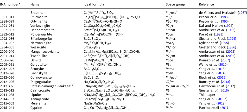
The Kalahari Manganese Fields in the North Cape Province of South Africa hosts the largest manganese resource on Earth, ~4 billion tons, accounting for 80% of the mineable manganese ore on land (Beukes, Reference Beukes, Trendall and Morris1983). There are two types of manganese ores present. The low-grade Mamatwan type, making up ~97% of the total reserve, is composed of microcrystalline braunite and kutnohorite lutite and occurs as stratiform beds in the Hotazel Formation of the Paleoproterozoic Transvaal Supergroup. The high-grade Wessels-type ore, constituting mainly of hausmannite, bixbyite, braunite-II, and manganite, was formed from structurally controlled hydrothermal enrichment around 1.0–1.25 Ga (Cairncross and Beukes, Reference Cairncross and Beukes2013; Gutzmer and Beukes, Reference Gutzmer and Beukes1996; Dixon, Reference Dixon1989). Since 1981, 24 new minerals discovered in the Kalahari Manganese Fields have been approved by the International Mineralogical Association Commission on New Minerals, Nomenclature and Classification (IMA–CNMNC) (Pasero, Reference Pasero2019) (Table 1), most of which are from the ores of the high-grade Wessels type.
List of new minerals discovered in the Kalahari Manganese Field.

*The data for IMA approval number are from the official website of IMA-CNMNC (Pasero, Reference Pasero2019).
**Potassic-mangani-leakeite was previously named as kornite by Armbruster et al. (Reference Armbruster, Oberhänsli, Bermanec and Dixon1993) and redefined by Hawthorne et al. (Reference Hawthorne, Oberti, Harlow, Maresch, Martin, Schumacher and Welch2012)
The new mineral, lipuite, is named in honour of the late Chinese geochemist and petrologist, Professor Pu Li (1911–1968). Professor Li received his BSc degree from the Southwest Union University in China in 1942 and PhD from the University of Cambridge in England in 1950. He then became a Professor at the Beijing Institute of Geology and later at the Guiyang Institute of Geochemistry, Chinese Academy of Sciences. Professor Li was the founder and pioneer of isotope geochemistry in China. He also made significant contributions to the development of Chinese geology in general, and petrology in particular. Both the new mineral and its name have been approved by IMA–CNMNC (IMA2014–085, Yang et al., Reference Yang, Gu, Xie, van Nieuwenhuizen, Evans and Downs2015b). The cotype sample has been deposited in the collections of the Mineral Museum of the University of Arizona, Tucson, Arizona, USA (UAMM #20010) and the RRUFF Project with deposition number R140946 (http://rruff.info/R140946).
Occurrence and paragenesis
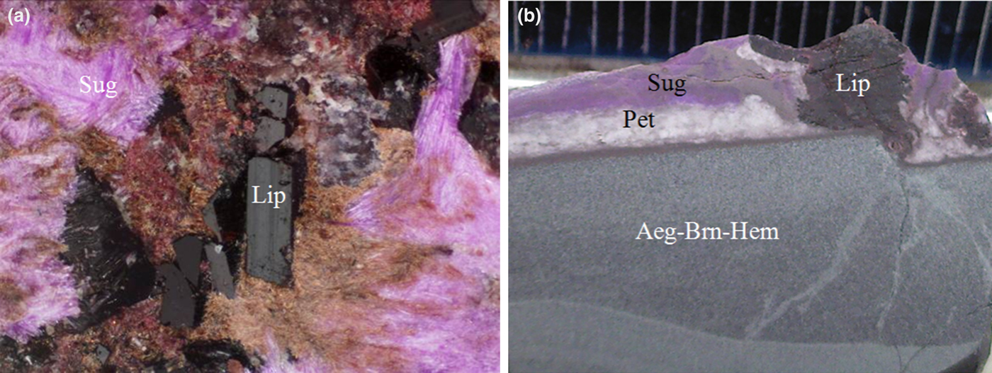
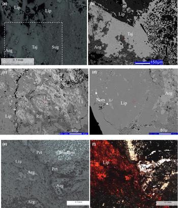
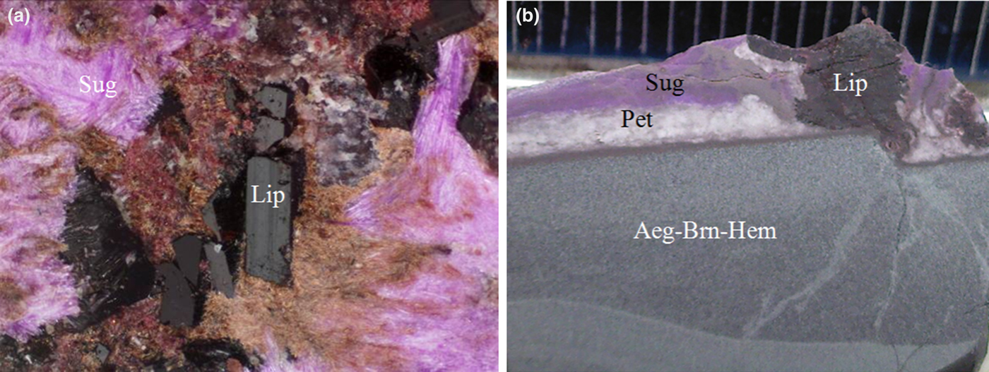
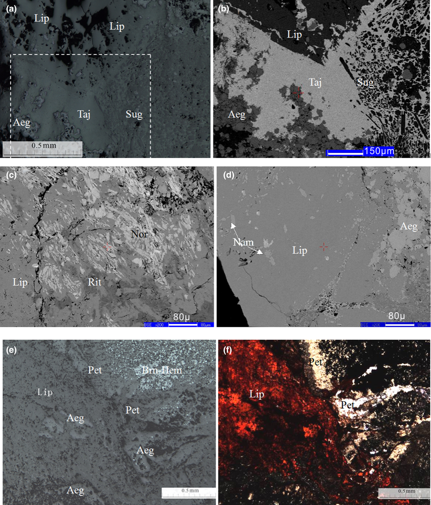
Lipuite was found on a number of specimens collected from the N'Chwaning III mine (part of the Wessels Mine), 27°7′51″S, 22°50′29″E. In the specimens, a crust composed of sugilite–lipuite–pectolite–taniajacoite, 2–5 mm thick, was found on a matrix of aegerine–braunite-II–hematite (Fig. 1). Associated minerals include Mn-bearing sugilite, taniajacoite, pectolite and richterite (Fig. 2a,b). Norrishite and namansilite are occasionally observed using microscopy as tiny grains in lipuite (Fig. 2c,d). Aegirine, hematite and braunite-II are commonly replaced by lipuite (Fig. 2e,f). The hydrothermal event that formed the observed mineral assemblage has been estimated to be at conditions in the range of 270–420°C at 0.2–1.0 kbar (Kleyenstuber Reference Kleyenstuber1984; Gutzmer and Beukes Reference Gutzmer and Beukes1996).
Occurrence and crystal morphology of lipuite: (a) euhedral platy dark crystals of lipuite (Lip) with pink-coloured sugilite (Sug), image width = 2 cm, type specimen R140946 stored in the Mineral Museum of the University of Arizona; (b) lipuite–sugilite–pectolite (Pet) crust on microcrystalline aegirine–braunite II–hematite agglomerate (Aeg–Brn–Hem), section vertical to the specimen surface, image width = 2 cm, specimen used to make lpblock07, lpblock08, lpblock09.
Photomicrographs and back-scattered electron images (BSE) of lipuite showing the mode of occurrence and optical properties: (a) assemblage of lipuite (Lip), sugiilite (Sug) and taniajacoite (Taj) with relics of aegirine (Aeg), parallel nicols, reflected light; (b) BSE image of the framed area in (a); (c) tiny crystals of norrishite (Nor) and richterite (Rit) in lipuite (BSE image); (d) namansilite (Nam) and aegirine in lipuite (BSE image); (e) lipuite with pectolite (Pet), aegirine, hematite (Hem) and braunite-II (Brn), parallel nicols, reflected light; and (f) characteristic red colour of lipuite, the same view as (e), parallel nicols, transmitted light.
Physical and optical properties
Lipuite occurs as platy and granular crystals, 1–3 mm long, or as dark veined agglomerates in the specimens (Fig. 1a), in which lipuite shows a dark brown colour with vitreous lustre. The mineral is brittle with uneven fracture. The Mohs hardness is estimated to be ~5 as tested on the specimen by apatite and orthoclase standards, and the streak is red. The measured density by the method of flotation in heavy liquids is 2.83(3) g/cm3, which is in good agreement with the calculated density of 2.86 g/cm3. Lipuite is transparent in thin section and features a characteristic red colour with weak pleochroism from bright red to dark red in parallel nicols (Fig. 2f). It is biaxial (+) and the interference colour in crossed nicols is also red, probably due to the strong influence of the intrinsic colour as seen in parallel nicols. The measured refractive indices are: α =1.635(1), β = 1.653(1) and γ = 1.670(1), with refractive dispersion of v > r. The measured 2V angle is 86(2)°. The calculated compatibility index (0.009) using the constants of Mandarino (Reference Mandarino1981) yields a superior compatibility with the Gladstone–Dale relationship.
Chemical composition
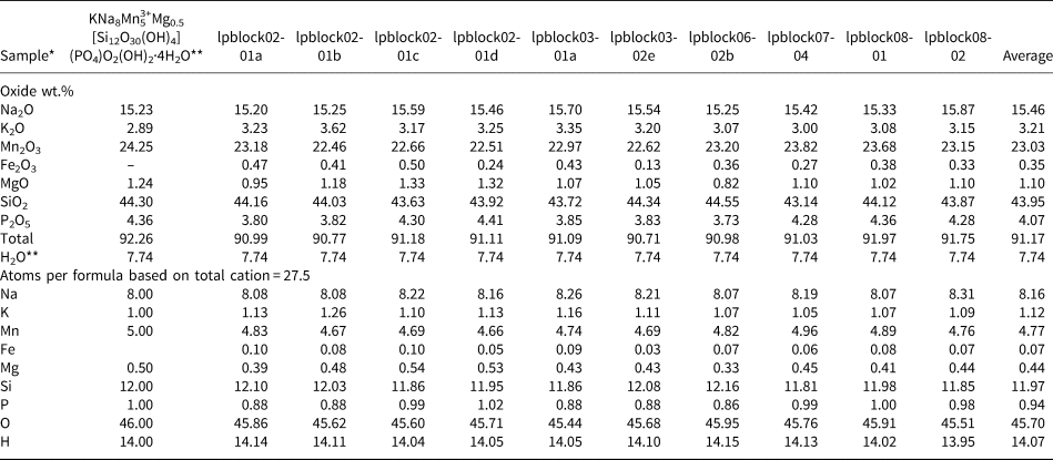
The chemical composition of lipuite was determined on a Shimadzu EPMA-1720 microprobe in the School of Geosciences and Infophysics of Central South University by wavelength-dispersive spectroscopy with operating conditions of accelerating voltage = 15 kV, beam current = 10 nA and beam size = 1 μm. Qualitative analysis revealed dominant Na, K, Mn, Si, P, O and minor Mg and Fe. An intensive X-ray photoelectron spectrometer (XPS) scan of the Li-1s electron was made to determine whether Li was present using a Thermo ESCALAB 250Xi XPS in the Modern Analysis and Testing Centre of Central South University; no peaks were detected in the binding energy range characteristic of Li (53–58 eV). Quantitative analysis was carried out using the following standards (and lines): SiO2 (SiKα); Ca5(PO4)3F (PKα); MgO (MgKα); MnSiO3 (MnKα); Fe2O3 (FeKα); NaAlSi2O6 (NaKα); and KAlSi3O8 (KKα). The ZAF3 correction program supplied with the instrument was used for the correction calculation. During analysis, the sample stage was moved around to prevent beam damage to the sample, which was observed to have significantly affected the quantitative analysis. All Fe was assumed to be Fe3+ because of the association with trivalent Mn. The theoretical content of 7.74 wt.% of H2O were used for calculating the number of atoms in the empirical formula. Representative data (Table 2) yield an empirical formula for the average based on total 27.5 cations and according to the structural analysis: K1.12Na8.16(Mn4.77Fe0.07)Σ4.84Mg0.44[Si11.97O30(OH)4](PO4)0.94O2(OH)2·4H2O. The ideal formula is KNa8Mn3+5Mg0.5[Si12O30(OH)4](PO4)O2(OH)2·4H2O.
Chemical composition of lipuite from electron microprobe analyses.

*Several analyses made on one grain are labelled as 01a, 01b, 01c, 01d etc., and one analysis on a single grain is labelled as 01, 02 etc. without letter a, b etc.
**Ideal formula for comparison; ***Theoretical ideal H2O contents are used for all analyses; ‘–’ = not present.
Raman spectrum
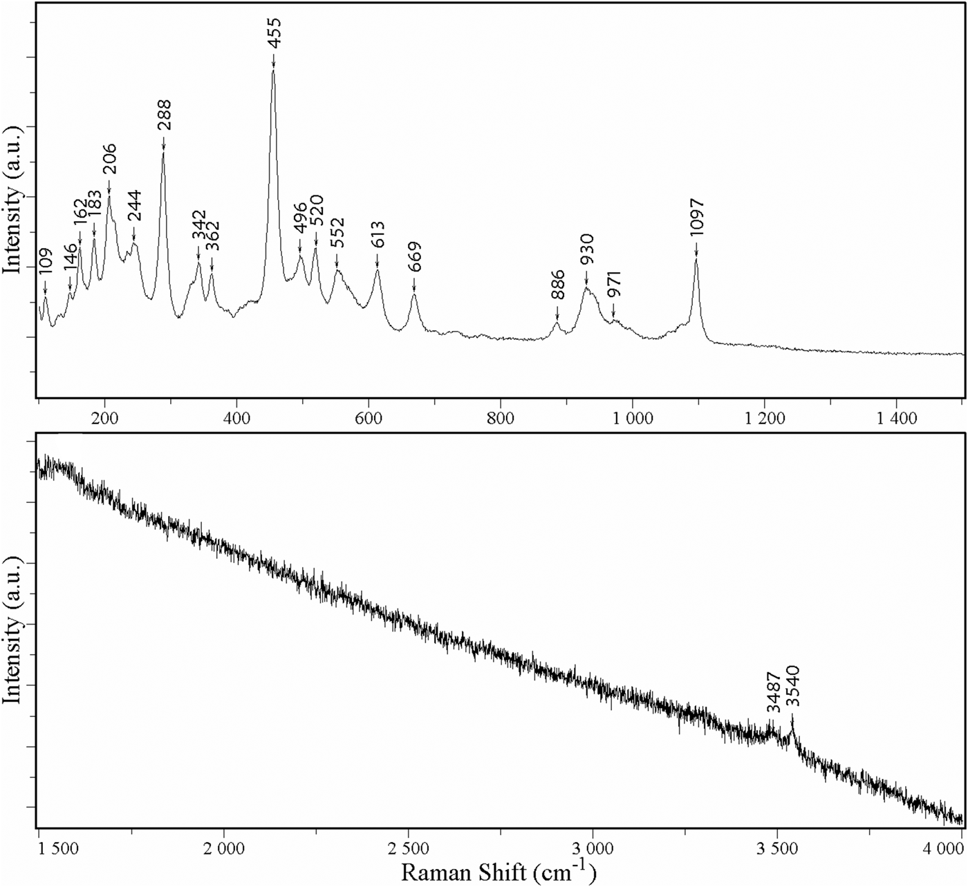
The Raman spectra of lipuite from 12 spots in 4 polished sections were recorded with a Horiba Labram Aramis Raman spectrometer in the State Key Laboratory of Powder Metallurgy, Central South University. The laser beam (He-Ne laser, 632.8 nm, laser power = 2 mW) was focused to 1 μm with a 100× objective in an Olympus microscope. The time of each scan in the range 100–4000 cm–1 was 20 min with a resolution of 2 cm–1. A representative spectrum is shown in Fig. 3. The spectrum is composed of over 21 bands at 109, 146, 162, 183, 206, 244, 288, 342, 362, 455, 496, 520, 552, 613, 669, 886, 930, 971, 1097, 3487 and 3540 cm–1. The bands between 880 and 1097 cm–1 can be assigned to P–O and Si–O stretching vibrations of the PO4 and SiO4 groups (e.g. Litasov and Podgornykh, Reference Litasov and Podgornykh2017; Chakhmouradian et al., Reference Chakhmouradian, Cooper, Ball, Reguir, Medici, Abdu and Antonov2014). Bands from 430 to 700 cm–1 are attributable to O–P–O and O–Si–O bending. The bands below 400 cm–1 are associated mainly with rotational and translational modes of PO4 and SiO4 tetrahedra, as well as Mn–O interactions and lattice modes. The bands between 3487 and 3540 cm–1 are due to O–H stretching.
Raman spectrum of lipuite (sample No. lpblock02-01a in Table 2)

X-ray crystallography and structure determination
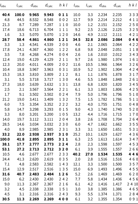
The powder X-ray diffraction data were collected on a Rigaku D/Max Rapid IIR micro-diffractometer in School of Geosciences and Infophysics of Central South University at 40 kV and 250 mA using a 0.1 mm collimator and exposure time of 60 min. The diffraction data are listed in Table 3. The refined unit-cell parameters are: a = 9.0772(6), b = 12.2231(8), c = 17.095(2) Å, V = 1896.7(1) Å3 and Z = 2.
Powder X-ray diffraction data for lipuite.

The strongest peaks are given in bold
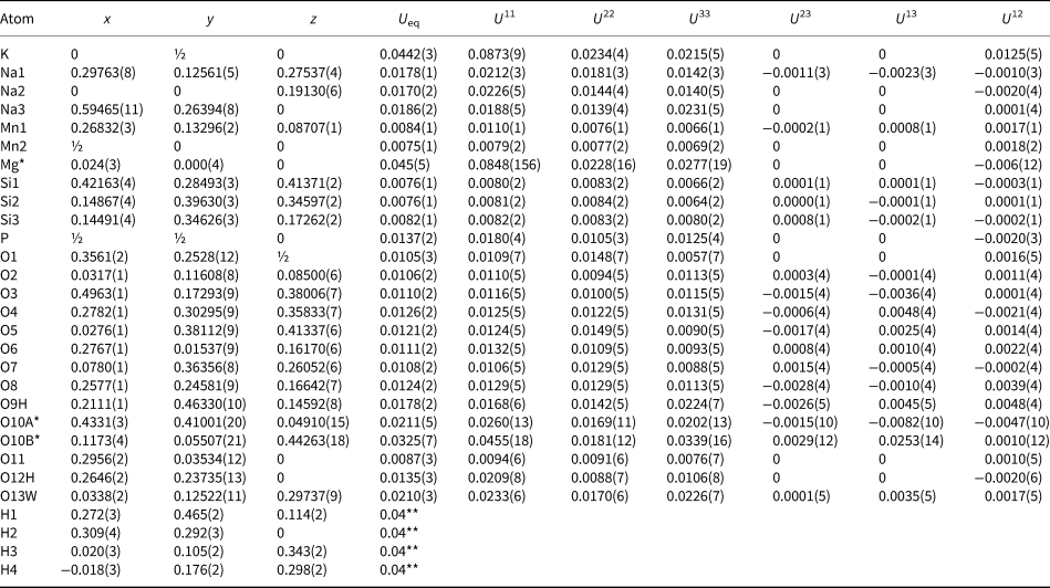
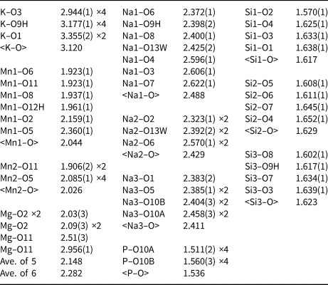
Single-crystal X-ray diffraction data of lipuite were collected on a Bruker X8 APEX2 CCD X-ray diffractometer equipped with graphite-monochromatised MoKα radiation in the Department of Geosciences, University of Arizona, and frame widths of 0.5° in ω and 30 s counting time per frame. All reflections were indexed on the basis of an orthorhombic unit cell (Table 4). The intensity data were corrected for X-ray absorption using SADABS (Bruker, Reference Bruker2001). The systematic absences of reflections suggest the possible space group Pnn2 or Pnnm. The crystal structure was solved and refined using SHELX-2014 (Sheldrick, Reference Sheldrick2015) based on the space group Pnnm, because it yielded better refinement statistics in terms of bond lengths and angles, atomic displacement parameters, and R factors. There are two possible models for Mg in lipuite, both resulting in the same R factors and partial site occupancies. In Model A, Mg is situated at the (0,0,0) site with ~40% occupancy and exhibits a (4 + 2) coordination (four O2 atoms at 2.051 Å and two O11 atoms at 2.718 Å). This model, nevertheless, produces an enormously elongated displacement ellipsoid along the a axis. In model B, Mg occupies a split site at (0.0265, 0.0003, 0) (Table 5), with a (4 + 1) coordination (two O2 atoms at 2.03 Å, two O2 atoms at 2.09 Å, one O11 atom at 2.09 Å, and one O11 atom at 2.956 Å) (Table 6), similar to that observed in calcioferrite (Lafuente et al., Reference Lafuente, Downs, Yang and Jenkins2014). This model results in a total Mg site occupancy similar to that in Model A, but a relatively more reasonable displacement ellipsoid. Consequently, Model B is adopted in this study.
Summary of crystal data and refinement results for lipuite.

Atomic coordinates and displacement parameters for lipuite.

*The site occupancies Mg, O10A, and O10B are all 0.5. ** The values for the H atoms are U iso values.
Selected bond distances (Å) in lipuite.

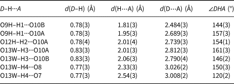
All four H atoms were located from the difference-Fourier synthesis. The positions of all atoms were refined with anisotropic displacement parameters, except for H atoms, which were refined with a fixed U iso value of 0.04. For simplicity, the ideal chemistry was assumed during the structure refinements. The final coordinates and displacement parameters of atoms, selected bond distances, and the hydrogen-bond scheme for lipuite are given in Tables 5, 6 and 7, respectively. The crystallographic information files have been deposited with the Principal Editor of Mineralogical Magazine and are available as Supplementary material (see below).
Hydrogen bonds in lipuite.

D – donor; A – acceptor.
Crystal structure description and discussion
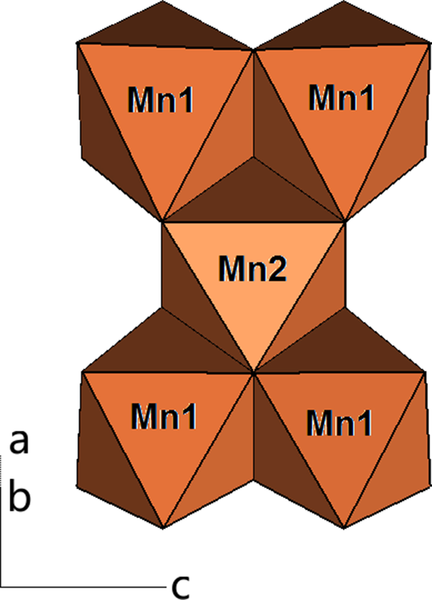
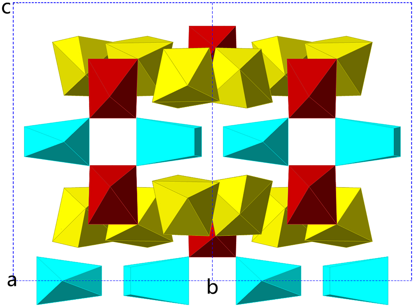
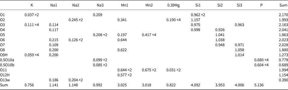
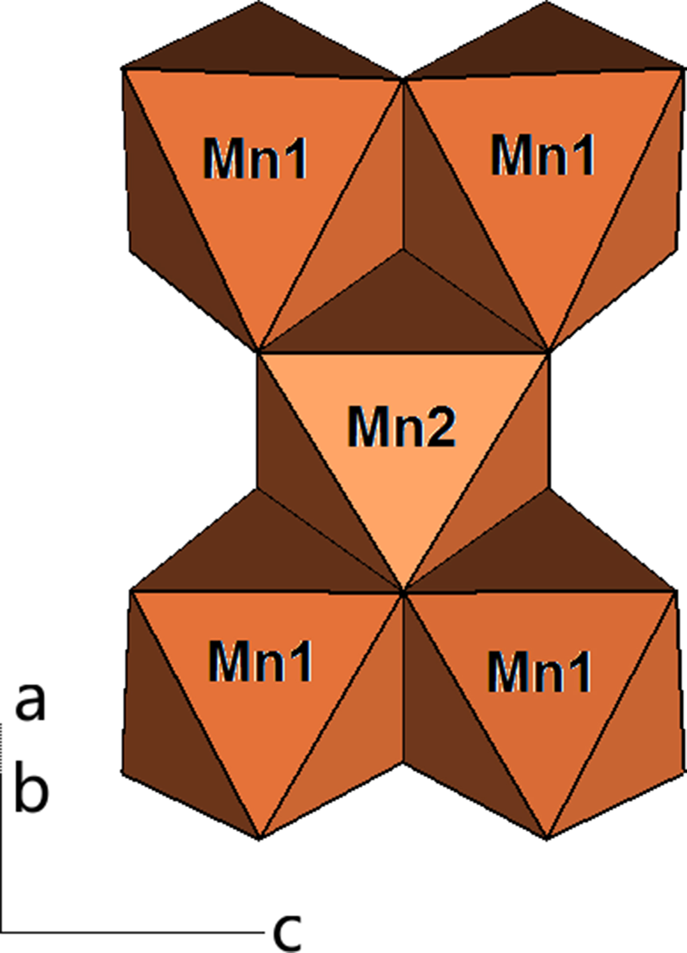
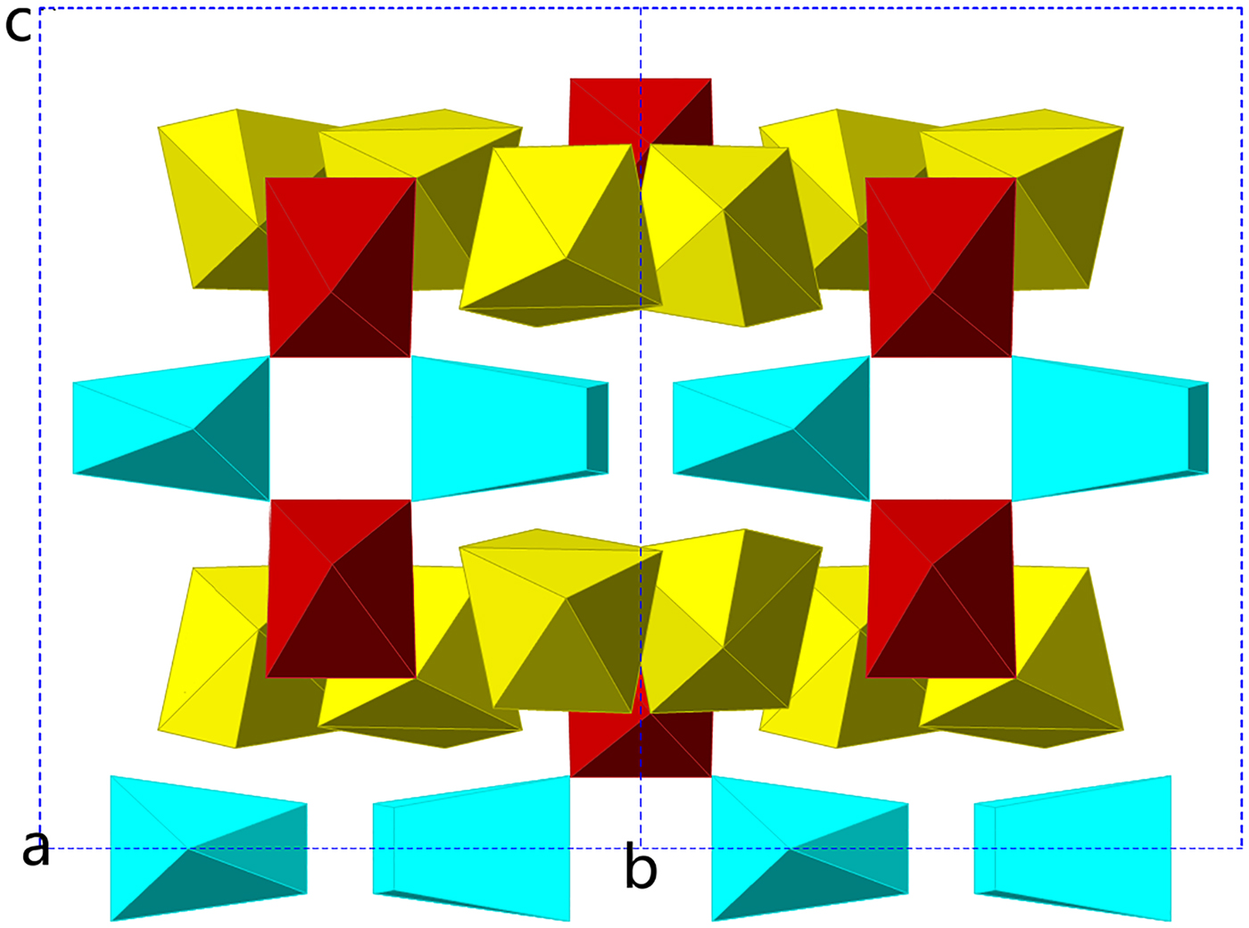
The crystal structure of lipuite is characterised by sheets of SiO4 tetrahedra that are linked together along [010] by K+, Na+, Mn3+, Mg2+ and P5+ cations, as well as hydrogen bonds (Fig. 4). These tetrahedral sheets consist of 14-membered rings of SiO4 tetrahedra that zigzag along [100] (Figs 4 and 5). Calculation of bond-valence sums (Table 8) indicates that one of the O atoms bonded to Si3 is protonated (O9–H) (Table 6). The two independent Mn3+ cations are both octahedrally coordinated and they form five-membered, edge-shared octahedral clusters (Fig. 6) between the SiO4 tetrahedral sheets. Among three distinct Na cations, Na1 and Na3 are 7-coordinated, whereas Na2 is 6-coordinated (Table 4, Fig. 7).
Crystal structure of lipuite, showing the zigzagged SiO4 tetrahedral sheets stacking along the b axis, linked together by K+, Na+, Mn3+, Mg2+ and P5+, which are represented by larger turquoise, smaller turquoise, brown, red and small yellow spheres, respectively.

SiO4 tetrahedral sheet consisting of 14-membered rings in lipuite: Si1, Si2 and Si3 tetrahedra are represented by green, red and yellow colours, respectively.

A five-membered cluster of edge-shared Mn-octahedra in lipuite
7-coordinated polyhedra (Na1 and Na3) and 6-coordinated polyhedra (Na2) in lipuite.
Bond-valence sums for lipuite*.

* Parameters for calculations of bond-valence sums were taken from Brese and O'Keeffe (Reference Brese and O'Keeffe1991).
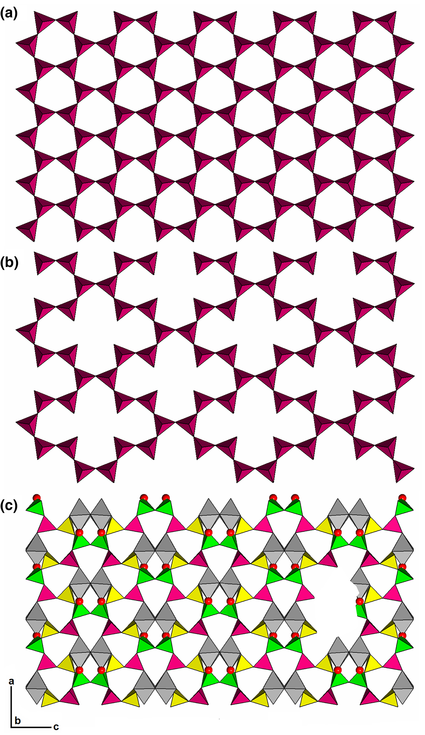
Lipuite represents a unique structure type. Nevertheless, its silicate tetrahedral sheets can be considered a derivative of the silicate sheets in mica, as illustrated in Fig. 8. A silicate sheet consisting of 6-membered rings in mica is shown in Fig. 8a. If two tetrahedra are removed from every four 6-membered rings in the sheet, as illustrated in Fig. 8b, then we get a silicate sheet that is topologically identical to that in lipuite (Fig. 5). Furthermore, if we replace the two ‘removed’ SiO4 tetrahedra with two edge-sharing MnO6 octahedra, we obtain the polyhedral sheet found in lipuite, which consists of two types of 6-membered rings, with one made of four tetrahedra and two octahedra and the other of five tetrahedra and one octahedra (Fig. 8c). The Mn1–O2 bonds are the cause for the kinking of the silicate sheets in lipuite (Fig. 4). Therefore, lipuite may be classified into Class 72 – Two-Dimensional Infinite Sheets with other than six-membered rings in the Dana system or VIII/HX Unclassified Strunz Phyllosilicates in the Strunz system.
Comparison of tetrahedral sheets in mica (a) and lipuite (b) and the location of Mn octahedra (c). Si1, Si2 and Si3 tetrahedra are represented by green, purple and yellow colours, respectively, and Mn octahedra by grey.
Interestingly, the O atoms bonded to P5+ are disordered over two distinct sites (O10A and O10B) with each half occupied, giving rise to an apparent distorted cubic coordination for P5+. This disorder may be partly explained by the weak bonding environments around the PO4 group, which is only bonded to Na+ and H+ (hydrogen bonds). Similar disorder of the O atoms in the PO4 group has also been observed in the eulytine-type compound Sr3La(PO4)3 (Barbier et al., Reference Barbier, Greedan, Asaro and McCarthy1990). A different kind of disorder of PO4 groups has been observed in whitlockite-type minerals and compounds (Zatovsky et al., Reference Zatovsky, Strutynska, Baumer, Shishkin and Slobodyanik2007; Hughes et al., Reference Hughes, Jolliff and Rakovan2008 and references therein), in which the P position in the PO3OH or PO3F group is split; it is randomly located either just above or below the midplane, leading the apical OH– or F– anion to point either along + c or –c.
There are two unique OH groups and one unique H2O in lipuite. The Raman measurements reveal two weak bands in the O–H stretching region, one at 3487 and the other 3540 cm–1 (Fig. 3). According to the correlation between O–H stretching frequencies and O–H···O hydrogen bond lengths in minerals (Libowitzky, Reference Libowitzky1999), these two bands would correspond to an O–H···O distance of 2.8–3.0 Å, agreeing with O–H···O distances for the hydrogen bonds formed by H2O (Table 5).
Acknowledgements
This study was funded by the Science Foundation Arizona and partial support from National Science Foundation of China (Grant No. 41172042).
Supplementary material
To view supplementary material for this article, please visit https://doi.org/10.1180/mgm.2019.12