Introduction
Haemozoin (HZ) or malaria pigment is a crystal made of ferriprotoporphyrin IX dimers derived from the catabolism of haemoglobin by intraerythrocytic malaria parasites (Pagola et al., Reference Pagola, Stephens, Bohle, Kosar and Madsen2000; Egan, Reference Egan2008). HZ is released in the circulation during the intraerythrocytic cycle of parasites and can modulate the functions of different host cell types (Tyberghein et al., Reference Tyberghein, Deroost, Schwarzer, Arese and Van den Steen2014; Deroost et al., Reference Deroost, Pham, Opdenakker and Van den Steen2016). On endothelial cells, HZ modulates the expression of adhesion molecules and the production of inflammatory mediators such as cytokines, chemokines and metalloproteinases (Taramelli et al., Reference Taramelli, Basilico, De Palma, Saresella, Ferrante, Mussoni and Olliaro1998; Basilico et al., Reference Basilico, Parapini, Sisto, Omodeo-Salè, Coghi, Ravagnani, Olliaro and Taramelli2010, Reference Basilico, Corbett, D’ Alessandro, Parapini, Prato, Girelli, Misiano, Olliaro and Taramelli2017). In complicated Plasmodium falciparum malaria, such as cerebral and placental malaria, activation of endothelial cells contributes to the cytoadherence of infected red blood cells to the microvascular endothelium, leading to micro-circulatory obstruction and tissue hypoxia (Beeson et al., Reference Beeson, Reeder, Rogerson and Brown2001; Brown et al., Reference Brown, Rogerson, Taylor, Tembo, Mwenechanya, Molyneux and Turner2001; Andrews and Lanzer, Reference Andrews and Lanzer2002; Pongponratn et al., Reference Pongponratn, Turner, Day, Phu, Simpson, Stepniewska, Mai, Viriyavejakul, Looareesuwan, Hien, Ferguson and White2003; Khaw et al., Reference Khaw, Ball, Golenser, Combes, Grau, Wheway, Mitchell and Hunt2013; Guiguemde et al., Reference Guiguemde, Hunt, Guo, Marciano, Haynes, Clark, Guy and Golenser2014).
Free haem is able to induce brain endothelial cells apoptosis contributing to the destruction of endothelial integrity in cerebral malaria (Liu et al., Reference Liu, Dickinson-Copeland, Hassana and Stiles2016). No data on the cytotoxic effect of haemozoin on endothelial cells are available.
The MTT [3-(4,5-Dimethylthiazol-2-yl)-2,5-diphenyltetrazolium bromide] assay is a commonly used test for in vitro cytotoxicity evaluation (Mosmann, Reference Mosmann1983; Berridge et al., Reference Berridge, Herst and Tan2005). The MTT tetrazolium salt is taken up by viable cells by endocytosis and it is reduced mostly in the mitochondria forming the blue formazan that reflects viable cell number. MTT is also internalized and reduced to formazan inside endosomes (Liu et al., Reference Liu, Peterson, Kimura and Schubert1997). Formazan is then transported to the cell surface forming needle-like MTT-formazan crystals. It has been reported that β-amyloid peptides, chloroquine, cholesterol and silica nanoparticles increase the exocytosis of formazan crystals (Abe and Saito, Reference Abe and Saito1998; Claus et al., Reference Claus, Jahraus, Tjelle, Berg, Kirschke, Faulstich and Griffiths1998; Liu and Schubert, Reference Liu and Schubert1998; Isobe et al., Reference Isobe, Michikawa and Yanagisawa1999; Fisichella et al., Reference Fisichella, Dabboue, Bhattacharyya, Saboungi, Salvetat, Hevor and Guerin2009).
The aim of the present work was to study the effect of malaria pigment, HZ, on the process of exocytosis in human endothelial cells. This analysis was stimulated by a preliminary observation that the reduction of MTT by endothelial cells treated with HZ did not correspond to the actual viability of the cells and that needle-like structures were seen outside endothelial cells monolayers early after the addition of MTT.
Materials and methods
Materials
All materials were from Sigma-Aldrich, unless otherwise stated. The MCDB-131 medium was from Life Technologies; RPMI 1640 medium was from EuroClone; fetal bovine serum was from HyClone; epidermal growth factor was from Cell Signalling Technology.
Cells and Plasmodium falciparum cultures
A long-term cell line of dermal microvascular endothelial cells (HMEC-1) immortalized by SV 40 large T antigen (Ades et al., Reference Ades, Candal, Swerlick, George, Summers, Bosse and Lawley1992) was kindly provided by the Center for Disease Control (Atlanta, GA). Cells were maintained in MCDB-131 medium supplemented with 10% fetal bovine serum, 10 ng mL−1 of epidermal growth factor, 1 µg mL−1 of hydrocortisone, 2 mm L-glutamine, 100 units mL−1 of penicillin, 100 µg mL−1 of streptomycin (EuroClone) and 20 mm Hepes buffer (EuroClone), pH 7.4. Plasmodium falciparum parasites (D10 and W2 strain; mycoplasma free) were kept in culture as described (D'Alessandro et al., Reference D'Alessandro, Corbett, Ilboudo, Misiano, Dahiya, Abay, Habluetzel, Grande, Gismondo, Dechering, Koolen, Sauerwein, Taramelli, Basilico and Parapini2015) at 5% hematocrit (human type A+ RBCs) at 37 °C in RPMI 1640 medium supplemented with 10% heat-inactivated A+ human plasma, 20 mm Hepes buffer, pH 7.4, in a standard gas mixture consisting of 1% O2, 5% CO2, 94% N2.
Preparation of HZ
To isolate HZ, Plasmodium-infected erythrocytes (4–8% parasitemia) were washed twice with serum-free culture medium, resuspended to 25% hematocrit and fractionated on a discontinuous Percoll/4% sorbitol (wt vol−1) gradient (0, 40, 80%) (Omodeo-Sale et al., Reference Omodeo-Sale, Motti, Basilico, Parapini, Olliaro and Taramelli2003). After centrifugation at 1075g, HZ was collected at the top of the 0–40% gradient interphase, washed three times with PBS and stored at −20 °C. The haem content of a weighed amount of HZ dissolved in 0.1 M NaOH was determined by reading the absorbance at 405 nm and by using a standard curve of hemin dissolved in 0.1 M NaOH.
HMEC-1 treatment and CXCL8 determination
HMEC-1 cells were seeded in complete medium at 105 cells well−1 in 24-well flat bottom tissue culture plates or at 104 cells well−1 in 96-well plates. After overnight incubation to allow the cells to adhere, monolayers were exposed to HZ (5–20 µg mL−1) or Lipopolysaccharide (LPS, 100 ng mL−1) in a humidified CO2/air-incubator at 37 °C for 2 or 24 h. In some experiments, after 24 h of HZ treatment, cells were incubated with genistein, 50 µm for 1 h and the MTT assay was performed afterward. In other experiments, cells were incubated 24 h with HZ 10 µg mL−1 in the presence of the mitochondrial protectors polydatin 25 µ m or cyclosporine A 5 µ m. All the experiments were performed in serum-free medium. At the end of treatment, supernatants were collected and CXCL8 levels were measured by DuoSet ELISA Kit (R&D System), following the manufacturer's instructions.
MTT assay
Cell viability was evaluated using the 3-(4,5-dimethylthiazol-2-yl)-2,5-diphenyltetrazolium bromide (MTT) assay. After HMEC-1 treatment, 20 µL of a 5 mg mL−1 solution of MTT in PBS were added to the cells for 3 additional hours at 37 °C in the dark. The supernatants were then discarded and the dark blue formazan crystals dissolved using 100 µL of lysis buffer containing 20% (wt vol−1) sodium dodecylsulfate, 40% N,N-dimethylformamide (pH 4.7 in 80% acetic acid). The plates were then read on a Synergy 4 (Biotek®) microplate reader at a test wavelength of 550 nm and at a reference wavelength of 650 nm. In some experiments, MTT reaction was stopped after 15, 30, 60, 120 min of incubation. To check the presence of needle-like crystals on cells surface, pictures were taken following incubation with MTT using a Nikon Ti-S microscope and a Nikon DS-FI1C COOLED camera. The percentage of cells exocytosing MTT formazan was established by counting 200–250 cells by light microscopy at 400 × magnification, in multiple fields after the different time of incubation with MTT, as described (Liu et al., Reference Liu, Peterson and Schubert1998).
LDH assay
The potential cytotoxic effect of HZ was measured as the release of lactate dehydrogenase (LDH) from HMEC-1 into the extracellular medium using the LDH Cytotoxicity Assay kit following the manufacturer's instructions. LDH was measured both in the extracellular medium and in the cells pellet. Briefly, cells were incubated for 24 h with HZ (10 µg mL−1) in a humidified CO2/air-incubator at 37 °C. Then, cell supernatants were collected and centrifuged at 13 000g for 2 min. Cells were washed with PBS and 0.5 mL of Triton × 100 (2% final concentration) were added to lyse the cells. One hundred microlitres of this solution or 100 µL of supernatant were mixed with 100 µL of LDH reaction mix, containing the LDH substrate, and incubated for 10 min at room temperature in the dark. Absorbance was then read at 490 nm with a reference wavelength of 650 nm using Synergy 4 (Biotek®) microplate reader. Percent cytotoxicity was calculated following the manufacturer's instructions.
Cell count by trypan blue
After HZ treatment for 24–48–72 h, cells were detached by trypsin treatment and counted in a Neubauer chamber by trypan blue exclusion. Data are expressed as a number of live cells per well. The number of dead cells never exceeded 5% of the total in all the conditions.
Apoptosis determination
HMEC-1 cells were treated for 24 h with HZ (10 µg mL−1) or exposed to UV-B irradiation for 30 min and then incubated for 24 h (positive control of apoptosis). The percentage of apoptotic cells was evaluated using FITC-conjugated annexin V and propidium iodide (PI, BD Biosciences, Franklin Lakes, USA) staining, according to the manufacturer's protocol. FITC Annexin V staining precedes the loss of membrane integrity of the latest stages of cell death, thus early apoptotic cells are PI negative and FITC Annexin V positive, whereas cells in late apoptosis or already dead (thus permeable to PI because of the loss of membrane integrity) are both FITC Annexin V and PI positive. Viable cells are FITC Annexin V and PI negative. The analyses were performed using a FACSCalibur and CellQuest software (BD Biosciences, Franklin Lakes, USA).
Cell cycle analysis
HMEC-1 cells were treated with HZ (10 µg mL−1) for 24 h. Cells were harvested, washed with cold PBS and fixed with 70% ethanol at 4 °C for 30 min. The fixed cells were centrifuged and washed twice with cold PBS containing 5% fetal bovine serum. Cells were then resuspended in 0.5 mL of PBS containing 1 mg mL−1 RNase A and 1% NP-40 at 37 °C for 30 min and stained with 1 mg mL−1 propidium iodide (BD Biosciences) at 4 °C overnight. The cellular DNA content was measured using FACSCalibur and CellQuest software (BD Biosciences).
Acridine orange exocytosis assay
HMEC-1 cells were seeded at 104 cells well−1 in 96-well flat bottom tissue culture plates. After an overnight incubation at 37 °C for adhesion, cells were treated for 24 h with HZ (10 µg mL−1). Cells were then treated with Acridine Orange (AO) 40 µ m in PBS for 15 min at 37 °C to allow AO to enter the cells. Fluorescence was evaluated in the supernatants and on cells monolayer by using a Synergy 4 (Biotek®) microplate reader with an excitation wavelength of 535 nm and an emission wavelength of 590 nm. After incubation of cells with AO, the supernatants were discarded. The cell monolayer was read at time 0 (T0), then 100 µL PBS were added to the cells and the plate incubated for 30 min at 37 °C. The supernatants were transferred in a new plate and read. The cell monolayer was read again (T30). The release of AO was expressed as fluorescence units (FU) in the supernatants and as the difference between the fluorescence of cells monolayer before and after PBS incubation (T0–T30).
Statistical analysis
All data were obtained from at least three independent experiments. Results are shown as means ± standard deviation. Differences between groups were analysed by two-tailed Student's t-test or by one-way or two-way ANOVA analysis and posthoc multiple comparisons tests (Sidak, Tukey), using the software GraphPad Prism6.
Results
Effect of HZ on MTT reduction by HMEC-1
HMEC-1 were treated with different doses of HZ for 24 h and viability was measured by LDH and MTT assays. A direct toxicity of HZ on HMEC-1 was not observed since the release of LDH was not significantly different in control HMEC-1 or in HZ-treated cells (Fig. 1A). However, a decrease in MTT reduction, a measure of cell respiratory capacity and viability, was observed in HZ treated cells, at the end of 24 h experiments. (Fig. 1B). A high dose (50 µg mL−1) of the antimalarial drug, dihydroartemisinin (DHA) was used as a control for cytotoxicity.
HZ treatment does not induce the release of LDH, but alters MTT metabolism. HMEC-1 were treated with different doses of HZ (2.5, 5, 10 µg mL−1), for 24 h. At the end of incubation, the percentage of cytotoxicity was measured by the release of LDH (A), whereas the percentage of cell viability was measured by MTT assay (B). DHA at 50 µg mL−1 was used as positive control. The results are the mean and standard deviation of three independent experiments. Ordinary one-way ANOVA, Tukey's multiple comparisons test: *P < 0.05 ***P < 0.01 ****P < 0.001 vs. control.

Compared with control, MTT reduction decreased to 80.3, 59.6 and 47.8% by 2.5, 5 or 10 µg mL−1 of HZ, respectively. To solve the discrepancy between the results obtained by the MTT and the LDH assays, other methods for measuring cell viability were used. The actual number of cells after 24–48–72 h of HZ treatment, was counted by the trypan blue exclusion method. At the end of the 72 h incubation, the cell number doubled in both control and HZ treated cells, with less than 5% mortality. On the contrary, DHA used as a control, induced 50% decrease in viability and cell number. (Fig. 2A).
HZ treatment does not induce HMEC-1 cell death and does not alter mitochondrial activity. HMEC-1 cells were treated with HZ and cells number (A), apoptosis (B) or cell cycle (C) were measured. (A) Viable cells were counted by trypan blue exclusion method. DHA at 50 µg mL−1 was used as positive control. (B) Apoptosis was determined by FACS analysis. Viable cells are both Annexin V and PI negative; cells in early apoptosis are Annexin V positive and PI negative; cells in late apoptosis or already dead are both Annexin V and PI positive. (C) cell cycle was measured by FACS analysis and PI staining. (D) HMEC-1 cells were treated with HZ in the presence of polydatin or cyclosporin A and MTT assay was performed. The results are the mean and standard deviation of three independent experiments. Two-way ANOVA, Tukey's multiple comparisons test *P < 0.05 **P < 0.001, vs. control.

Both early apoptosis (FITC Annexin V positive and PI negative cells) and late apoptosis (FITC Annexin V and PI positive cells) were investigated. UV irradiation was used as positive control. As shown in Fig. 2B, differently from UV irradiation, HZ did not induce signs of early or late apoptosis in endothelial cells.
Since resting cells, in contrast to proliferating cells, can be metabolically quiescent and reduce low amounts of MTT, the cell cycle was analysed to establish if the inhibition of MTT reduction was dependent on a shift from the proliferative to the resting status. As shown in Fig. 2C, the percentage of cells in the different phases of the cell cycle was the same in control and in HZ-treated cells indicating that HZ did not induce an alteration in the cell cycle.
To verify if the inhibition of MTT reduction by HZ on endothelial cells was mediated by suppression of mitochondrial activity, the mitochondrial protectors polydatin and cyclosporine A were also used. None of these compounds recovered the MTT metabolism (Fig. 2D).
HZ enhanced the formation of needle-like MTT formazan crystals at cell surface
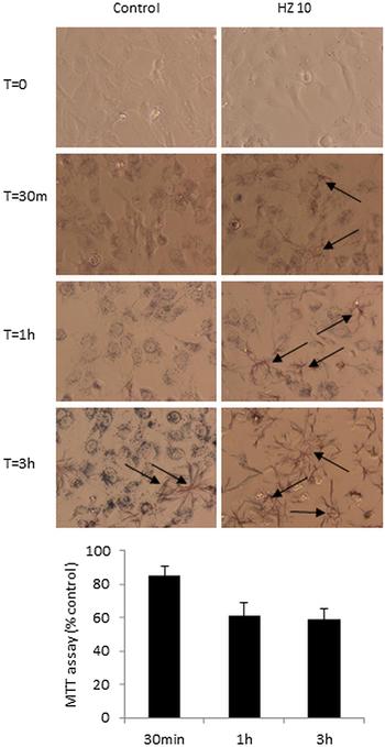
Needle-like formazan crystals were observed by microscopy after 30 min of MTT addition in HZ treated cells and increased over time (Fig. 3, arrows). In control cells, the majority of formazan crystals remained localized in intracellular granules and only after 180 min, some crystals appeared on the surface of control cells. At the same time points, the MTT decreased, as well (Fig. 3, histogram). The percentage of cells exocytosing MTT formazan is shown in Table 1. A significant increase above control was seen already after 30 min (69.2%) and reached a plateau after 120 min (82.7%). In the control, the percentage of cells exocytosing MTT reached 3.2% and 5.1% after 120 or 180 min, respectively. These results suggested an accelerated exocytosis of formazan crystals induced by HZ. To verify that HZ alone was unable to directly induce needle-like formazan crystals, HZ was incubated with MTT in a cell-free system. HZ did not induce crystals formation, indicating that the phenomenon depends on metabolically active cells.
Accelerated formation of MTT-formazan needle-like crystals in HMEC-1 cells treated with HZ (10 µg mL−1) for 24 h. After HZ treatment, MTT was added to the cells and light microscopy pictures of the cells (magnification 400 × ) were taken after 30 min, 1 h and 3 h of incubation at 37 °C Arrows show needle-like crystals. The histogram shows the decrease of MTT reduction over time (the data are expressed as % of untreated control).

Effect of haemozoin on MTT formazan exocytosis by HMEC-1 cells

The percentage of cells exocytosing MTT formazan was determined at different times by counting 200 cells in multiple fields under a light microscope. Data are mean ± s.d. of at least three different experiments.
* Significantly different from control (P < 0.05) by two-tailed Student's t-test.
a HMEC-1 cells were treated with 10 µg mL−1 of haemozoin for 24 h before the addition of MTT.
MTT-formazan exocytosis in genistein-treated cells
Genistein, a compound known to inhibit MTT-formazan exocytosis, was used with the aim to counterbalance HZ effects (Liu et al., Reference Liu, Peterson, Kimura and Schubert1997; Fisichella et al., Reference Fisichella, Dabboue, Bhattacharyya, Saboungi, Salvetat, Hevor and Guerin2009). Genistein added to the cells after incubation with different concentrations of HZ, was able to inhibit the enhanced MTT exocytosis induced by HZ and a recovery of MTT reduction was observed (Fig. 4A). Similarly, the percentage of cells exocytosing MTT formazan decreased from 87.6% ± 10.6 of HZ treated HMEC-1 to 28.4% ± 7.9 in the presence of Genistein, as shown in the microscopic pictures (Fig. 4, panel B). Chloroquine at 50 µg mL−1 was used as positive control since it has been reported that it enhances MTT-formazan exocytosis in rat astrocytes cells (Isobe et al., Reference Isobe, Michikawa and Yanagisawa1999). A significant decrease in MTT reduction was also observed in CQ-treated cells compared with controls and genistein partially restored the decrease of MTT induced by CQ. (Fig. 4A).
Inhibition of MTT-formazan exocytosis by genistein. (A) After 24 h treatment with different doses of HZ (2.5, 5, 10 µg mL−1) or CQ (50 µg mL−1), HMEC-1 cells were incubated with genistein (50 µ m) for 1 h and MTT assay was performed. Data are expressed as percent of control untreated cells. (B) Pictures were taken after 180 min of incubation with MTT of HMEC-1 cells treated with 10 µg mL−1 of HZ alone or with genistein. The arrows show intracellular formazan crystals. The results are the mean and standard deviation of at least three independent experiments. Two-way ANOVA, Tukey's multiple comparisons test **P < 0.001 vs. control.

Accelerated release of preformed CXCL8 and AO exocytosis by HMEC treated with HZ
HMEC-1 were treated with HZ or LPS for 2 and 24 h and CXCL8 levels were measured in cells supernatants. As shown in Fig. 5A, 2 h of HZ, but not of LPS treatment induced significant release of CXCL8 indicating that HMEC-1 respond to HZ inducing exocytosis with a rapid release of preformed CXCL8. After 24 h of treatment, both HZ and LPS induced much stronger activation of HMEC-1 as demonstrated by the significant increase in the release of CXCL8 suggesting a transcriptional effect and the novo chemokine synthesis.
CXCL8 and acridine orange release by HMEC-1 treated with HZ. (A) HMEC-1 were treated for 2 and 24 h with HZ (10 µg mL−1) or LPS (100 ng mL−1). At the end of the treatment cell supernatants were collected and assayed for the presence of CXCL8. (B) HMEC-1 cells were incubated with HZ 10 µg mL−1 for 24 h and subsequently loaded with AO 40 µ m for 15 min (T0). AO release was expressed as fluorescence units (FU ex λ = 535 nm; em λ = 590 nm) and evaluated both in the supernatants and in the cells monolayer at T0 (immediately after AO load) and at T30 (after 30 min of incubation to allow AO exocytosis), and calculating the difference in FU between T0 and T30. The results are expressed as means ± s.d. of three independent experiments. Two-way ANOVA, Tukey's multiple comparisons test *P < 0.05, ***P < 0.0001 vs. control.

Since CXCL8 can be exocytosed by Weibel–Palade bodies (WPBs), which are lysosome-related secretory organelles of endothelial cells, AO, a fluorescent dye that once protonated, is nonspecifically trapped into acidic vesicles (Traganos and Darzynkiewicz, Reference Traganos and Darzynkiewicz1994) was used to evaluate acidic vesicle exocytosis. A slight increase, although not significant, in the release of AO was observed both in the supernatants of HZ treated cells compared with control and also by evaluating the loss of cell-associated AO at different times (T0 – T30) (Fig. 5B).
Beta-Hematin, but not other HZ components increased MTT exocytosis
In order to evaluate which HZ component contributed to the decrease in MTT reduction and accelerated crystal exocytosis, synthetic HZ (beta-haematin, BH, constituted by the sole haem backbone), 4-HNE or 15-HETE (two lipoperoxidation derivatives generated by HZ from arachidonic acid), host fibrinogen (which is always associated with HZ) were also used to treat HMEC-1. The doses of each HZ component used were comparable with those able to induce biological activities in other in vitro models, as reported (Giribaldi et al., Reference Giribaldi, Ulliers, Schwarzer, Roberts, Piacibello and Arese2004; Barrera et al., Reference Barrera, Skorokhod, Baci, Gremo, Arese and Schwarzer2011; Polimeni et al., Reference Polimeni, Valente, Aldieri, Khadjavi, Giribaldi and Prato2013; Basilico et al., Reference Basilico, Corbett, D’ Alessandro, Parapini, Prato, Girelli, Misiano, Olliaro and Taramelli2017). The results shown in Fig. 6, confirmed a significant decrease in MTT metabolism induced by BH, whereas 4HNE, 15-HETE and fibrinogen did not significantly interfere.
BH is the HZ component involved in the increase of MTT – formazan exocytosis. HMEC-1 cells were treated for 24 h with different HZ components: BH at 10 µg mL−1, 4-hydroxynonenal (HNE) at 100 nm, 15-HETEs at 10 µ m, fibrinogen (FG) at 200 µg mL−1. The MTT assay was performed at the end of treatment and the data are expressed as percent of control. The results are the mean and standard deviation of three independent experiments. Ordinary one-way ANOVA, Tukey's multiple comparisons test: **P < 0.01 vs. control.

Discussion
Endothelial activation is a common feature observed in severe malaria. HZ is present at the site of parasite sequestration where can contribute to the endothelial activation. In vitro experiments have shown that HZ is able to induce the production of metalloproteinases and chemokines by endothelial cells (Prato et al., Reference Prato, D'Alessandro, Van den Steen, Opdenakker, Aresel, Taramelli and Basilico2011; Basilico et al., Reference Basilico, Corbett, D’ Alessandro, Parapini, Prato, Girelli, Misiano, Olliaro and Taramelli2017) and, when phagocytized by monocytes/macrophages, can induce both inhibitory and stimulatory effects (Basilico et al., Reference Basilico, Tognazioli, Picot, Ravagnani and Taramelli2003; Khadjavi et al., Reference Khadjavi, Giribaldi and Prato2010; Prato and Giribaldi, Reference Prato and Giribaldi2011).
Here, we show that HZ was not cytotoxic on human endothelial cells, but was able to affect MTT metabolism accelerating formazan exocytosis.
Firstly, our results indicate that the MTT assay cannot be used for measuring cell viability in the presence of HZ. Alternatively, an accurate selection of the incubation time in the presence of MTT has to be done, since up to 30 min the difference between control and treated cells was not significant. After 1 or 3 h of incubation with MTT, despite an apparent reduction in MTT metabolism, cells were alive, as demonstrated by the release of LDH in the supernatants, a measure of cell cytotoxicity, or by cell count with the vital dye trypan blue. In parallel, a significant increase in the percent of cells exocytosing MTT formazan crystals was observed in HZ treated cells, suggesting that the decrease in MTT metabolism was not due to cell death but most likely to modification of the exocytic pathways. The ability to accelerate formazan-MTT exocytosis is not specific for HZ, but it was described for different agents, such as β-amyloid peptides, chloroquine, cholesterol and silica nanoparticles (Abe and Saito, Reference Abe and Saito1998; Claus et al., Reference Claus, Jahraus, Tjelle, Berg, Kirschke, Faulstich and Griffiths1998; Liu and Schubert, Reference Liu and Schubert1998; Isobe et al., Reference Isobe, Michikawa and Yanagisawa1999; Fisichella et al., Reference Fisichella, Dabboue, Bhattacharyya, Saboungi, Salvetat, Hevor and Guerin2009). Moreover, the kinetic of formazan exocytosis and needle-like crystals formation varies depending on the cell type (Molinari et al., Reference Molinari, Tasat, Palmieri and Cabrini2005). These observations suggest to carefully define, for each cell type, the time of incubation with MTT assay. These results also suggest that when new compounds are tested for cytotoxicity using the MTT assay, it is important to exclude that the reduction of MTT is due to an accelerated formazan exocytosis to avoid false positive results.
It has been described that HZ can induce apoptosis in type II pneumocytes and erythroid precursors and can interfere with cell cycle progression of erythroid cells (Lamikanra et al., Reference Lamikanra, Theron, Kooij and Roberts2009; Skorokhod et al., Reference Skorokhod, Caione, Marrocco, Migliardi, Barrera, Arese, Piacibello and Schwarzer2010; Maknitikul et al., Reference Maknitikul, Luplertlop, Chaisri, Maneerat and Ampawong2018). However, here we show that HZ did not induce apoptosis or modification of the cell cycle in endothelial cells, indicating that HZ can induce different effects depending on the cell type. Moreover, in our experiments HMEC-1 cells were stimulated with a concentration of HZ relevant to physiological conditions, using a single treatment for 24 h. We cannot exclude that higher doses, longer time of incubation or repeated treatments with HZ could be toxic for endothelial cells, as well.
In the MTT assay, the reduction of MTT, a yellow tetrazolium dye, to purple formazan crystals, has been mainly attributed to the mitochondrial dehydrogenases (Mosmann, Reference Mosmann1983). Thus, it is commonly accepted that the MTT assay is a suitable indicator of mitochondrial activity (Berridge and Tan, Reference Berridge and Tan1993; Berridge et al., Reference Berridge, Tan, Mccoy and Wang1996). However, the hypothesis that HZ could interfere with MTT metabolism in HMEC-1 by suppression of mitochondrial activity was excluded, since mitochondrial protectors, such as polydatin and cyclosporine A, were unable to recover MTT metabolism. Moreover, the presence of needle-like crystals on the surface of the cells after only 30 min of incubation with MTT supported the fact that an acceleration of MTT-formazan exocytosis occurred in HZ treated cells.
Exocytosis is a process in which intracellular vesicles fuse to the plasma membrane and release different molecules. WPBs are endothelial specific secretory organelles containing bioactive molecules involved in inflammation and hemostasis (Metcalf et al., Reference Metcalf, Nightingale, Zenner, Lui-Roberts and Cutler2008). Their membranes express the late-endosome/lysosome marker CD63 (Vischer and Wagner, Reference Vischer and Wagner1993). Activation of endothelial cells by different stimuli results in WPBs exocytosis and in the release of peptides such as von Willebrand Factor, Angiopoietin 2 and Interleukin 8 (CXCL8) (Utgaard et al., Reference Utgaard, Jahnsen, Bakka, Brandtzaeg and Haraldsen1998). It has been reported that in P. falciparum malaria, plasma levels of von Willebrand factor, Angiopoietin 2 and CXCL8 are higher in patients with severe vs non-severe disease (Hollestelle et al., Reference Hollestelle, Donkor, Mantey, Chakravorty, Craig, Akoto, O'Donnell, van Mourik and Bunn2006; Yeo et al., Reference Yeo, Lampah, Gitawati, Tjitra, Kenangalem, Piera, Price, Duffull, Celermajer and Anstey2008; Ayimba et al., Reference Ayimba, Hegewald, Ségbéna, Gantin, Lechner, Agosssou, Banla and Soboslay2011). In pediatric cerebral malaria, high levels of CXCL8 have also been observed in the cerebrospinal fluid (Armah et al., Reference Armah, Wilson, Sarfo, Powell, Bond, Anderson, Adjei, Gyasi, Tettey, Wiredu, Tongren, Udhayakumar and Stiles2007). Here, we showed that HZ rapidly induced the release of CXCL8 from HMEC-1 suggesting exocytosis of the chemokine rather than de novo synthesis. On the contrary, LPS, a potent microbial stimulus did not induce the release of CXCL8 after 2 h, but only after 24 h of incubation. This is consistent with the reported observation that LPS is unable to induce Weibel–Palade exocytosis by human aortic endothelial cells (Into et al., Reference Into, Kanno, Dohkan, Nakashima, Inomata, Shibata, Lowenstein and Matsushita2007).
WPBs can be classified as lysosome-related organelles, thus AO exocytosis was evaluated in HZ treated cells. In our experiments, HZ induced only a slight, not significant enhancement of AO exocytosis indicating that even if the WPBs could be involved in exocytosis induced by HZ, trafficking of other acidic compartments, such as endothelial lysosomes, seems not to be altered. Moreover, CXCL8 is stored in WPBs, while the precise localization of the reduced formazan product inside the cells is still unclear. It has been reported that MTT formazan granules co-localize with lysosomes/endosomes (Liu et al., Reference Liu, Peterson, Kimura and Schubert1997; Liu and Schubert, Reference Liu and Schubert1997), mitochondria or, more recently, in lipid droplets (Diaz et al., Reference Diaz, Melis, Musin, Piludu, Piras and Falchi2007; Stockert et al., Reference Stockert, Blázquez-Castro, Cañete, Horobin and Villanueva2012). The result on AO also suggests that: (i) formazan crystals do not completely co-localize with lysosomes; (ii) HZ does not, or only partially, perturb intracellular vesicles trafficking of acidic vesicles; (iii) further studies are needed to better understand which compartments are involved in MTT-formazan or CXCL8 exocytosis by HZ.
HZ activates endothelial cells, but little is known on the mechanisms involved in this process. The accelerated MTT formazan exocytosis suggests that HZ, perturbing the intracellular trafficking, could stimulate also the exocytosis of WPBs, contributing to the release of different vasoactive molecules, as demonstrated with CXCL8. Genistein, used as an inhibitor of exocytosis (Liu and Schubert, Reference Liu and Schubert1997; Fisichella et al., Reference Fisichella, Dabboue, Bhattacharyya, Saboungi, Salvetat, Hevor and Guerin2009), was able to decrease the MTT-formazan exocytosis induced by HZ. Interestingly, it has been reported that genistein is also able to block the VEGF-induced von Willebrand factor exocytosis by human aortic endothelial cells (Matsushita et al., Reference Matsushita, Yamakuchi, Morrell, Ozaki, O'Rourke, Irani and Lowenstein2005).
HZ is a ferriprotoporphyrin-IX crystal bound to lipids, DNA, and plasma proteins such as fibrinogen and it can generate lipoperoxidation products (15-HETE and 4-HNE) (Goldie et al., Reference Goldie, Roth, Oppenheim and Vanderberg1990; Schwarzer et al., Reference Schwarzer, Kuhn, Valente and Arese2003; Barrera et al., Reference Barrera, Skorokhod, Baci, Gremo, Arese and Schwarzer2011). All the different components of HZ are responsible for endothelial cells and monocytes/macrophages activation. Some effects of HZ are indeed mediated by the ferriprotoporphyrin-IX moiety, others by 15-HETE and 4-HNE or by parasite DNA or host fibrinogen associated with HZ (Omodeo-Salè et al., Reference Omodeo-Salè, Basilico, Folini, Olliaro and Taramelli1998; Taramelli et al., Reference Taramelli, Recalcati, Basilico, Olliaro and Cairo2000; Schwarzer et al., Reference Schwarzer, Kuhn, Valente and Arese2003; Jaramillo et al., Reference Jaramillo, Godbout and Olivier2005; Parroche et al., Reference Parroche, Lauw, Goutagny, Latz, Monks, Visintin, Halmen, Lamphier, Olivier, Bartholomeu, Gazzinelli and Golenbock2007; Prato et al., Reference Prato, Gallo, Giribaldi and Arese2008; Shio et al., Reference Shio, Kassa, Bellemare and Olivier2010; Skorokhod et al., Reference Skorokhod, Caione, Marrocco, Migliardi, Barrera, Arese, Piacibello and Schwarzer2010; Barrera et al., Reference Barrera, Skorokhod, Baci, Gremo, Arese and Schwarzer2011). This study indicates that accelerated MTT-formazan exocytosis in endothelial cells is mediated by the Fe(III)protoporphyrin IX moiety and not by other HZ components. We found indeed that both the native HZ and the synthetic HZ enhance MTT-formazan exocytosis, as in other studies where both the native and synthetic HZ showed similar activity (Jaramillo et al., Reference Jaramillo, Godbout and Olivier2005).
In conclusion, we demonstrated that HZ was not cytotoxic on human microdermal endothelial cells, but accelerated the MTT-formazan exocytosis through the action of its haem moiety. Thus, MTT assay is not suitable to measure cell viability in the presence of compounds that alter MTT formazan exocytosis. Moreover, HZ, by inducing perturbation of intracellular vesicles trafficking, could contribute to the pathogenesis of severe malaria through the release of bioactive molecules in the circulation.
Acknowledgements
Thanks are due to Laura Galastri, Tiziana Bianchi and Paola Verducci from Associazione Volontari Italiani Sangue (AVIS Comunale Milano) for providing fresh blood to culture Plasmodium falciparum.
Financial support
This work was supported by the Ministero dell'Istruzione, dell'Università e della Ricerca (PRIN 2015.4JRJPP_004) and by the Università degli Studi di Milano (Piano Sviluppo linea B 2016).
Conflict of interest
None
Ethical standards
Not applicable