Introduction
Uranyl sulfate minerals are quite common and widely occurring alteration products formed by oxidation–hydration weathering of pitchblende and associated with sulfides such as pyrite or chalcopyrite (Plášil, Reference Plášil2014; Finch and Murakami, Reference Finch and Murakami1999; Krivovichev and Plášil, Reference Krivovichev, Plášil, Burns and Sigmon2013), resulting from acid-mine drainage phenomena (Fernandes et al., Reference Fernandes, Veiga, Franklin, Prado and Taddei1995; Brugger et al., Reference Brugger, Burns and Meisser2003; Plášil et al., Reference Plášil, Sejkora, Škoda and Škácha2014b). Zippeite-related uranyl sulfates are among the most common supergene uranium minerals, occurring under low to weakly acidic pH. ‘Zippeites’ have been known since the first half of the 19th century, with the earliest descriptions closely linked to the Jáchymov deposit (John, Reference John1821; Haidinger, Reference Haidinger1845). Nonetheless, the official establishment of the zippeite group or family is still lacking. Previous studies of zippeite-related minerals indicate that they are hydrated uranyl sulfates that can accommodate hydroxyl groups within their structure units (uranyl sulfate layers of the zippeite topology with the characteristic U:S ratio = 2:1) and have in the interlayer mono- (Na+, K+, NH4+), di- (Mg2+, Ca2+, and Me2+ of the transition metal elements) or trivalent cations (Y3+, REE 3+) and molecular H2O. There are significant differences in the degree of ordering in the zippeite-type structures. Usually, those that are highly hydrated (with structural units not directly linked via bridging metal cations) are more ordered and have lower symmetry (P  $\overline 1 $, but not exclusively) than the less hydrated (usually monoclinic). The crystal-structure properties of synthetic counterparts of known minerals were first reported by Burns et al. (Reference Burns, Deely and Hayden2003), and there have been numerous recent structural studies dealing with natural minerals that have provided valuable data (e.g. Brugger et al., Reference Brugger, Burns and Meisser2003; Plášil and Škoda, Reference Plášil and Škoda2015; Plášil et al., Reference Plášil, Dušek, Novák, Čejka, Císařová and Škoda2011a,Reference Plášil, Mills, Fejfarová, Dušek, Novák, Škoda, Čejka and Sejkorab, Reference Plášil, Fejfarová, Wallwork, Dušek, Škoda, Sejkora, Čejka, Veselovský, Hloušek, Meisser and Brugger2012, Reference Plášil, Fejfarová, Škoda, Dušek, Marty and Čejka2013, Reference Plášil, Dušek, Čejka and Sejkora2014a, Reference Plášil, Škácha, Sejkora, Kampf, Škoda, Čejka, Hloušek, Kasatkin, Pavlíček and Babka2017; Plášil, Reference Plášil2015; Kampf et al., Reference Kampf, Plášil, Olds, Nash and Marty2018; Olds et al., Reference Olds, Plášil, Kampf, Burns, Nash, Marty, Rose and Carlson2018; Kampf et al., Reference Kampf, Plášil, Olds, Emproto, Ma, Sejkora, Adams and Marty2025; Plášil, Reference Plášil2026). A brief overview of the informal zippeite family of minerals has been provided by Sejkora et al. (Reference Sejkora, Dolníček and Plášil2023).
Pseudojohannite was originally described from Jáchymov by Ondruš et al. (Reference Ondruš, Veselovský, Gabašová, Hloušek and Šrein2003) as triclinic Cu5(UO2)6(SO4)3(OH)16·14H2O with a zippeite-type structure, and it was redefined by Brugger et al. (Reference Brugger, Wallwork, Meisser, Pring, Ondruš and Čejka2006) based on a synchrotron high-resolution powder diffraction study as triclinic (P1 or P  $\overline 1 $) Cu6.5[(UO2)4O4(SO4)2]2(OH)5·25H2O, with a = 10.027(1) Å, b = 10.822(1) Å, c = 13.396(1) Å, α = 87.97(1)°, β = 109.20(1)°, γ = 90.89(1)°, V = 1371.9(5) Å3. Later, Plášil et al. (Reference Plášil, Fejfarová, Wallwork, Dušek, Škoda, Sejkora, Čejka, Veselovský, Hloušek, Meisser and Brugger2012) provided a structure solution and refinement from the single-crystal X-ray data on a sample from the Widowmaker mine in White Canyon, USA (R 1 = 0.0347 for 2664 observed reflections), showing that pseudojohannite has the space group P
 $\overline 1 $, with a = 8.6744(4), b = 8.8692(4), c = 10.0090(5) Å, α = 72.105(4)°, β = 70.544(4)°, γ = 76.035(4)°, and V = 682.61(5) Å3 and the chemical formula Cu3(OH)2[(UO2)4O4(SO4)2](H2O)12 (Z = 1). According to that study, the structure of pseudojohannite is based on the uranyl sulfate sheets of the zippeite topology identical to those found in zippeite-type structures containing Mg2+, Co2+ or Zn2+. Plášil (Reference Plášil2015) showed that the pseudojohannite structure from the type locality, Jáchymov in the Czech Republic, is identical to that presented in the previous study.
In this contribution, a new dataset on a crystal from Jáchymov is presented, including a rigorous determination of H-atom positions from X-ray diffraction data and providing a detailed discussion on the nature of the interlayer complex based on the bond-valence approach.
Sample
The pseudojohannite crystal (Fig. 1) used here is the same as that used in the structural study of pseudojohannite by Plášil (Reference Plášil2015). The sample from which it had been recovered is deposited in the mineralogical collection of the National Museum in Prague. The specimen, measuring 6 cm × 4 cm × 1.5 cm, originates from the Červená vein at the Rovnost mine, Jáchymov, Czech Republic (see Plášil et al., Reference Plášil, Sejkora, Škoda and Škácha2014b). It represents a fragment of the ore vein, containing U- and Cu-ore mineralization (pitchblende, chalcopyrite, tennantite, and chalcocite). Pseudojohannite forms well-developed crystals up to 0.2 mm across (Fig. 1). They have a typical lens- or discoid morphology, characteristic of the zippeite-group minerals (cf. Plášil et al., Reference Plášil, Mills, Fejfarová, Dušek, Novák, Škoda, Čejka and Sejkora2011b, Reference Plášil, Dušek, Čejka and Sejkora2014a).
Pseudojohannite crystals (used for the structure study; Pjh) growing on a strongly oxidized matrix consisting of pitchblende, chalcocite and chalcopyrite ore. Minerals associated: brochantite (Bct), cuprosklodowskite (Cskl), antlerite (Atl), Cu-rich compreignacite (Cgn). Locality: Cross of Červená and Radiová veins, Rovnost shaft, Daniel level, Jáchymov, Czech Republic. FOV is 1 mm, photo: Stephan Wolfsried.

Figure 1 Long description
A cluster of bright green pseudojohannite crystals adjacent to yellow compreignacite. Crystals of dark green brochantite are present with pale green cuprosklodowskite. Some patches of turquoise antlerite are also on the specimen. The pseudojohannite crystals are in a cluster approximately 250 by 200 micrometres
X-ray crystallography and structure refinement
Single-crystal XRD data for pseudojohannite, with approximate dimensions 0.14 mm × 0.05 mm × 0.02 mm, were collected on an XtaLAB Synergy-DW system equipped with a HyPix-Arc150 detector, using monochromatic MoKα radiation from a microfocus rotating anode. An Oxford Instruments CryoJet was used for low-temperature data collection to minimize atomic thermal vibrations. Integration of the diffraction data was carried out using the CrysAlis RED software (Rigaku, 2026). A multi-scan absorption correction was applied to the data in ABSPACK3 (Rigaku, 2026); the R int of the dataset was 4.5%. Starting with the atomic coordinates reported by Plášil (Reference Plášil2015), all structural refinements were carried out using Jana2020 (Petříček et al., Reference Petříček, Palatinus, Plášil and Dušek2023) with the full-matrix least-squares algorithm based on F 2. Anisotropic displacement parameters (ADP) for all non-hydrogen atoms were applied. All of the H atoms of the six independent H2O molecules and the OH group were located from difference-Fourier maps and refined with restraints on O–H distances and H–O–H angles (0.8±0.01 Å, 108±0.3°). The U iso of the H atoms were set to 1.5×U eq of the donor O atoms. The final refinement converged to R 1 = 0.0198 for 6029 unique reflections with I > 3σ(I) and wR 2 = 0.0699 for all 6924 reflections. The crystal was modelled as a split crystal (non-crystallographic twin) with a subordinate volume of the second domain; nevertheless, despite the small number, they still affected F obs somewhat, as some reflections overlapped. Jana2020 discarded 205 observed reflections due to an incomplete overlap during refinement. Crystallographic data, as well as experimental and refinement details, are summarized in Table 1. Atomic parameters are given in Table 2. Selected bond distances and geometrical measures are reported in Tables 3, 4 (U, S, Cu; polyhedral measures calculated using Vesta from Momma and Izumi, Reference Momma and Izumi2008) and Table 5 (H bonds). The bond-valence analysis, done following Brown’s (Reference Brown2002) standard procedure, is presented in Table 6. The crystallographic information files have been deposited with the Principal Editor of Mineralogical Magazine and are available as Supplementary material (see below).
Data collection and structure refinement details for pseudojohannite at 100 K

Table 1 Long description
Pseudojohannite, Cu3(H2O)10(OH)2[(UO2)4O4(SO4)2](H2O)2; Temperature=100 K; Space group=P–1 (#2); =a = 8.6697(4) Å; =b = 8.8712(5) Å; =c = 9.9825(6) Å; =α = 71.775(6)°; =β = 70.663(5)°; =γ = 76.011(5)°; V=679.88(7) Å3; Z=1; Dcalc (g cm-3)=4.340; Wavelength=MoKα, 0.71073 Å; Crystal dimensions=0.14 × 0.05 × 0.02 mm; Collection mode=ω scans to fill an Ewald sphere; Frame width and time=0.5, 50 s; Limiting θ angles=2.24–38.95°; Limiting hkl indices=–14<h<14, –15<k<15,–17<l<16; No. of reflections measured, unique, observed [I > 3σ(I)]=61748, 6924, 6029; μ (mm–1), absorption correction method=36.32, multi-scan (CrysAlis; ABSPACK3); Tmin/Tmax=0.436/1.000; Rint, Completeness to θmax=0.0446, 96%; Parameters, constraints, restraints=237, 13, 19; R1, wR2 (obs)=0.0198, 0.0673; R1, wR2 (all)=0.0267, 0.0699; GOF (obs, all)=1.047, 1.013; Δρmin, Δρmax (e.Å–3)=–0.61, 0.46; Twin matrix=-0.9978, 0.0092, -0.6217; =-0.0088, -1.0021, -0.5596; =0,0,1; Twin fractions=0.914(4)/0.086(4)
Atom coordinates and displacement parameters (as equivalent or isotropic*; in Å2) for pseudojohannite at 100 K

Table 2 Long description
The table presents the atomic coordinates and displacement parameters for pseudojohannite at a temperature of 100 Kelvin. It lists the fractional coordinates x/a, y/b, and z/c, along with the equivalent or isotropic displacement parameters (Uiso/Ueq) in Ångström squared for various atoms, including uranium, copper, sulfur, and oxygen. Also included are hydrogen atoms.
Selected bond distances (Å) for pseudojohannite at 100 K in comparison with the data from Plášil (Reference Plášil2015) collected at 298 K

Table 3 Long description
The table compares bond distances in pseudojohannite at 100 Kelvin and 298 Kelvin. For atoms at 100K, 298 K; U1–O11u= 1.831(3), 1.815(8); U1–O16u= 1.809(3), 1.781(9); U1–O8e= 2.246(3), 2.236(9); U1–O8ei= 2.285(3), 2.269(9); U1–O9e= 2.310(3), 2.319(8); U1–O12eii= 2.477(3), 2.464(10); U1–O17ei= 2.477(2), 2.482(8); = 1.820, 1.798; = 2.359, 2.354; U2–O3u= 1.817(3), 1.793(8); U2–O5u= 1.839(3), 1.809(8); U2–O8e= 2.271(2), 2.292(8); U2–O9e= 2.226(3), 2.236(9); U2–O9eiii= 2.288(3), 2.263(9); U2–O13eiv= 2.495(2), 2.495(8); U2–O14= 2.505(3), 2.495(11); = 1.828, 1.801; = 2.357, 2.356; S1–O12e= 1.475(4), 1.476(12); S1–O13e= 1.478(3), 1.465(9); S1–O14e= 1.475(3), 1.472(8); S1–O15e= 1.474(2), 1.459(8); = 1.476, 1.468; Cu1–O1w= 1.955(4), 1.953(12); Cu1–O1wv= 1.955(4), 1.953(12); Cu1–O4w= 2.523(3), 2.538(8); Cu1–O4wv= 2.523(3), 2.538(8); Cu1–O6w= 1.972(2), 1.950(8); Cu1–O6wv= 1.972(2), 1.950(8); = 2.150, 2.147; Cu2–O2oh= 1.935(3) , 1.923(9); Cu2–O2ohvi= 1.943(2), 1.948(9); Cu2–O4w= 2.415(4), 2.420(11); Cu2–O10w= 1.948(3) , 1.962(10); Cu2–O11wi= 3.138(3), 3.167(11); Cu2–O15w= 1.973(2) , 1.972(9); = 2.225, 2.232.
Subscripts u, e, oh and w denote uranyl, equatorial, OH and water O atoms, respectively.
Selected polyhedral measures for pseudojohannite at 100 K and 298 K (Plášil, Reference Plášil2015)

Table 4 Long description
The table compares polyhedral measures of pseudojohannite at two temperatures: 100 K and 298 K. For 100 K, 298 K; ΔS1= 0.001, 0.004; ECoNS1= 3.999, 3.998; VCu1= 12.914, 12.837; ECoNCu1= 4.156, 4.123; ΔCu1= 0.1158, 0.1213; VCu2= 13.571, 13.620; ECoNCu2= 4.164, 4.147; ΔCu2= 0.1651, 0.1658.
Notes: V Mx – metal-cation polyhedral volume [Å3]; ΔMx – distortion index (on bond length) (Baur, Reference Baur1974); ECoNMx – effective coordination number (Hoppe, Reference Hoppe1979)
Hydrogen-bond geometry (Å, °) for pseudojohannite at 100 and 298 K. H2O groups were modelled with constrained geometry (O–H = 0.80 Å, H–O–H = 108°)

Table 5 Long description
The table gives hydrogen-bond geometry in pseudojohannite at 100 K and 298 K, focussing on bond lengths and angles. It has values for bond lengths for the hydrogen to acceptor bonds and the oxygen to acceptor bonds at 100 K and 298 K. It also has angles for O-H-A at 100 K and calculated. For full discussion of the implications see text.
a Notes: H-bond types: 1 = direct interlayer to layer; 2 = intralayer donation OH– to interstitial Oequatorial; 3 = between interlayer H2O molecules; ‘Layer’ refers to the [(UO2)4O4(SO4)2] sheets. b refers to the inferred hydrogen bonds of pseudojohannite from Jáchymov reported by Plášil (Reference Plášil2015). c Colmenero et al. (Reference Colmenero, Plášil and Sejkora2022); calculated. Symmetry codes: (none) x, y, z; (iii) –x+1, –y+1, –z+1; (iv) –x+1, –y+2, –z+1; (vi) –x+2, –y+2, –z; (vii) –x+2, –y+1, –z; (viii) x, y, z–1. Subscripts u, e, oh and w denote uranyl, equatorial, OH and water O atoms, respectively.
Bond-valence analysis for pseudojohannite at 100 K; values are given in valence units, vu*

Table 6 Long description
The table analyses bond-valence for pseudojohannite at 100 K. U1 and U2 have bond-valence sums of 5.86 and 5.84, respectively, and S1 = 5.95. O3u and O5u have values of 1.62 and 1.55 for U2.
* Notes: Bond-valence parameters are from Gagné and Hawthorne (Reference Gagné and Hawthorne2015). sumC – sum of bond-valences for cations; sumA – the sum of the BV for anions; HBD – number of donated H-bonds (of the theoretical strength 0.8 vu); HBA – number of accepted H-bonds (of the theoretical strength 0.2 vu); assig. – assignment. Subscripts u, e, oh and w denote uranyl, equatorial, OH and water O atoms, respectively.
Description of the structure
The structure refinement of pseudojohannite from Jáchymov at 100 K confirms the model proposed by Plášil et al. (Reference Plášil, Fejfarová, Wallwork, Dušek, Škoda, Sejkora, Čejka, Veselovský, Hloušek, Meisser and Brugger2012) for the crystal from the Widowmaker mine in Utah (USA) and by Plášil (Reference Plášil2015), and now includes hydrogen atom positions and hydrogen-bonding details that had been unresolved. Based on the crystal structure of pseudojohannite at T = 100 K, the general structural architecture will be reviewed briefly, and then the focus will shift towards the nature and type of H2O present in the structure.
Pseudojohannite at T = 100 K
In the structure of pseudojohannite (Fig. 2), there are two symmetrically unique U atoms, two symmetrically unique Cu atoms, one S atom, and 17 O atoms, six of which belong to H2O molecules and one to an OH– group. The structure consists of uranyl sulfate sheets (structural units) and an interlayer complex comprising hydrated Cu2+–Φ polyhedra and an additional H2O molecule (Figs 2, 3a,b). Previous studies have provided details on connectivity within the sheet; however, it should be noted that this concerns the uranyl sulfate sheet of the zippeite anion topology (see Burns et al., Reference Burns, Deely and Hayden2003; Burns, Reference Burns2005; Lussier et al., Reference Lussier, Lopez and Burns2016), and with the composition [(UO2)4O4(SO4)2]4–. The interlayer complex comprises an infinite chain of Cu-polyhedra, with one Cu site linked to six ligands (H2O molecules), forming a strongly tetragonally distorted octahedron with C 4v symmetry (4+2) (Table 3). The second Cu site is coordinated by five ligands (3 H2O + 2 OH), comprising one long and four shorter bonds, characteristic of the square pyramidal coordination (4+1). Both are typical of the Jahn-Teller effect on Cu(II) (Jahn and Teller, Reference Jahn and Teller1937; Burns and Hawthorne, Reference Burns and Hawthorne1995; Hawthorne and Schindler, Reference Hawthorne and Schindler2000). The infinite chain extends along [100] (Fig. 2). In total, there are six H2O molecules and one OH– group in the unit cell of pseudojohannite. This was clear from previous studies; the current dataset, however, enabled a reliable determination of the positions of H atoms in the structure. Three of the H2O groups (six by symmetry), O1, O4 and O6, are bonded to the Cu1 site (Fig. 3a), with Cu–O bond lengths between 1.955 and 2.523 Å. Each of them donates two H bonds to adjacent structural units, mostly the terminal uranyl oxygen atoms (Table 6). The O···O distances of these H bonds are in the range 2.595–2.889 Å. O1 and O4 of those mentioned H2O molecules are not involved in further bonds and can be considered as [3]-coordinated, transformer H2O groups (see the discussion below for details). The O6 site is [4]-coordinated and is a non-transformer H2O group.Three of the H2O groups, O4 (bridges the Cu1 and Cu2 sites), O10, O11 and O15 are bonded to the Cu2 site (Figs 4, 3a), with Cu–O bond lengths of between 1.945 and 2.415 Å (3.138 Å = O11; the ‘distant’ vertex of the tetragonal bipyramid). Each donates two H bonds to adjacent structural units (Table 6). The O···O distances of these H-bonds are in the range 2.681 to 2.990 Å. These three H2O molecules mentioned are not involved in further bonding and can be considered 3-coordinated, transformer H2O groups. Two additional ligands of the Cu2 site are two symmetrically related O2 atoms from the OH– groups (Table 3). The O···O distances of these H bonds within the OH– are 2.780 Å. The isolated O7 atom of the H2O molecule is bonded to two H atoms (Fig. 3b). One H bond emanates towards the adjacent structural unit (acceptor is the O3 atom), and the second one is accepted by the O6 (H2O) (Table 6). The O7 atom receives two H bonds from O4 and O10. It is thus 4-coordinated and is a non-transformer H2O group. Therefore, in the structure of pseudojohannite, there are eight transformer H2O groups, four non-transformer H2O (two of which are not directly linked to any metal cation site), and two OH– groups. The structure formula of pseudojohannite can thus be expressed as Cu3(H2[3]O)8(H2[4]O)2(OH)2[(UO2)4O4(SO4)2](H2[4]O)2 (Z = 1).
Crystal structure of pseudojohannite at 100 K showing two structural units: [(UO2)4O4(SO4)2] sheets (UO7 = yellow, SO4 = pale yellow, translucent) and interlayer complex (copper = pale blue, O = red, H = grey) connected by H bonds (dashed blue lines). Unit-cell edges outlined in black lines.

Figure 2 Long description
The image shows the crystal structure of pseudojohannite, featuring two main structural units: uranyl sulfate sheets and interlayer complexes. The uranyl sulfate sheets are depicted as yellow polyhedra, while the interlayer complexes are shown with copper atoms labeled as Cu1 and Cu2. These copper atoms are connected by dashed lines representing hydrogen bonds. The structure is outlined with black lines indicating the unit-cell edges. The orientation is marked with axes labelled 'a' and 'c'.
Interlayer complex in pseudojohannite structure at 100 K. (a) Projection down [001] showing three infinite Cu-based chain segments (extending along [100]) with an O7w (H2O) group located between them. (b) The local environment around O7w, displaying bonding within the interlayer (O4w–>O7w; O10w–>O7w).

Figure 3 Long description
The image A shows a projection of the pseudojohannite structure down the [001] axis, with three infinite Cu-based chain segments extending along [100]. Key elements include Cu1 and Cu2 sites, various O atoms (e.g., O1w, O3u, O4w) and bonding interactions. The image B illustrates the local environment around O7w, displaying bonding within the interlayer, such as O4w to O7w and O10w to O7w. Both diagrams include axes for orientation.
Infinite chain of Cu-polyhedra in the structure of pseudojohannite at 100 K running parallel to [100]. The long (weak; 0.02 vu) Cu2–O11 bond is omitted, and Cu2 is displayed as a tetragonal pyramid. Thermal ellipsoids of O atoms are given at the 75% probability level. Unit-cell edges outlined in black lines.

Figure 4 Long description
The diagram shows a chain of Cu-polyhedra in the structure of pseudojohannite. The polyhedra are connected by lines representing bonds. Two Cu sites, labeled Cu1 and Cu2, are displayed as green polyhedra. Several oxygen atoms are labelled, including O2h, O4w, O1w, O15w, O10w and O6w. The unit-cell edges are outlined in black lines. An axis indicator at the bottom left shows directions labelled 'a' and 'b'.
Comparison to other pseudojohannite structures
Several previous structural studies have described pseudojohannite. First among them, the study by Plášil (Reference Plášil2015) is of particular interest, as it was carried out on the same crystal as the current study but at room temperature. Therefore, a direct comparison between the current structure and the aforementioned refinement is useful (see Tables 3, 4, 5 and 7). The study by Plášil et al. (Reference Plášil, Fejfarová, Wallwork, Dušek, Škoda, Sejkora, Čejka, Veselovský, Hloušek, Meisser and Brugger2012) examined the same pseudojohannite crystal from White Canyon, USA, and apparently, the crystal used differs in unit-cell metrics (Table 7). Furthermore, Colmenero et al. (Reference Colmenero, Plášil and Sejkora2022) conducted a theoretical study using periodic DFT calculations and reported theoretical results on hydrogen bonding in pseudojohannite and on its mechanical properties. At first glance, we observe small but significant differences in the unit-cell volumes of pseudojohannite at 100 K and 298 K. The dataset at 100 K clearly yielded a larger unit cell for pseudojohannite than at 298 K, with a similar trend observed in some refined parameters, namely the polyhedral measures of Cu polyhedra (Table 4). The study by Colmenero et al. (Reference Colmenero, Plášil and Sejkora2022) reported a small but universal anisotropy (A u) of 3.19. They argued that it is of much smaller magnitude than in uranopilite (6.65; Colmenero et al., Reference Colmenero, Plášil, Timón and Čejka2020) and thus did not predict any particularly interesting features, such as negative linear compressibility, for pseudojohannite. Nevertheless, it seems that some interesting behaviour of pseudojohannite may occur at lower temperatures, which might be associated with the Cu polyhedra. The study by Colmenero et al. (Reference Colmenero, Plášil and Sejkora2022) also provided some theoretical insights into the hydrogen bonding in pseudojohannite. Despite some differences between the computational results from periodic DFT and the current, highly satisfactory refinement based on XRD data, it is necessary to emphasize that the two H bond schemes are similar. There are differences in the labelling of the atoms; the current study retained the same labelling scheme as that used by Plášil (Reference Plášil2015) for clarity. The comparison is provided in Table 5. The main differences lie in the refined/calculated <(O–H···A)> bond angles, which are highly sensitive to the methodology and await comparison with XRD data and theoretical DFT calculations. It should be emphasized that, despite the limited accuracy of the H-atom positions determined and refined from X-ray data, the current structure refinement provides the best available full-structural model for pseudojohannite, which represents a well-ordered zippeite-type structure. It is the only model of the zippeite-type structures that includes reliable, unambiguously refined H atoms. The model proposed by Plášil et al. (Reference Plášil, Mills, Fejfarová, Dušek, Novák, Škoda, Čejka and Sejkora2011b) included H atoms as well; however, because the interlayer complex is affected by positional and occupational disorder, the model’s validity is questionable. A well-argued description of hydrogen bonding in the zippeite-type mineral marécottite has been given by Plášil and Škoda (Reference Plášil and Škoda2015); however, their analysis is based solely on bond-valence considerations, as H atoms were not determined.
Comparison of unit-cell parameters for pseudojohannite from various localities at 100 K and room temperature and theoretical results

Table 7 Long description
The table compares unit-cell parameters of pseudojohannite from different localities at 100 Kelvin and room temperature, alongside theoretical calculations. The study at 100 Kelvin shows a volume of approximately 680 cubic angstroms, while Jáchymov and White Canyon at room temperature have similar volumes around 679 and 683 cubic angstroms, respectively. Theoretical calculations using PBE and DFT-D2 methods predict larger volumes, with PBE showing the largest at 720 cubic angstroms. The angles and cell dimensions vary slightly between experimental and calculated data.
1 Notes: Plášil (Reference Plášil2015); 2Plášil et al. (Reference Plášil, Fejfarová, Wallwork, Dušek, Škoda, Sejkora, Čejka, Veselovský, Hloušek, Meisser and Brugger2012); 3Colmenero et al. (Reference Colmenero, Plášil and Sejkora2022)
Discussion: bond-valence approach to the interlayer complexes in pseudojohannite and other related uranyl sulfate minerals
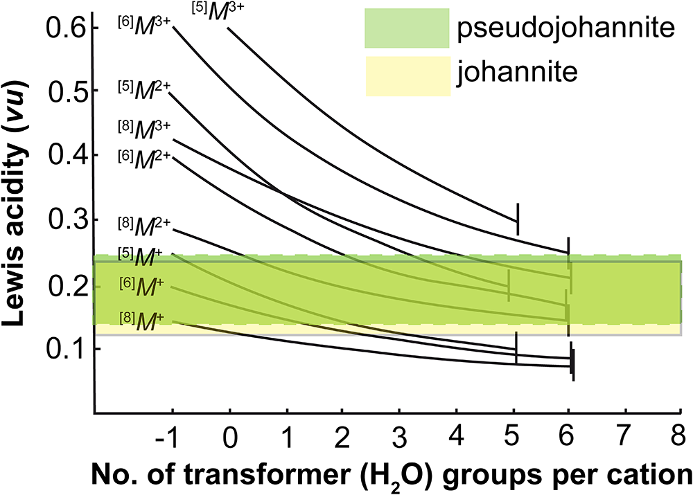
To evaluate weakly bonded constituents of oxysalt minerals (such as molecular water), a so-called bond-valence approach has been developed by Frank Hawthorne and co-workers (e.g., Hawthorne, Reference Hawthorne1992, Reference Hawthorne2012, Reference Hawthorne2015; Hawthorne and Schindler, Reference Hawthorne and Schindler2009; Schindler and Hawthorne, Reference Schindler and Hawthorne2008). The fundamentals of the theory lie in the rigorous base of the bond-valence theory, one of the complementary theories of chemical bonding (Brown, Reference Brown2023). Using the bond-valence approach, we can inspect, evaluate and predict how and why the particular structural units and interlayer/interstitial complexes are organised and interacting in terms of bonding. One of the fundamental principles of bond-valence theory states that stable crystal structures form when the Lewis basicity (LB) of the structural unit (SU) matches the Lewis acidity (LA) of the interstitial complex (IN). Both values are affected by several facts; nevertheless, we must emphasize the specific roles of hydrous species (H2O and OH–) present within the SU. The presence of H2O (and, namely, that of so-called ‘transformer’ H2O, with [3]O) affects both the dimensionality of the oxysalt structures and effectively reduces the LA of the cation to which it is bonded. The bond-valence approach enables the prediction of the total number of H2O molecules in the structure based on bond-valence requirements and enables typological distinctions. In the following paragraphs, we use the specific terminology of the bond-valence approach. For those unfamiliar with it and the calculations, refer to the original papers by Frank Hawthorne and co-workers. Some of the features of the zippeite-related minerals have already been discussed in the paper by Schindler and Hawthorne (Reference Schindler and Hawthorne2008). The zippeite-related structures (e.g. the overview given by Sejkora et al., Reference Sejkora, Dolníček and Plášil2023) possess several general distinct types of sheets, which do, however, have similar ranges in Lewis basicities: [(UO2)4O3(SO4)2(OH)]3– (0.14–0.25 vu), [(UO2)8O5(SO4)4(OH)3]5– (0.14–0.25 vu), [(UO2)2O2(SO4)2]2– (0.14–0.25 vu), [(UO2)8O7(OH)(SO4)4] (0.15–0.28 vu) and [(UO2)2O(OH)(SO4)] (0.15–0.24 vu). There are certain complications/irregularities concerning mainly the natural zippeite (see Plášil et al., Reference Plášil, Mills, Fejfarová, Dušek, Novák, Škoda, Čejka and Sejkora2011b, Reference Plášil, Petříček, Mills, Favreau and Galea-Clolus2018), where the situation among the OH distribution on the zippeite-type sheets is less resolved; thus, they are not considered further below. The overview of the most important characteristics of each type of sheet and their presence in the zippeite-like minerals is given in Table 8, along with a comparison to johannite. Pseudojohannite has a unprotonated [(UO2)2O2(SO4)2]2– sheet, otherwise found both in natural (Plášil et al., Reference Plášil, Fejfarová, Škoda, Dušek, Marty and Čejka2013) and synthetic magnesiozippeite (Burns et al., Reference Burns, Deely and Hayden2003), and in other M 2+-containing synthetic (Spitsyn et al., Reference Spitsyn, Kovba, Tabachenko, Tabachenko and Mikhaylov1982) or ‘natural zippeites’. The interlayer complex, Cu3(H2[3]O)8(H2[4]O)2(OH)2(H2[4]O)2, present in pseudojohannite, is somewhat unique among the zippeite-like structures. It is, along with the infinite chain of Mg-octahedra, {Mg2(H2O)5(Oyl)6}, found in natural magnesiozippeite, the only one of the most polymerized interlayer complexes. The charge deficiency per anion (CDA) of the structural unit of pseudojohannite is 0.20 vu. The CDA is an important measure of the average bond valence per O atom contributed by the interstitial species and adjacent structural units. It correlates strongly with the average O-coordination number in the interlayer complex, [CN in]. The CDA pseudojohannite matches the CDA range for structural units of uranyl sulfates listed by Plášil et al. (2023): 0.15–0.25 vu. Now, following the approach of Schindler and Hawthorne (Reference Schindler and Hawthorne2008), we can calculate the Lewis basicity (LB) of the structural unit of the pseudojohannite, which is in the range 0.15–0.25 vu. From the valence-matching principle of the bond-valence theory, we know that from the possible interstitial complexes, only those matching the same range will form the stable structures; the Lewis acidity (LA) of interstitial complexes should lie in the range of 0.15–0.25 vu. The graphical representation of the LA–LB correspondence principle (the valence matching principle) is shown in Fig. 5. In the case of pseudojohannite (LB of the SU is highlighted in yellow), it is clear that [6]Cu2+ has to bond at least to the two transformer H2O groups to obey the principle (the overlap of the yellow LB field with the particular curve). More precisely, it should bond to two to six H2O groups on the transformer. Since we know well, based on the results of the current study, the number of transformer (8) and non-transformer (4) H2O groups and the number of (OH)– groups and the coordination number of the O atom within the (OH)– group, we can calculate directly the value of the Lewis acidity of the interlayer complex in pseudojohannite, which is 0.20 vu. This value is: (1) in line with the range of LB of the SU in pseudojohannite; and (2) corresponds (from Fig. 5) with five transformer H2O groups per [6]-coordinated divalent cation. This calculated result is consistent with the structure: there are four transformer H2O groups + two non-transformer bonded to Cu1, and four transformer H2O groups + 2 (OH)– bonded to Cu2. We can make some further conclusions. It is interesting to check whether there are clear, specific criteria that can distinguish pseudojohannite from chemically similar but structurally distinct johannite. Both are supergene uranyl sulfate minerals that commonly occur together. Johannite, triclinic Cu(UO2)2(SO4)2(OH)2·8H2O (Mereiter, Reference Mereiter1982), unlike pseudojohannite, is not of the zippeite-type, but contains sheets based on phosphuranylite topology with (OH)– within the sheets. The CDA value for johannite is somewhat lower than that for pseudojohannite, = 0.20 vu. The LA of the SU in johannite is also slightly lower, ∼0.13–0.24 vu. According to the LB–LA principle, we need at least two H2O groups to coordinate to [6]Cu2+ in johannite to achieve a stable structure (Fig. 5). The H atom positions in the structure of johannite are not known, but based on the study of Mereiter (Reference Mereiter1982), we have a clear idea of the interlayer complex constitution: the Cu2+ is bonded to four transformer H2O groups (plus two bridging Oyl atoms of adjacent layers). The calculated value of the LA of the interlayer complex in johannite is of the same magnitude as in pseudojohannite, = 0.20 vu (!). The stereochemistry of the uranyl sulfate sheets and interlayer complexes in pseudojohannite and johannite is similar (!); johannite has slightly lower values of CDA. It could suggest that those properties will not be the most important aspects when these two minerals precipitate from aqueous solutions. The key role will probably be played by the activity of Cu (and the distinction of solubility products of pseudojohannite and johannite). Pseudojohannite has a much higher Cu/U ratio than johannite = 3:4 vs 1:2. It means that the pseudojohannite structure concentrates much more Cu than johannite does. At the same time, the lower CDA value for johannite (and the presence of OH within the sheets) can imply that johannite might prefer to precipitate from solutions under a more acidic pH. Hence, let’s adopt an easy mechanistic approach. If there is enough Cu in the resting solution after precipitation of johannite, the volume of the resting solution will be smaller, and the concentration of Cu will increase (along with the pH), so that the precipitation of pseudojohannite can proceed. Of course this is only speculative. In the majority of specimens from Jáchymov, pseudojohannite occurs as microcrystalline grape-shaped aggregates, composed of thousands of discoid crystals, unlike johannite, which is always macrocrystalline. This might indicate that pseudojohannite forms readily from the solution without ‘much time to grow in larger crystals’.
The variation in Lewis acidity with the number of transformer (H2O) groups for different interstitial-cation charges and coordination numbers of a general interlayer complex; the ranges in Lewis basicity of the structural units (SU) of pseudojohannite and johannite (which overlap significantly) are marked in light green and yellow, respectively. Where the lines of variable Lewis acidity overlap the range of Lewis basicity of the SU, the valence-matching principle is obeyed.

Figure 5 Long description
A graph showing Lewis acidity in valence units on the y-axis and the number of transformer water groups per cation on the x-axis. The graph includes curves labelled with different interstitial-cation charges and coordination numbers. The ranges for pseudojohannite and johannite are highlighted in light green and yellow, respectively. The graph illustrates how Lewis acidity varies with the number of transformer water groups, with overlapping regions indicating where the valence-matching principle is obeyed.
Selected uranyl sulfates and some of their bond-valence characteristic; all values given in valence units (vu)

Table 8 Long description
The table compares selected uranyl sulfates based on their charge deficiency per anion, Lewis basicity range, calculated Lewis acidity, interlayer complex, and structural references. Pseudojohannite has a CDA of 0.20 valence units, while Hubbardite has a slightly higher value at 0.22. Natrozippeite shows the widest range of Lewis basicity, spanning from 0.14 to 0.28 valence units. The calculated Lewis acidity values are generally close, with Pseudojohannite and Johannite both at 0.20 valence units. The interlayer complexes vary, as Pseudojohannite has a copper-based complex and Natrozippeite a sodium-based one. The data is sourced from various studies.
Notes: CDA – charge deficiency per anion; LB – Lewis basicity; LA – Lewis acidity.
* – The number of inverse-transformer H2O groups is unclear; they are not expected to be present as the structure does not contain monovalent cations.
Supplementary material
The supplementary material for this article can be found at https://doi.org/10.1180/mgm.2026.10212.
Acknowledgements
The helpful comments of two reviewers, the Associate Editor and the Principal Editor are greatly appreciated. This study was supported by the CzechNanoLab Research Infrastructure, supported by MEYS CR (LM2023051) (JP).
Competing interests
The authors declare none.