Down syndrome, caused by trisomy 21, is the most common chromosomal abnormality, with an incidence of one case in every 650 live birthsReference Rubens, del Pozzo, Pablos, Calderón and Castrejón 1 ; however, this rate varies according to mother’s age. In mothers who are 45 years of age or older, the incidence reaches one in every 30 live births.Reference Rubens, del Pozzo, Pablos, Calderón and Castrejón 1
Down syndrome is strongly associated with structural heart disease due to abnormalities in heart development and with functional abnormalities, which constitute the main cause of mortality during the first 2 years of life in this population. The incidence of congenital heart defects in newborns with Down syndrome is 40–60%.Reference Cabeza- Ruiz, Beas-Jiménez, Centeno-Prada and Naranjo-Orellana 2 – Reference Roizen, Magyar and Kuschner 5 The most common heart defects are atrioventricular septal defect, atrial septal defect, ventricular septal defect, and patent ductus arteriosus. These cardiac abnormalities may be accompanied by changes in the heart axis evidenced by QRS axis deviations in the ECG.Reference Rubens, del Pozzo, Pablos, Calderón and Castrejón 1 , Reference Cabeza- Ruiz, Beas-Jiménez, Centeno-Prada and Naranjo-Orellana 2 , Reference Tubman, Shields, Craig, Mulholland and Nevin 4 As these conditions are generally detected and treated early during the perinatal period, life expectancy has increased in these patients from 12 years in the 1940s to 60 years presently.Reference Yang, Rasmussen and Friedman 6 , Reference van der Bom, Zomer and Zwinderman 7 In the Swedish registry, mortality has considerably decreased in the last decade.Reference Englund, Jonsson, Zander, Gustafsson and Annerén 8 The same trend was observed in a recent registry from the United States of America, showing a decrease in admissions of adult patients with Down syndrome.Reference Baraona, Gurvitz, Landzberg and Opotowsky 9
In the absence of structural heart disease, the incidence of primary electrical diseases may present similar distribution than in patients without Down syndrome. Data on patients with Down syndrome and electrical disease and without structural heart disease are limited or unavailable.Reference Cabeza- Ruiz, Beas-Jiménez, Centeno-Prada and Naranjo-Orellana 2 , Reference Raveau, Lignon and Nalesso 3 Early identification of electrical disturbances in this population is crucial to reduce cardiovascular events. It has been demonstrated that echocardiography can detect subtle cardiac abnormalities – that is diastolic dysfunction – even in Down syndrome patients with normal surface ECGs.Reference Al-Biltagi, Serag, Hefidah and Mabrouk 10
The ECG is an easily accessible diagnostic tool that is widely available and has low cost. The thorough analysis of the ECG could be very useful for the initial screening of these conditions, which in some cases present ECG abnormalities either with specific patterns, such as long-QT interval or Wolff-Parkinson-White or pre-excitation patterns – several times mentioned in the literature but scarce “real case data” – or through indirect signs produced by anatomical abnormalities with ECG manifestations, such as QRS axis deviations or signs of hypertrophy.
The aim of this nonsystematic review was to provide a succinct overview of the most common electrocardiographic findings and rhythm disturbances associated with Down syndrome.
Methods
The study was a nonsystematic review consisting in a bibliographic search using PubMed and EMBASE. MESH terms were “Down syndrome”; “21 trisomy”; “electrocardiography”; and “electrocardiogram”. Original articles, review articles, and cases reports published between 1981 and 2012 in either English or Spanish were included. Manuscripts were selected by two investigators (Milagros Caro, Diego Conde) and confirmed by an expert electrocardiologist (Adrian Baranchuk). Manuscripts were included at the discretion of the coauthors.
Results
Down syndrome and impaired automaticity
Patients with Down syndrome may commonly exhibit impaired cardiac automaticity even in the absence of structural heart disease, which would manifest with inadequate heart rate response to exercise or chronotropic incompetence and impaired heart rate variability.Reference Fernhall and Otterstetter 11 , Reference Iellamo, Galante and Legramante 12
Recent studies have proposed an imbalance of the autonomic nervous system with reduced sympathetic activity and persistent vagal tone during exercise, and impaired baroreflex sensitivity.Reference Figueroa, Collier, Baynard, Giannopoulou, Goulopoulou and Fernhall 13 , Reference Heffernan, Baynard and Goulopoulou 14 In a study published by Fernhall et al,Reference Fernhall and Otterstetter 11 which evaluated blood pressure and heart rate responses during treadmill exercise test, handgrip test, and cold pressure test in 24 patients – 12 patients with Down syndrome and 12 without it – the maximal heart rate achieved and blood pressure response were reduced in the Down syndrome group (p<0.05). All patients with Down syndrome exhibited markedly reduced haemodynamic responses regardless of excessive weight. These findings are similar to those published by Figueroa et alReference Figueroa, Collier, Baynard, Giannopoulou, Goulopoulou and Fernhall 13 who evaluated heart rate variability and systolic blood pressure response during 2-minute isometric handgrip and during recovery in 13 patients with Down syndrome as against 14 patients without it. The increases in heart rate and systolic blood pressure and the decrease in the high frequency component were significantly greater in controls than in individuals with Down syndrome (p<0.05). The attenuated heart rate and systolic blood pressure response to handgrip test in patients with Down syndrome was attributed to impaired vagal modulation. In another study, Iellamo et alReference Iellamo, Galante and Legramante 12 evaluated whether patients with Down syndrome but without structural heart disease exhibited impaired autonomic cardiac regulation. Blood pressure and R-R interval variability at rest and during active orthostatism in 10 patients with Down syndrome and in 10 healthy controls were compared. During standing, the decrease in the R-R interval was significantly lower in the Down syndrome group compared with the control group. These data indicate that patients with Down syndrome exhibited reduced heart rate response to orthostatic stress associated with blunted sympathetic activation and reduced vagal withdrawal and with a lesser reduction in baroreflex sensitivity in response to active orthostatism.
Conduction disturbances in Down syndrome
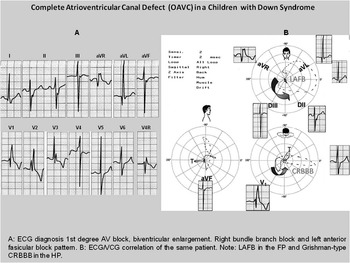
Atrioventricular conduction disturbances, especially complete atrioventricular block, occur in Down syndrome associated with atrioventricular septation defects. Patients with Down syndrome and complete or partial atrioventricular canal defects frequently present atrioventricular conduction disturbances varying from first-degree atrioventricular block to complete atrioventricular block,Reference Seale and Shinebourne 15 , Reference Blom, Ottenkamp, Deruiter, Wenink and Gittenberger-De Groot 16 which may be present since birth or may develop immediately or late after corrective surgery. In Figure 1, a classic 12-lead ECG of a child with atrioventricular canal defect can be seen. The accompanying vectorcardiogram illustrates the classic ECG disorder.
12-lead ECG and vectorcardiogram (VCG) of a child with Down syndrome and complete atrioventricular canal defect.

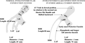
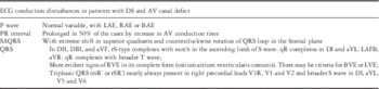
In atrioventricular septal defects, the conduction system has anatomical abnormalities in the atrioventrcular node, which is posteriorly displaced, and in the bundle branches producing a combination of proximal and distal conduction disorders (Figs 1 and 2). Combined distal conduction defect – left anterior fascicular block+right bundle branch block – are frequently seen (Table 1).
Anatomical comparison of the normal conduction system and the conduction system of a patient with atrioventricular endocardial cushion defect.

ECG characteristics of patients with AV canal defects.

AV=atrioventricular; BAE=biatrial enlargement; BVE=biventricular enlargement; DS=Down syndrome; LAE=left atrium enlargement; LAFB=left anterior fascicular block; LVE=left ventricular enlargement; RAE=right atrium enlargement; RVE=right ventricular enlargement
Electrophysiologic studies of non-surgical Down syndrome patients with atrioventricular block and atrioventricular septal defects demonstrate a high incidence of intra-atrial and atrioventricular nodal conduction delay that in 50% of cases or greater are manifested as first-degree atrioventricular block.Reference Blom, Ottenkamp, Deruiter, Wenink and Gittenberger-De Groot 16
Fournier et alReference Fournier, Young, García, Tamer and Wolff 17 evaluated 32 children with atrioventricular canal: 18 underwent preoperative electrophysiologic studies and 14 underwent postoperative studies. In the preoperative group, the mean age was 3 years (range 6 months to 16 years). First-degree atrioventricular block was observed in five patients due to internodal conduction delay and in one patient due to atrioventricular nodal conduction delay. A total of four patients had internodal conduction delay but normal PR interval in the surface ECG, and sinus node disease was seen in one patient. In the postoperative group, the following abnormalities were observed: first-degree atrioventricular block in nine patients – due to atrioventricular nodal conduction delay in two, His-Purkinje system conduction delay in one, upper normal intracardiac intervals in three, and unidentified in three – prolongation of the right ventricular apical activation time was observed in 11 of the 13 patients with complete right bundle branch block. Sinus node dysfunction was detected in three patients and abnormal atrioventricular nodal function in four patients. Left ventricular systolic function was normal in both groups. This study reflects that postoperative conduction disease in patients with repaired atrioventricular canal can be due to either the original condition, the postoperative damage of the conduction system, or both.
Complete atrioventricular block after corrective surgery of congenital heart defects may occur during the early postoperative period or late after surgery. Early postoperative atrioventricular blocks resolve spontaneously within 9 days after surgery in 60% of cases.Reference Blom, Ottenkamp, Deruiter, Wenink and Gittenberger-De Groot 16 , Reference Kugler, Gillette, Gutgesell and McNamara 18 Late atrioventricular block develops between 2 days and 25 years after surgery.
In a retrospective analysis, Banks et alReference Banks, Jenson and Kugler 19 analysed the patients included in the Pediatric Cardiology Database between 1977 and 2000, who had undergone corrective congenital heart surgery and had presented postoperative atrioventricular blocks of different degrees requiring implantation of a permanent pacemaker. In all, 53 patients were eligible and were divided into two groups: group I included patients with early atrioventricular block and permanent pacemaker implant before discharge and group II included patients with late atrioventricular block. Group I included 45 patients, five with Down syndrome.
There were eight patients with late atrioventricular block; six had Down syndrome and developed symptomatic atrioventricular block between 5 days and 12 years after surgery. These patients presented sinus heart rate between 75 and 166 beats/minute and a PR interval for the conducted beats between 140 and 260 milliseconds. Of these patients, six had complete or incomplete right bundle branch block and was associated with left anterior fascicular block in two patients. First-degree atrioventricular block associated with paroxysmal complete atrioventricular block was present in four patients. The remaining four patients presented high-degree atrioventricular block or complete atrioventricular block. The R-R interval measured at the moment of the diagnosis ranged between 1.1 and 25.8 seconds. Documented escape rhythms had a heart rate between 24 and 75 beats/minute. This study suggests that, in patients with Down syndrome, late atrioventricular block after corrective heart surgery for congenital heart defects is more common (5/45 versus 6/8 patients; p<0.001).
Supraventricular arrhythmias in Down syndrome
Some registries of patients with Down syndrome have reported a high incidence of Wolff-Parkinson-White syndrome, but there are no studies including a large number of patients to establish the real prevalence of pre-excitation syndromes in this population.
Cabeza-Ruiz et alReference Cabeza- Ruiz, Beas-Jiménez, Centeno-Prada and Naranjo-Orellana 2 analysed the electrocardiographic findings in 22 athletes with Down syndrome and found that 14 of the 22 had ECG abnormalities, whereas 8 (36.4%) did not. Among the 14 individuals with abnormal ECG, three presented Wolff-Parkinson-White pattern, one had congenital long-QT syndrome, three had increased S-wave amplitude, one had increased S-wave and R-wave amplitude, one had T-wave inversion in 3-lead ECG, and one had sinus bradycardia.
Data on Down syndrome and atrial fibrillation are scarce. They come from few case reports of children with 21 trisomy mosaic and stroke. As the usual studies for cryptogenic stroke – echocardiogram, 24-hour Holter monitoring – did not demonstrate the cause, implantation of loop recorder, to establish symptom–rhythm correlation, was suggested.Reference Stöllberger, Weiss, Zlabinger and Finsterer 20
Repolarisation abnormalities in Down syndrome (long-QT syndrome)
Some data suggest that the incidence of long-QT interval is increased in Down syndrome; however, we have not found publications with statistical information to estimate its real frequency. Patients with Down syndrome usually have conditions associated with prolonged long-QT interval, such as hypothyroidism or congenital heart defects and pulmonary hypertension.Reference Tisma-Dupanovic, Gowdamarajan, Goldenberg, Huang, Knilans and Towbin 21 In a case report published by Tisma-Dupanovic et al, a 13-year-old girl with Down syndrome and atrioventricular canal defect presented with QT prolongation and non-specific T-wave changes and pronounced U-waves. A diagnosis of concomitant Andersen–Tawil syndrome was genetically confirmed. The first ECG performed at 13 days of age demonstrated normal sinus rhythm, right atrial enlargement, superior QRS axis, and right ventricular hypertrophy. No arrhythmias associated with long QT were observed.Reference Tisma-Dupanovic, Gowdamarajan, Goldenberg, Huang, Knilans and Towbin 21 , Reference Rodríguez and Reyes 22
Down syndrome and congenital defects: its impact on the ECG
The Atlanta Down Syndrome ProjectReference Freeman, Taft and Dooley 23 reported that among 227 Down syndrome infants with congenital heart defects, 45% had atrioventricular canal defects, 35% had ventricular septal defect, 8% had ostium secundum atrial septal defect, 7% had persistent patent ductus arteriosus, 4% had isolated tetralogy of Fallot, and 1% had other. Each defect will have its own ECG expression depending on the type and magnitude of the defect. In atrioventricular canal defects, the QRS axis in the frontal plane is directed superiorly with extreme right or left axis deviation.Reference Seale and Shinebourne 15
Conduction disturbances are common, particularly first-degree atrioventricular block frequently seen in partial or complete atrioventricular canal defect, due to altered organisation of the atrioventricular node anatomy and bundle of His branches and due to a conduction defect in the trabecular myocardiumReference Raveau, Lignon and Nalesso 3 , Reference Seale and Shinebourne 15 (Figs 1 and 2). Fragmented QRS complex has been identified as a predictor of arrhythmias, a common complication of Down syndrome patients with congenital heart defects. However, no specific data on this association have been published.
In a study published by Narchi,Reference Narchi 24 who studied the value of ECG within the first 48 hours of life to diagnose congenital heart disease in 37 neonates with Down syndrome, 24 had no clinical evidence of congenital heart disease and had normal ECGs and normal echocardiograms and 13 had congenital heart defects. The ECG was normal in seven neonates with congenital heart disease: four with atrial septal defect, two with tetralogy of Fallot, and one with ventricular septal defect. A left QRS axis deviation was found in six neonates: five with complete atrioventricular septal defect and one with ventricular septal defect and mitral valve prolapse. The study concluded that surface ECG is useful to detect atrioventricular canal defects but has a low sensitivity for other congenital heart defects.
Tubman et alReference Tubman, Shields, Craig, Mulholland and Nevin 4 conducted a prospective study to evaluate the effectiveness of clinical examination, chest radiography and electrocardiography compared with echocardiography in detecting heart disease early in the life of children with Down syndrome. Among 81 patients, 34 had congenital heart disease detected by echocardiography: 13 had atrioventricular septal defects, seven had ostium secundum atrial septal defect, six had solitary patent ductus arteriosus, five had isolated ventricular septal defect, and three had combinations of heart defects. Individual examination methods had low sensitivity – clinical examination 53%, chest radiography 44% and ECG 41% – but high specificity – clinical examination 94%, chest radiography 98% and ECG 100% – for diagnosis, although sensitivity improved when the three techniques were combined – the sensitivity increased to 71%, with a specificity of 91%.
A total of 66 babies had a normal ECG, but 19 of them had congenital heart disease – two had atrioventricular septal defects, four had ventricular septal defect, five had ostium secundum atrial septal defect, five had patent ductus arteriosus and three had a combination of defects.
Patients with Down syndrome and partial atrioventricular canal defects reaching adulthood usually have ostium primum atrial septal defect and cleft mitral valve or surgically repaired atrioventricular canal. These patients present conduction disturbances due to the anatomical displacement of the atrioventricular node and also have signs of hypertrophy in the ECG depending on the dominant physiology and on the development of Eisenmenger’s syndrome.
After surgery, about 5% of patients may present episodes of atrial fibrillation or atrial flutter.Reference Khairy and Marelli 25 , Reference Daliento, Rizzoli and Marchiori 26 In addition, about 30% may develop ventricular arrhythmias,Reference Khairy and Marelli 25 , Reference Daliento, Rizzoli and Marchiori 26 particularly ventricular premature beats. Complex arrhythmias are more common in the presence of left ventricular dysfunction.
In adults with Down syndrome and partial atrioventricular septal defects, first-degree atrioventricular block is the most distinctive ECG finding in 50% of cases or greater, generally due to intra-atrial conduction delays. In these patients, the right bundle branch is particularly long and emerges from a bundle of His inferiorly displaced with hypoplasia of the anterior fascicle resulting in rSr’ ECG pattern in the right precordial leads and extreme left axis deviation (Fig 2). Q-waves can be seen in leads I and aVL, and S-waves in leads II, III and aVF with notched QRS.Reference Banks, Jenson and Kugler 19
Atrial septal defect can also occur in Down syndrome, depending on the series, in about 33% of cases.Reference Rubens, del Pozzo, Pablos, Calderón and Castrejón 1 Ostium secundum is the most common form. The ECG in unrepaired ostium secundum atrial septal defect usually shows sinus rhythm, but atrial fibrillation and atrial flutter are not uncommon and its incidence increases with age.Reference Gatzoulis, Freeman, Siu, Webb and Harris 27 , Reference Attie, Rosas, Granados, Zabal, Buendia and Calderon 28 Atrioventricular node conduction is usually preserved, but first-degree atrioventricular block can be seen in 6–19% of cases, with rare progression to high-grade atrioventricular block.Reference Daliento, Rizzoli and Marchiori 26 , Reference Anderson, Rogers, Canent and Spach 29 Typically, QRS morphology shows a rSr’ pattern or rsR’ pattern in the right precordial leads.Reference Daliento, Rizzoli and Marchiori 26 , Reference Rodriguez-Alvarez, Martinez De Rodriguez and Goggans 30 QRS duration is usually slightly prolonged at the expense of delay in the right bundle.
Patent ductus arteriosus is relatively common as presented by Rubens et al.Reference Rubens, del Pozzo, Pablos, Calderón and Castrejón 1 Its ECG manifestations depend on various factors such as the duration of the disease, the size of the ductus, and its association with other malformations or not.
When patent ductus arteriosus is small, it has no electrocardiographic expression. In moderate size patent ductus arteriosus, electrocardiographic manifestations are non-specific and are mainly due to atrium and left ventricle overload. First-degree atrioventricular block can be seen.Reference Daliento, Rizzoli and Marchiori 26 , Reference Mirowski, Arevalo, Medrano and Cisneros 31 The QRS axis remains unchanged and deep S-waves in lead V1 and tall R-waves in the precordial leads V5 and V6 as secondary repolarisation changes can be seen.Reference Mirowski, Arevalo, Medrano and Cisneros 31 , Reference Marquis, Miller, McCormack, Matthews and Kitchin 32
Ventricular septal defect may also occur in patients with Down syndrome. The most common form is perimembranous and its ECG manifestations depend on the size of the defect and the degree of volume overload and pressure in the ventricles. ECG manifestations vary from normal to florid abnormalities such as long and notched P-waves in leads I and II and large negative terminal P-wave in lead V1 due to right atrial overload. The QRS axis usually has a slight deviation to the right but between 3% and 15% of patients may have left axis deviation.Reference Daliento, Rizzoli and Marchiori 26 , Reference Mirowski, Arevalo, Medrano and Cisneros 31 , Reference Moller, Patton, Varco and Lillehei 33
Signs of biventricular overload can be found in 23–61% of cases and complete right bundle branch block can be seen in up to 60% of the cases.Reference Daliento, Rizzoli and Marchiori 26 , Reference Riggs, Mehta, Hirschfeld, Borkat and Liebman 34 , Reference Kidd, Driscoll and Gersony 35
Arrhythmias may be seen as isolated premature ventricular beats and couplets. Episodes of non-sustained and sustained ventricular tachycardia may occur in about 6% of the cases, especially associated with increased pulmonary artery pressure.Reference Daliento, Rizzoli and Marchiori 26 , Reference Kaufman, Alfayyadh, Hordof and Apfel 36
Limitations
The major limitation of this review is the lack of specific literature on this topic. Most of the data provided in this review come from articles that did not specifically address electrical problems associated with Down syndrome. Information on ventricular arrhythmias is quite limited and it seems its prevalence after surgery does not differ from other forms of congenital heart diseases.
Conclusions
Down syndrome is associated with a broad spectrum of cardiovascular abnormalities, which may present isolated or in combination. Its correct and early diagnosis is essential to achieve impact in the quality of life and survival in these patients.
Acknowledgements
None.
Financial Support
This research received no specific grant from any funding agency, commercial or not-for-profit sectors.
Conflicts of Interest
None.