Introduction
Medulloblastomas (MBs) are malignant brain tumors that arise in the posterior fossa (PF), and occur in 1 per 200,000 in the United States (Habrand & De Crevoisier, Reference Habrand and De Crevoisier2001). Although MBs can occur at any age, they are typically diagnosed before the age of 10, with a peak incidence around 5 years of age, and occur 1.5–2 times as frequently in males as compared to females (Gottardo & Gajjar, Reference Gottardo and Gajjar2006). Treatment for MBs includes maximal surgical resection, chemotherapy, radiation therapy to the PF or tumor bed, and, because of its tendency to disseminate, with cranial-spinal radiation (CSR) as well. The 5-year survival rates for MB are now 70–85% (Gajjar et al., Reference Gajjar, Chintagumpala, Ashley, Kellie, Kun, Merchant and Gilbertson2006). Unfortunately, many survivors experience long-term impairments in their quality of life and cognitive development, including intellectual and learning abilities (e.g., Dennis, Spiegler, Hetherington, & Greenberg, Reference Dennis, Spiegler, Hetherington and Greenberg1996; Edelstein et al., Reference Edelstein, Spiegler, Fung, Panzarella, Mabbott, Jewitt and Hodgson2011; George et al., Reference George, Kuehn, Vassilyadi, Richards, Parlow, Keene and Ventureyra2003; Mabbott, Penkman, Witol, Strother, & Bouffet, Reference Mabbott, Penkman, Witol, Strother and Bouffet2008; Padovani, Andre, Constine, & Muracciole, Reference Padovani, Andre, Constine and Muracciole2012; Palmer, Reference Palmer2008; Palmer et al., Reference Palmer, Goloubeva, Reddick, Glass, Gajjar, Kun and Mulhern2001; Ris, Packer, Goldwein, Jones-Wallace, & Boyett, Reference Ris, Packer, Goldwein, Jones-Wallace and Boyett2001; Spiegler, Bouffet, Greenberg, Rutka, & Mabbott, Reference Spiegler, Bouffet, Greenberg, Rutka and Mabbott2004). It has been suggested that these difficulties may be the result of core cognitive deficits in speed of processing (Mabbott et al., Reference Mabbott, Penkman, Witol, Strother and Bouffet2008), working memory, attention, and declarative memory. That is, relative declines in IQ may reflect a failure to learn new information at a developmentally appropriate rate (Mulhern, Merchant, Gajjar, Reddick, & Kun, Reference Mulhern, Merchant, Gajjar, Reddick and Kun2004).
Several studies have shown that MB is associated with declines in both verbal and non-verbal memory (George et al., Reference George, Kuehn, Vassilyadi, Richards, Parlow, Keene and Ventureyra2003; Kieffer-Renaux et al., Reference Kieffer-Renaux, Bulteau, Grill, Kalifa, Viguier and Jambaque2000; Mulhern et al., Reference Mulhern, Palmer, Reddick, Glass, Kun, Taylor and Gajjar2001; Nagel et al., Reference Nagel, Delis, Palmer, Reeves, Gajjar and Mulhern2006). However, the pathophysiology underlying the relation between MB and memory difficulties in humans is unclear. Past work has focused on the impact of MB treatment on the volume and structure of white matter (WM) across the whole brain and/or within specific regions of the brain (Khong et al., Reference Khong, Kwong, Chan, Sham, Chan and Ooi2003, Reference Khong, Leung, Fung, Fong, Qiu, Kwong and Chan2006; Mabbott, Noseworthy, Bouffet, Rockel, & Laughlin, Reference Mabbott, Noseworthy, Bouffet, Rockel and Laughlin2006; Mulhern et al., Reference Mulhern, Palmer, Reddick, Glass, Kun, Taylor and Gajjar2001, Reference Mulhern, Reddick, Palmer, Glass, Elkin, Kun and Gajjar1999; Mulhern, White, et al., Reference Mulhern, White, Glass, Kun, Leigh, Thompson and Reddick2004; Qiu, Leung, Kwong, Chan, & Khong, Reference Qiu, Leung, Kwong, Chan and Khong2006; Reddick et al., Reference Reddick, Glass, Palmer, Wu, Gajjar, Langston and Mulhern2005, Reference Reddick, White, Glass, Wheeler, Thompson, Gajjar and Mulhern2003; Shan et al., Reference Shan, Liu, Glass, Gajjar, Li and Reddick2006). However, while reduced WM volume has been associated with declines in overall intellectual abilities (Khong et al., Reference Khong, Leung, Fung, Fong, Qiu, Kwong and Chan2006; Mabbott et al., Reference Mabbott, Noseworthy, Bouffet, Rockel and Laughlin2006; Mulhern et al., Reference Mulhern, Palmer, Reddick, Glass, Kun, Taylor and Gajjar2001), it has not been associated with lower learning and memory abilities (Reddick et al., Reference Reddick, White, Glass, Wheeler, Thompson, Gajjar and Mulhern2003). It is possible that, while general indices of WM are good predictors of global cognitive abilities such as IQ, indices of discrete WM tracts may be a better predictor of specific cognitive abilities such as learning and memory.
In children and adults, declarative memory relies predominantly on specific regions of the brain such as the prefrontal cortex and medial temporal lobe (Eichenbaum, Reference Eichenbaum2013; Demaster & Ghetti, Reference Demaster and Ghetti2012; Ghetti, DeMaster, Yonelinas, & Bunge, Reference Ghetti, DeMaster, Yonelinas and Bunge2010; Isaacs et al., Reference Isaacs, Vargha-Khadem, Watkins, Lucas, Mishkin and Gadian2003; Ofen, Chai, Schuil, Whitfield-Gabrieli, & Gabrieli, Reference Ofen, Chai, Schuil, Whitfield-Gabrieli and Gabrieli2012). In support of this, Mabbott and colleagues (Mabbott, Rovet, Noseworthy, Smith, & Rockel, Reference Mabbott, Rovet, Noseworthy, Smith and Rockel2009) found that the structure of the uncinate fasciculus (UF), the major WM tract connecting the anterior temporal lobe with the medial and lateral orbitofrontal cortex (Catani, Howard, Pajevic, & Jones, Reference Catani, Howard, Pajevic and Jones2002), was predictive of audio-verbal memory performance in healthy children. To date, the effects of MB treatment on the UF have not been investigated.
While the relation between MB treatment and the UF remains speculative, there is a wide literature linking CSR with damage to the hippocampus in animal models (e.g., Madsen, Kristjansen, Bolwig, & Wortwein, Reference Madsen, Kristjansen, Bolwig and Wortwein2003; Monje & Palmer, Reference Monje and Palmer2003; Raber et al., Reference Raber, Fan, Matsumori, Liu, Weinstein, Fike and Liu2004; Winocur, Wojtowicz, Sekeres, Snyder, & Wang, Reference Winocur, Wojtowicz, Sekeres, Snyder and Wang2006). The hippocampus is a critical structure for declarative memory (Eichenbaum, Reference Eichenbaum2013; Squire, Reference Squire1992) and in humans, it continues to develop and increase in volume throughout adolescence (Pfluger et al., Reference Pfluger, Weil, Weis, Vollmar, Heiss, Egger and Hahn1999). Despite the importance of the hippocampus for declarative memory and its vulnerability to CSR, there is only one study that directly examines MB treatment on hippocampal volume in children. In a longitudinal study by Nagel and colleagues (Nagel et al., Reference Nagel, Palmer, Reddick, Glass, Helton, Wu and Mulhern2004), children treated for MB exhibited progressive declines in hippocampal volume for approximately 2 years after diagnosis before returning to a positive growth pattern. However, since the researchers focused on changes in raw hippocampal size, it is not possible to ascertain whether these changes reflect overall declines in brain size or a specific vulnerability of the hippocampus to treatment.
One way to examine whether treatment-mediated changes to hippocampal volume is the result of changes to the whole brain or a specific vulnerability of the hippocampus is to correct hippocampal size by total intracranial volume (ICV). If changes in hippocampal volume remain after removing variance due to ICV, this suggests that treatment results in specific or preferential damage to the hippocampus, rather than global damage to the entire brain. Furthermore, it would be important to include a comparative sample of healthy children to determine whether alterations in hippocampal volume following treatment are different from typical development. To our knowledge, no study has fully examined treatment-mediated effects on specific hippocampal vulnerability in pediatric MB.
The primary aim of this study was to examine the effects of MB treatment on structures within the brain that have been associated with learning and memory performance. Specifically, we examined differences between survivors of MB (and a single survivor treated for recurrent astrocytoma with comparable CSR) and healthy controls in the structure of the UF using diffusion tensor imaging (DTI; Mabbott et al., Reference Mabbott, Rovet, Noseworthy, Smith and Rockel2009) and the size of the hippocampus using volumetric magnetic resonance imaging (MRI). We predicted that, as compared to healthy children, survivors would exhibit insult to the UF and smaller hippocampal volumes.
A secondary aim was to examine whether or not treatment-mediated effects on memory structures were related to performance on a standardized test of learning and memory. We predicted that measures of UF structure and hippocampal volume would be significantly correlated with learning and memory, whereas global brain measures [i.e., total WM and gray matter (GM) volume] would not be correlated.
Materials and Methods
Participants
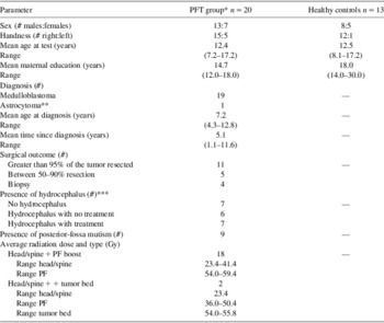
The current sample included 20 survivors with a history of posterior fossa tumors (MB = 19, recurrent astrocytoma = 1) and treated with surgery, chemotherapy, and CSR (“PFT Group”), and 13 age- and sex-matched controls (“healthy controls”) who were free of any previous or current neurological or clinical disorders (Table 1). Participants in the PFT group were recruited from brain tumor programs as part of a larger study at three hospitals: Alberta Children's Hospital in Calgary (ACH), British Columbia Children's Hospital in Vancouver (BCCH), and The Hospital for Sick Children in Toronto (SickKids). Healthy controls were recruited through advertisement in newspapers and within the Hospitals, as well as through families (e.g., siblings). While the rate of sibling participation at ACH and BCCH was not available, no siblings served as controls at SickKids. Overall, our recruitment approach allowed us to solicit participants who came from similar backgrounds as our PFT group, and also across a broad range of races, ethnicities, and socioeconomic (SES) backgrounds. However, data regarding participants’ race and SES were not collected because they are classified as personally identifying by our Research Ethics Board (REB) and are only collected when it is critical for research. All cases were scanned between 2007 and 2011. The protocol for this study was approved by the REB at each participating site. All participants were provided written informed consent or assent, and parental consent was obtained where applicable.
Demographic and clinical characteristics of the posterior fossa tumor (PFT) group and healthy controls

*All participants in the PFT Group were diagnosed between 2000–2009. When compared with the overall MB population diagnosed at SickKids during this time (n = 53), the two groups did not differ in terms of sex (χ2(1) = .16, p = 0.69) or age at diagnosis (t(56.2) = .73, p = .47).
**This participant was included because other than tumor pathology, they resembled individuals with medulloblastoma, i.e., tumor occurred along the vermis of the posterior fossa and treatment included surgical resection, CSR and adjuvant chemotherapy. Exclusion of this participant from the analyses did not change the results.
***Hydrocephalus was not a significant covariate in the analyses and it did not change the results.
The specific chemotherapy used depended on the brain tumor protocol and included different combinations of cisplatin, vincristine, etoposide, ifosfamide, carboplatin, and cyclophosphamide. The PFT group did not differ from the healthy controls in terms of sex (χ2(1) = 0.04; p = .84), handedness (χ2(1) = 1.59; p = .21), or age at assessment (t(31) = .08; p = .94). However, the PFT group had lower maternal education as compared to healthy controls (t(31) = −3.25; p = .003). To control for group differences in maternal education, this was included as a covariate in all analyses. However, since it was not a significant factor nor did it moderate any of the result patterns, these results were not reported.
MR Imaging and Post-processing
MRI measurements were performed at SickKids using a GE LX 1.5 Tesla (T) MRI scanner with an eight-channel head coil (GE Healthcare, Milwaukee, WI) and at the BCCH and ACH using a Siemens 1.5T MRI scanner with an eight-channel channel head coil (Siemens Canada Ltd., Mississauga, ON). The scanning protocol included a three-dimensional (3D)-T1 FSPGR gradient echo, inversion recovery-prepared sequence (IR time = 400 ms; echo time/repetition time [TE/TR] = 4.2/10.056 ms; 116–124 contiguous axial slices; number of excitations [NEX] = 1; 256 × 192 matrix, interpolated to 256 × 256; FOV = 24 × 24 cm; rbw = 162.734 kHz; slice thickness = 1.5 mm) and a diffusion-weighted sequence (single shot spin echo DTI sequence with EPI readout: 25–31 directions; b = 1000 s/mm2; TE/TR = 85.5/15000 ms; 45–50 contiguous axial slices; NEX = 1; 128 × 128 matrix, interpolated to 256 × 256; FOV = 24 × 24 cm; rbw = 1953.12 kHz; slice thickness = 3 mm).
To evaluate inter-site consistency in MRI measurements, one individual (Research Coordinator) was scanned at all three hospitals. Signal-to-noise ratios for the b = 0 diffusion tensor images from SickKids, BCCH, and ACH were 24.7, 43.6, and 39.1, respectively, and signal-to-noise ratios for the b = 1000 diffusion tensor images were 14.0, 17.4, and 16.6, respectively. As ratios were higher for BCCH and ACH relative to SickKids, site of imaging (BCCH/ACH vs. SickKids) was included as a covariate in subsequent analyses of volumetric MRI and DTI data.
Intracranial Volume
MRI post-processing of the 3D-T1 FSPGR involved extraction of the brain from the skull and extra-cerebral cerebrospinal fluid (CSF). All T1 images were aligned along the anterior and posterior commissure axis and ICVs were obtained for each participant. Each ICV was segmented using FSL to obtain total WM and GM volume.
DTI Processing for the Uncinate Fasciculus
For DTI post processing, seed/waypoint placement and probabilistic tractography were conducted in FMRIB Software Library (Smith et al., Reference Smith, Jenkinson, Woolrich, Beckmann, Behrens, Johansen-Berg and Matthews2004; Woolrich et al., Reference Woolrich, Jbabdi, Patenaude, Chappell, Makni, Behrens and Smith2009) using the same procedures as those outlined in Law and colleagues (Law et al., Reference Law, Bouffet, Laughlin, Laperriere, Briere, Strother and Mabbott2011). MRI data were corrected for inhomogeneity and eddy current, and DTI maps (FA, mean diffusivity [MD], λ1, λ2, and λ3) were calculated. The DTI model generates quantitative measures that index tissue structure based on water molecule displacement. Fractional anisotropy (FA) is a ratio ranging from 0 to 1, derived from λ1, λ2, λ3, and it is an index of the primary direction of water diffusion. MD (λ1 + λ2 + λ3/3) is a measure of the magnitude of water diffusion in all directions. Axial diffusivity (AD; λ1) and radial diffusivity (RD; λ2 + λ3/2) measures diffusion parallel and perpendicular to WM pathways, respectively. MD, AD, and RD express the rate of diffusion (mm2s−1). High FA and/or low MD values indicate restricted or anisotropic movement, which suggests high coherence of the WM bundles. Low FA and/or high MD values indicate unrestricted or isotropic movement, which suggests WM insult.
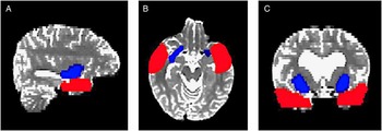
Second, the probability of connection between all voxels was calculated and served as a basis for fiber tracking analysis (Behrens, Berg, Jbabdi, Rushworth, & Woolrich, Reference Behrens, Berg, Jbabdi, Rushworth and Woolrich2007). A standardized template was used to generate regions of interest for tractography of the left and right UF. Briefly, the external capsule (seed) and anterior temporal lobe (waypoint; Catani & Thiebaut de Schotten, Reference Catani and Thiebaut de Schotten2008) were manually traced on the b=0 diffusion tensor image of 10 healthy control participants. The regions of interest were then non-linearly registered into the DTI space of an eleventh healthy control participant, and combined using the Image Calculator function in Analyze 9.0 (Mayo Clinic, Rochester, MN). The completed template was then applied to both hemispheres for every participant (Figure 1). Each tract was thresholded and visually inspected. If necessary, a mask was used to exclude outlier voxels that were not consistent with the known UF anatomy.
A standardized template with the external capsule (blue; seed) and anterior temporal lobe (red; waypoint) was applied to each participant's b = 0 diffusion tensor image for probabilistic tractography of the uncinate fasciculus. An example for one participant is shown in sagittal (A), axial (B), and coronal (C) plane.

Hippocampal Volume
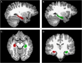
For hippocampal tracing, one of the authors (J.S.) manually traced the left and right hippocampus on a T1 image using Analyze. Tracing followed criteria modified from Pruessner and colleagues (2000) and Duvernoy (2005). Specifically, hippocampal boundaries were outlined slice by slice by a trackball-driven cursor in the sagittal plane to delineate the hippocampus inferiorly from the white matter of the parahippocampal gyrus, anteriorly by the alveus, and superiorly by the lateral ventricle. Further boundary verification was conducted in the coronal plane, such that designation began at the rostral end when the hippocampal head first appeared below the amygdala and ended when the crura of the fornices depart from the hippocampal tail at the caudal end. Volumes were further inspected in the axial plane (Figure 2).
Sagittal (A, B), axial (C), and coronal (D) views of left (green) and right (red) hippocampal traces in Analyze 9.0. Boundaries of the hippocampal formation included the parahippocampal gyrus, alveus, and lateral ventricle.

One-third of the participants were randomly selected (six from the PFT group, three controls) and retraced by J.S. and a second rater (F.L.) for intra- and inter-rater reliability, respectively. Reliability was assessed using Cronbach's alpha. Intra-rater reliability was .98 for the right hippocampus and .95 for the left hippocampus. Inter-rater reliability was .88 for the right hippocampus and .89 for the left hippocampus. Both raters were blind to the group status of each participant.
Raw hippocampal volumes were calculated using Analyze and corrected for ICV using a regression-based technique (Arndt, Cohen, Alliger, Swayze, & Andreasen, Reference Arndt, Cohen, Alliger, Swayze and Andreasen1991; Free et al., Reference Free, Bergin, Fish, Cook, Shorvon and Stevens1995). Specifically, the volume of each region of interest (ROI; i.e., right and left hippocampal volume) was regressed on ICV (collapsed across groups) such that the residual value (i.e., the structure's actual size minus its predicted value based on the individual's ICV) is accounted for in each ROI for each individual. This technique removes variance in hippocampal volume that is shared with overall brain size. All analyses of hippocampal volume used ICV adjusted values.
Statistical Analysis
Univariate analyses of variance (ANOVA) were used to assess group differences in ICV, and a multivariate ANOVA (MANOVA) was used to assess group differences in brain tissue volume (i.e., total WM and GM volume). Repeated-measures MANOVA was used to examine UF structure (FA, MD, AD, RD) with hemisphere (i.e., left, right) as a within-subjects variable. Hippocampal volume was examined using repeated measures ANOVA with hemisphere (i.e., left, right) as a within-subjects variable. Significant interactions were further examined using tests of simple effects. Effects were considered significant at p < .05 and marginally significant at p < .1.
Neuropsychological Testing
A subset of 10 participants with MB also underwent clinical assessments at SickKids, which included an assessment of learning and memory using the Children's Memory Scale (CMS). This assessment occurred within 2 months of the neuroimaging for seven survivors, and within 19 months for three survivors. The CMS includes subtests examining immediate and delayed memory for both verbal (i.e., stories and word pairs) and nonverbal information (i.e., visual-spatial patterns and faces). An overall age-normed index of memory can then be derived for each participant.
A correlation analysis was conducted to examine the relation between the CMS general memory index and measures of brain structure including: total GM volume, total WM volume, right hippocampal volume, left hippocampal volume, FA of the right UF, and FA of the left UF.
Results
Global Measures of Brain Structure
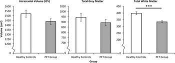
Analyses revealed that there were no group differences in ICV (F(1,30) = 2.93; p = .10; ηp 2 = .09). However, when ICV was segmented into WM and GM volume, there was a significant multivariate effect such that participants in the PFT group had smaller brain tissue volumes as compared to healthy controls (F(2,29) = 15.67; p = .0001; ηp 2 = .52). Univariate effects showed that while the PFT group had significantly reduced total WM volume as compared to healthy controls (F(1,30) = 24.56; p = .0001; ηp 2 = .45), there were no significant group differences for total GM volume (F(1,30) = 1.12; p = .30; ηp 2 = .04; Figure 3).
Survivors of posterior fossa tumors (PFT) exhibited significantly lower total white matter volume as compared to healthy controls and marginally smaller intracranial volume. There were no significant group differences in total grey matter volume.

Uncinate Fasciculus
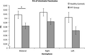
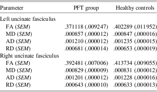
One healthy control participant was excluded from the analyses because the inferior temporal lobes were not imaged on DTI. The UF was produced bilaterally for all other participants with probabilistic tractography (Figure 4). All multivariate effects were non-significant (p's > .1). However, a significant univariate effect of group emerged for FA (F(1,29) = 5.05; p = .03; ηp 2 = .15); participants in the PFT group had significantly lower FA as compared to healthy controls (Figure 5). There were no significant effects of lateralization (F(1,29) = 1.00; p = .33; ηp 2 = .03) or interaction between group and hemisphere (F(1,29) = .55; p = .46; ηp 2 = .02). No significant univariate effects emerged for measures of AD, RD, and MD (p's > .1; Table 2).
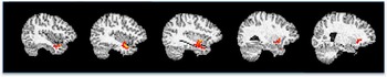
Sagittal view of the left uncinate fasciculus overlaid on T1 image from lateral to medial. The uncinate fasciculus (UF) connects anterior temporal lobe with the lateral and medial orbitofrontal cortex.

Survivors of posterior fossa tumors (PFT) exhibited significantly lower fractional anisotropy (FA) of the bilateral uncinate fasciculus. There was no significant main or interaction effect by hemisphere.

Means and standard error of means for DTI indices of the left and right uncinate fasciculus for the posterior fossa tumor (PFT) group and healthy controls

Note. All values are adjusted for scanner type.
Hippocampal Volume
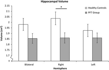
The main effect for hemisphere was not significant (F(1,30) = .17; p = .68; ηp 2 = .01). Although the main effect for group was only marginally significant (F(1,30) = 3.16; p = .09; ηp 2 = .10), there was a significant interaction between group and hemisphere (F(1,30) = 6.98; p = .01; ηp 2 = .19). Follow-up tests of simple effects revealed that the PFT group had significantly smaller right hippocampal volumes as compared to healthy controls (F(1,30) = 5.56; p = .03; ηp 2 = .16); there were no group differences for the left hippocampus (F(1,30) = 1.12; p = .30; ηp 2 = .04; Figure 6) or hemispheric differences for the PFT group (F(1,18) = 1.02; p = .33; ηp 2 = .05) or healthy controls (F(1,11) = .20; p = .67; ηp 2 = .02).
Survivors of posterior fossa tumors (PFT) had significantly smaller right hippocampus as compared to healthy controls; there were no effects of hemisphere for either group.

Given the previous finding of significant group differences in brain tissue volume (i.e., total GM and WM volume) but not in ICV, one could argue that smaller right hippocampal volumes in the PFT group may reflect treatment-mediated effects on brain tissue volume rather than a specific vulnerability of the hippocampus. However, even when tissue volume was entered as a covariate, the pattern of results remained the same. Specifically, there was a significant interaction between group and hemisphere (F(1,29) = 4.67; p = .04; ηp 2 = .14), with participants in the PFT group showing significantly smaller hippocampal volumes compared to healthy controls in the right (F(1,29) = 5.15; p = .03; ηp 2 = .15), but not in the left hemisphere (F(1,29) = 1.55; p = .22; ηp 2 = .05).
Relation Between Structural Measures and Memory Performance
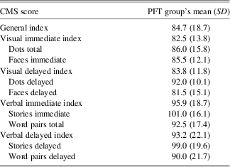
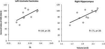
The subset of 10 survivors with MB exhibited significantly lower performance on the general index of the CMS as compared to the normative sample (t(8.6) = 2.58; p = .03; Table 3). In examining the relation between measures of brain structure and a general index of learning and memory, total GM volume emerged as a marginally significant correlate (R = .60; p = .06), while FA of the left UF (R = .64; p = .045) and volume of the right hippocampus (R = .71; p = .02) were significant correlates (Figure 7). Total WM volume was not significantly correlated with the general index of the CMS (R = .30; p > .1).
Means and standard deviation (SD) of a subset of the posterior fossa tumor (PFT) group (n = 10) standard scores on the Children's Memory Scale (CMS)

Note. Population means are 100 with an SD of 15.
In a correlational analysis of 10 survivors, FA of the left uncinate fasciculus (UF) and volume of the right hippocampus was significantly correlated with performance on the Children's Memory Scale (CMS).

Discussion
To our knowledge, the present study is the first to examine differences in memory structures between children with posterior fossa tumors (predominantly MB) and treated with CSR and healthy controls. Consistent with our predictions, PFT survivors exhibited lower FA of the UF and smaller hippocampal volumes as compared to healthy controls. Furthermore, we also found evidence linking measures of specific memory structures to learning and memory performance. Below, we discuss the relevance of these findings in context with prior literature.
FA of the Uncinate Fasciculus
While previous studies have focused on the effects of treatment for pediatric MB on the whole brain (Khong et al., Reference Khong, Kwong, Chan, Sham, Chan and Ooi2003, Reference Khong, Leung, Fung, Fong, Qiu, Kwong and Chan2006; Mabbott et al., Reference Mabbott, Noseworthy, Bouffet, Rockel and Laughlin2006; Mulhern et al., Reference Mulhern, Palmer, Reddick, Glass, Kun, Taylor and Gajjar2001; Mulhern et al., Reference Mulhern, Reddick, Palmer, Glass, Elkin, Kun and Gajjar1999; Mulhern, White, et al., Reference Mulhern, White, Glass, Kun, Leigh, Thompson and Reddick2004; Qiu et al., Reference Qiu, Leung, Kwong, Chan and Khong2006; Reddick et al., Reference Reddick, Glass, Palmer, Wu, Gajjar, Langston and Mulhern2005, Reference Reddick, White, Glass, Wheeler, Thompson, Gajjar and Mulhern2003; Shan et al., Reference Shan, Liu, Glass, Gajjar, Li and Reddick2006), a unique contribution of the present study is that we focused on treatment-mediated effects to a discrete WM tract, that is, the UF. The UF plays a significant role in the development and support of memory processes in healthy children (Mabbott et al., Reference Mabbott, Rovet, Noseworthy, Smith and Rockel2009), and continues to develop into adulthood (Lebel & Beaulieu, Reference Lebel and Beaulieu2011; Lebel, Walker, Leemans, Phillips, & Beaulieu, 2008). Given its protracted developmental course, it may be more vulnerable to the neurotoxic effects of MB treatment as compared to other WM tracts that reach maturity early in development, for example, the fornix, which is invariant between the age 5 and young adulthood (Lebel et al., Reference Lebel, Walker, Leemans, Phillips and Beaulieu2008). In future work, it may also be important to examine the structure of another WM tract in the brain that is not implicated in memory processing; this would allow one to ascertain whether memory tracts are especially vulnerable to the effects of treatment. It is conceivable that since MB treatment affects both WM and the hippocampus, WM tracts that connect the hippocampus to other regions of the brain may be disproportionately affected.
As mentioned earlier, DTI indices provide an indirect measure of WM architecture via measures of water diffusion; therefore, it is not known exactly what contributes to lower versus higher FA values of any WM tract. Specifically, the current finding of lower FA of the UF in the PFT group as compared to healthy controls may reflect disruptions in the myelination process, reduced cohesiveness and compactness of the fiber tracts, and/or increased extra-axonal space (Beaulieu, Reference Beaulieu2002; Nagy, Westerberg, & Klingberg, Reference Nagy, Westerberg and Klingberg2004). Despite this uncertainty, many researchers link lower FA to disruptions in the myelination process because WM is especially vulnerable to the toxic effects of CSR during rapid myelination (Moore, Reference Moore2005), and children are especially vulnerable to the effects of CSR (Pfefferbaum et al., Reference Pfefferbaum, Mathalon, Sullivan, Rawles, Zipursky and Lim1994).
Hippocampal Volume
In the present study, we also found that children treated for PFTs had smaller hippocampal volumes as compared to healthy control children. Critically, differences between the groups could not be attributed to differences in intracranial size or brain tissue volume, suggesting that the effects of treatment were specific to the hippocampus. In other words, the hippocampus may be especially vulnerable to neurotoxicity. In their longitudinal study, Nagel and colleagues (2004) found that MB treatment led to declines in hippocampal volume for the first 2 years after diagnosis before returning to a positive growth pattern. While the current study was not longitudinal, the average time from diagnosis of our PFT group was approximately 5 years. This suggests that even though hippocampal growth may return to a positive growth pattern 2 years after diagnosis, survivors do not catch up to their same-aged peers within 5 years.
It has been suggested that smaller hippocampal volumes may be the result of radiation-induced vascular compromise leading to hypoxic-ischemic injury to the hippocampus (Abayomi, Reference Abayomi1996). In addition, more recent research has focused on the effects of radiation on hippocampal neurogenesis. The hippocampus is one of the only two zones in the postnatal mammalian brain, including the subventricular zone, to generate new neurons (Luskin, Reference Luskin1993). Neurogenesis occurs throughout life and is thought to be important for memory functioning, especially for new learning (for review see: Castilla-Ortega, Pedraza, Estivill-Torrus, & Santin, Reference Castilla-Ortega, Pedraza, Estivill-Torrus and Santin2011; McEwen, Reference McEwen1999). In animal models, irradiation has been linked to decreased neurogenesis and impaired performance on hippocampally mediated tasks such as spatial and contextual learning (Andres-Mach, Rola, & Fike, Reference Andres-Mach, Rola and Fike2008; Ben Abdallah et al., Reference Ben Abdallah, Filipkowski, Pruschy, Jaholkowski, Winkler, Kaczmarek and Lipp2013; Saxe et al., Reference Saxe, Battaglia, Wang, Malleret, David, Monckton and Drew2006; Shors, Townsend, Zhao, Kozorovitskiy, & Gould, Reference Shors, Townsend, Zhao, Kozorovitskiy and Gould2002).
In humans, only one study has directly examined the effects of radiation on hippocampal neurogenesis. In a post-mortem study, Monje and colleagues (Monje et al., Reference Monje, Vogel, Masek, Ligon, Fisher and Palmer2007) found that cases (four MB, one leukemia) previously treated with CSR showed decreased hippocampal neurogenesis as compared to age- and sex-matched controls who died of non-neurological and non–neuro-oncological causes, with MB cases showing a 10-fold decrease. Of interest, this pattern of results was found even for those cases who had completed CSR treatment 23 years prior. This suggests that CSR may permanently decrease hippocampal neurogenesis, rather than temporarily disrupt it. However, given the study's small sample size (n = 5), more research is needed to clarify the above results. It would also be interesting for future research to focus on potential radiation-mediated volumetric differences in the neurogenic zone of the hippocampus, that is, the dentate gyrus. This type of analysis has been successfully applied to both clinical populations and healthy controls using high-resolution imaging focused on the medial temporal region with 3T MRI (Gold & Squire, Reference Gold and Squire2005; Olsen et al., Reference Olsen, Palombo, Rabin, Levine, Ryan and Rosenbaum2013; Pereira et al., Reference Pereira, Junque, Bartres-Faz, Ramirez-Ruiz, Marti and Tolosa2013). This type of analysis was not possible within the current paradigm, which used 1.5T scanners. Relative to a 3T scanner, a 1.5T scanner has reduced signal-to-noise ratio and spatial resolution.
PF Treatment and Laterality
The current results also showed that while group differences in the UF were bilateral, group differences in hippocampal volume were evident only in the right hemisphere. This suggests that the right hippocampus may be more sensitive to the effects of MB treatment than the left hippocampus. This may reflect slowed development and/or greater atrophy of the right versus left hippocampus. Future longitudinal studies are needed to clarify the mechanism by which such hemispheric differences exist between brain tumor survivors and healthy controls. Irrespective of the specific mechanism, this finding is somewhat consistent with the longitudinal results reported by Nagel and colleagues (Nagel et al., Reference Nagel, Palmer, Reddick, Glass, Helton, Wu and Mulhern2004); participants with MB exhibited a numerically steeper decline in right (estimated change: −.092) versus left (estimated change: −.065) hippocampal volume from the time of diagnosis to 1-year post-diagnosis. However, it is not clear why such hemispheric asymmetry in hippocampal volume or sensitivity would exist when MB treatment includes the whole brain. One possibility is that the left hippocampus, as part of the dominant hemisphere, may be more resilient to the effects of neuro-toxicity. Another possibility is that there may be differences in the developmental rate of the hippocampus; the right hippocampus may be more sensitive to the neurotoxic effects of treatment because it develops later than the left hippocampus. There is some evidence showing that right frontal-temporal regions of the brain develop later than homologous regions in the left hemisphere (Thatcher, Walker, & Giudice, Reference Thatcher, Walker and Giudice1987).
Linking Structure and Behavior
A secondary aim of the present study was to examine whether measures of memory structures, i.e., FA of the UF and volume of the hippocampus, would be correlated with memory performance. Consistent with prior studies, global WM volume was not significantly correlated with memory performance (Reddick et al., Reference Reddick, White, Glass, Wheeler, Thompson, Gajjar and Mulhern2003), although global gray matter volume was marginally correlated. Furthermore, higher scores on a learning and memory scale were associated with higher FA of the left UF and larger volumes of the right hippocampus. Although there appears to be a structure by function interaction (i.e., left UF vs. right hippocampal volume), it is important to note that the current sample size was quite small (n = 10) and such effects are likely unstable. Furthermore, FA of the left and right UF were marginally correlated with each other (R = .59; p = .07) and volume of the left and right hippocampus were significantly correlated with each other (R = .74; p = .02). This suggests that given a larger sample size, we may have observed correlations between bilateral UF and hippocampus and memory performance.
While a positive relationship between FA of the left UF and memory performance is consistent with the existing literature (Mabbott et al., Reference Mabbott, Rovet, Noseworthy, Smith and Rockel2009), the relationship between hippocampal volume and memory is somewhat controversial, and “bigger” has not always been found to be “better.” In a meta-analysis by Van Petten (Van Petten, Reference Van Petten2004), a negative relationship was found between hippocampal volume and memory performance among children, adolescents and young adults. Although this is in direct contrast to the current results, it is important to note that while Van Petten's meta-analysis concerned healthy participants, the current study examined memory performance in participants treated for PFTs. Thus, smaller hippocampal volumes may be advantageous up to a certain point in healthy children, but treatment likely leads to hippocampal volumes that are “too small”. This can be likened to a Goldilocks effect whereby optimal mnemonic processing requires the hippocampus to be not too big and not too small; it needs to be “just right.”
Limitations and Future Directions
The current findings need to be considered in the context of some limitations. First, we considered only the UF as a discrete WM tract implicated in memory processing, however, other WM tracts such as the fornix and the cingulum bundle are also implicated in learning and memory (for review see: Ghetti & Bunge, Reference Ghetti and Bunge2012). Second, the current study's sample size was relatively small. As a result, we were limited in addressing our secondary aims, that is, the relation between MB treatment, measures of brain structure, and memory performance. The current results provide some hints as to how such factors may be related to one and another, but more research is needed to explicate these findings. Furthermore, given the current sample size, our ability to detect small to medium effects was limited. Observed statistical power for detecting group differences was .73 for hippocampal volume, .58 for FA of the UF, 1.0 for WM volume, .18 for GM volume, and .38 for ICV. In other words, even though certain contrasts may not have reach statistical significance (e.g., group differences in ICV and GM), it does not necessarily mean that no such group differences exist in the population. It will be important for future research to conduct similar analyses with bigger sample sizes (i.e., >99) to clarify the present findings.
Third, the current study focused on the effects of treatment to the entire hippocampal volume, however, there is evidence to suggest that the hippocampus does not develop in a uniform manner. For example, it has been found that between the ages of 4 and 25, there is decreased volume in the anterior hippocampus and increased volume in the posterior hippocampus (Gogtay et al., Reference Gogtay, Nugent, Herman, Ordonez, Greenstein, Hayashi and Thompson2006; Insausti, Cebada-Sanchez, & Marcos, Reference Insausti, Cebada-Sanchez and Marcos2010). Nagel and colleagues (Nagel et al., Reference Nagel, Delis, Palmer, Reeves, Gajjar and Mulhern2006) also reported greater volumetric loss in the posterior versus anterior hippocampus after treatment, likely because posterior portions of the hippocampus are closer to higher-dose fields of CSR in the PF. Upon further examination of our current sample, we noted that the shape of the hippocampus in the PFT group appeared to be more distorted as compared to healthy controls. As a result, it would be difficult to ascertain where the “midline” point along the hippocampus lay along the anterior–posterior axis. Therefore, it may be helpful to examine treatment-mediated changes in the shape of the hippocampus using a deformation approach (e.g., Lerch et al., Reference Lerch, Yiu, Martinez-Canabal, Pekar, Bohbot, Frankland and Sled2011).
Lastly, it is important to note that while the current study examined differences between individuals treated for PFTs and healthy controls, it is not clear what factor(s) underlie these differences, that is, clinical (e.g., hydrocephalus, tumor location) and/or treatment factors (e.g., surgical resection, chemotherapy, CSR). While CSR has been identified as the best predictor of cognitive outcome (Copeland, deMoor, Moore, & Ater, Reference Copeland, deMoor, Moore and Ater1999; Lafay-Cousin et al., Reference Lafay-Cousin, Bouffet, Hawkins, Amid, Huang and Mabbott2009; Palmer, Reddick, & Gajjar, Reference Palmer, Reddick and Gajjar2007; Reddick et al., Reference Reddick, Russell, Glass, Xiong, Mulhern, Langston and Gajjar2000; Rutkowski et al., Reference Rutkowski, Bode, Deinlein, Ottensmeier, Warmuth-Metz, Soerensen and Kuehl2005), including memory, in pediatric MB (Kieffer-Renaux et al., Reference Kieffer-Renaux, Bulteau, Grill, Kalifa, Viguier and Jambaque2000), animal studies have also linked some of the chemotherapy agents used with impairments in learning and memory (e.g., Eijkenboom & Van Der Staay, Reference Eijkenboom and Van Der Staay1999; Shabani, Larizadeh, Parsania, Hajali, & Shojaei, Reference Shabani, Larizadeh, Parsania, Asadi Shekaari and Shahrokhi2012; Shabani, Larizadeh, Parsania, Asadi Shekaari, & Shahrokhi, Reference Shabani, Larizadeh, Parsania, Hajali and Shojaei2012). It will be important for future research to explore the contribution of different factors to the long-term outcome in brain tumor survivors. For example, to examine the effects of chemotherapy and CSR, future studies can include a control group with low-grade PF tumors, who also experience hydrocephalus, but are treated with surgery only.
Conclusions
To understand how brain tumor treatment leads to learning and memory impairments in children, we focused on changes to specific structures in the brain that has been implicated in learning and memory. We found that as compared to healthy controls, survivors of PFTs exhibited less coherent WM of the UF and a smaller right hippocampus. Furthermore, we also found that both the UF and the hippocampus were significantly related to performance on a standardized battery of learning and memory. In identifying and examining the effects of treatment on discrete and specific regions of the brain, we can better understand and predict the long-term cognitive outcome. This is critical not only for the ongoing modification of treatment protocols to reduce damage to brain tissue and to minimize long-term impairments, but also for the design and administration of targeted treatment, for example, animal models show that physical exercise can significantly boost hippocampal neurogenesis after irradiation (Wong-Goodrich et al., Reference Wong-Goodrich, Pfau, Flores, Fraser, Williams and Jones2010).
Acknowledgments
The authors do not have any conflicts of interest. This work was supported by grants from the C17 Research Network and the Canadian Cancer Society (D.J.M., grant number 18062), and a postdoctoral fellowship from the Canadian Institute of Health Research (L.R.).