




No CrossRef data available.
Asymmetry of the hippocampus and amygdala in MRI volumetric measurements of normal adults
Published online by Cambridge University Press: 01 September 2004
Abstract
Multiple studies have explored the relationship between MRI-based volumetric measurements of the hippocampus and amygdala, the degree of volumetric asymmetry of these structures, and symptom manifestation. However, considerable variability exists with regard to the reported volumetric values of these structures. The present study employed meta-analytic procedures to provide a systematic analysis of the normal population parameters of hippocampal and amygdala volumetric asymmetry as well as the absolute intrahemispheric volumes of these structures in normal adults. A literature review of studies published between 1990 and 2002 resulted in a representative sample of 82 studies (N = 3,564 participants) providing volumetric information of the hippocampus and 51 studies (N = 2,000 participants) providing volumetric information of the amygdala. Results revealed that both the hippocampus and the amygdala are reliably asymmetrical structures in normal adults, with larger right hippocampal (D = 0.21, p < .001) and right amygdala (D = 0.09, p < .01) volumes. Additional analyses indicated that differences in MRI magnet field strength and slice thickness values might differentially contribute to volumetric asymmetry estimates. These results expand on previous volumetric normative studies and may be relevant to investigators studying the clinical correlates of hippocampal and amygdala volumes. (JINS, 2004, 10, 664–678.)
Information
- Type
- Research Article
- Information
- Journal of the International Neuropsychological Society , Volume 10 , Issue 5 , September 2004 , pp. 664 - 678
- Copyright
- 2004 The International Neuropsychological Society
INTRODUCTION
The use of magnetic resonance imaging (MRI) to obtain volumetric measurements has allowed researchers the opportunity to explore in vivo differences between clinical groups and normal control participants in the volumes of particular neuroanatomical structures. The hippocampus and amygdala are of particular interest, with numerous studies focusing on the volumetric measurement of these structures in a variety of clinical conditions. In temporal lobe epilepsy (TLE), for example, hippocampal and amygdala volumetric measurements can provide an index of mesial temporal sclerosis that is useful for seizure onset lateralization, surgical decision-making, and outcome prediction after surgical resection (Breier et al., 1996; Gilmore et al., 1995; Radhakrishnan et al., 1998; Salanova et al., 1999; Watson et al., 1997b). The relevance of hippocampal and amygdala volumes has also been investigated in schizophrenia, with several meta-analyses suggesting that schizophrenia is associated with bilateral volume reductions in both structures (Lawrie & Abukmeil, 1998; Nelson et al., 1998; Wright et al., 2000). Reduced hippocampal volumes have been associated with mild cognitive impairment, thought to be a transitional state between normal aging and Alzheimer's disease (AD; Jack et al., 1997, 1999; Petersen et al., 2001), and measurement of hippocampal and amygdala volumes has been shown to improve the sensitivity of early AD diagnosis (Dickerson et al., 2001; Galton et al., 2001; Krasuski et al., 1998; Laakso et al., 1998).
Recently, Jack et al. (1995) highlighted the relevance of statistically characterizing volumetric measurement values in normal individuals. Specifying the range of normal values allows for greater sensitivity and specificity in clinical measurement and decision-making (O'Brien & Dyck, 1995). Valid normative volumetric information is essential for determining structural volume loss and changes in left–right asymmetry among patients. The presence of asymmetrical volumes in structures where symmetry is expected or the absence of asymmetry in structures where asymmetry is expected may be indicative of pathological processes and could provide insights into the biology of neuropsychiatric illnesses (Wang et al., 2001). The latter is clearly illustrated by Giedd et al. (1996) and Castellanos et al. (1996), who studied the developmentally normative volumes of several neuroanatomical structures in children and found that those with attention deficit hyperactivity disorder (ADHD) had a loss of the normal volumetric asymmetry of the caudate nucleus. More recently, Geroldi et al. (2000) reported a dose-dependent effect between the presence of the apolipoprotein ε4 allele and hippocampal asymmetry in AD, such that patients without the allele had a right greater than left asymmetry, those with one copy of the allele had no asymmetry, and those with two copies had a left greater than right asymmetry (cf. Bigler et al., 2002).
Most studies that have examined hippocampal and amygdala volumes in normal adult participants vary considerably with regard to the degree of volumetric asymmetry as well as the absolute intrahemispheric volumes reported (e.g., Bhatia et al., 1993; Bigler et al., 1997; Cendes et al., 1993; Convit et al., 1999; Kälviäinen et al., 1997a, 1997b; Szesko et al., 1999; Watson et al., 1992; Whitworth et al., 1998). Hippocampal asymmetry has received more attention among researchers than amygdala asymmetry, with most studies reporting either a larger right hippocampus or no left–right hippocampal asymmetry. Recently, Brierley et al. (2002) conducted a meta-analysis of MRI-based amygdala volumes and found no significant difference between left and right amygdala volumes in normal individuals. These in vivo results are in marked contrast to post-mortem examinations of amygdala volumes, which suggest that a right greater than left asymmetry exists in normal individuals (Gloor, 1997; Murphy et al., 1987). Unfortunately, Brierley et al. relied exclusively on significance tests and failed to provide effect size estimates to index the magnitude of the left–right volumetric difference. Statistical significance is largely dependent on sample size and its sole use in meta-analytic research has been previously questioned (Rosenthal, 1991; Rosenthal & DiMatteo, 2001). Furthermore, the authors' conclusions may have been limited by the absence of homogeneity tests, publication bias analysis, and exclusion of outliers (see Cooper & Hedges, 1994; Durlak & Lipsey, 1991; Hunter & Schmidt, 1990; Rosenthal, 1991).
It has been suggested that the variability in volumes found in previous studies may be due to differences in image acquisition techniques, hippocampal and amygdala boundary definitions, and image processing software across multiple centers (Jack et al., 1995). However, after examining preliminary unpublished data Jack et al. indicated that the pulse sequence parameters involved in image acquisition might not inherently produce systematic over- or under-estimates of volumes. This view has been challenged by Luft et al. (1998), who suggested that image acquisition parameters do indeed influence MRI-based volumetric measurements. Similarly, the relevance of imaging slice thickness to reliable volume acquisition has been previously debated (Cook et al., 1992; Jack et al., 1995; Laakso et al., 1997; Luft et al., 1998) and it remains to be determined whether differences in slice thickness contribute to overall measurement error or play an inconsequential role in volumetric determinations. Pruessner et al. (2000) offered an additional possible source of volumetric variability, namely, a low level of statistical power in most studies to detect what may be subtle volumetric asymmetries. In this manner, the ability to detect volumetric asymmetry of particular structures may be influenced by both the magnitude of the asymmetry as well as the number of participants included in the study.
Although recent efforts have attempted to delineate normative volumetric values within particular age and gender groups (e.g., Allen et al., 2002; Edland et al., 2002; Ge et al., 2002; Jack et al., 1999), the degree of left–right asymmetry and the absolute hippocampal and amygdala volumes in the normal population remain to be established. The present study was guided by three questions (Jack et al., 1995). First, is there a normal “relative” or interhemispheric volumetric asymmetry in the size of the hippocampi and/or amygdalae among normal adults? Second, if such an asymmetry exists, which side is larger? And third, what are the normal “absolute” or intrahemispheric left and right volumes?
To address these questions, the present study undertook a meta-analytic approach for examining the parametric values of hippocampal and amygdala asymmetry and absolute intrahemispheric volumes in normal adults. The use of meta-analytic procedures provides several distinct advantages. First, by aggregating data over many studies it is possible to evaluate specific hypotheses in a substantially larger pooled sample. As a result, meta-analysis provides a powerful methodology for detecting subtle effects that might otherwise be missed given the frequently smaller and practical sample sizes of most experimental studies. Second, the use of aggregated samples allows for increased reliability in the calculation of parametric values by reducing the confidence interval around the parameter estimate. Third, although previous investigations have employed other measures of asymmetry, such as laterality ratios and coefficients (e.g., Bernasconi et al., 1999), the use of an effect size index in the present study was considered superior because it takes into account the variance in obtained measurements across participants. Fourth, the use of meta-analytic procedures allows for the direct comparison of potential moderator variables and their influence on the obtained results. Consequently, it becomes possible to quantitatively evaluate particular MRI imaging parameters and sample characteristics in order to determine their potential influence on volume calculations.
METHODS
Literature Search
A systematic search was conducted using the Medline and PsycInfo computerized databases as well as exhaustively inspecting the reference sections of relevant studies. Studies were considered for inclusion if they met the following criteria: (1) they were published between 1990 and 2002; (2) volumetric information (reported as mean and standard deviation) was provided for either the hippocampus or amygdala in each hemisphere; (3) volumetric measurements were not corrected for variability in head size; (4) volumetric measurements were obtained through magnetic resonance imaging; (5) volumetric measurements were obtained from neurologically uncompromised participants in a “normal” or “control” group; and (6) these participants were at least 18 years of age.
The use of uncorrected volumetric measurements allows for a comparison of interhemispheric volume differences without the potentially confounding influence of diverse correction methods across studies. These correction methods may be necessary when comparing volumes among clinical groups given the substantial variability in absolute volume measurements across gender and age (Jack et al., 1995). However, such corrections are not necessary for the purposes of the present study since the primary unit of analysis is the magnitude of interhemispheric asymmetry. In this manner, the magnitude of asymmetry may be independent of fluctuations in absolute volumes among groups and is not assumed to covary with intracranial volume. On the other hand, the exclusion of studies that solely provide corrected volumetric measurements might result in a restricted range of samples and reduced overall variability if the studies reporting uncorrected measurements represent only a minor fraction of the total number of volumetric studies in this area. In fact, 83% of all the studies that provided volumetric measurement information reported either uncorrected volumetric values or both uncorrected and corrected volumetric values, whereas 17% only reported corrected volumetric values. For these reasons, it was deemed that the sample composition of the current analyses was sufficiently adequate and representative to address the primary goals of the study.
Two-hundred and eighteen studies were initially considered for the analyses. Of these, 51 studies did not provide mean volumetric values and were excluded. Twenty-nine additional studies were excluded because they only reported corrected volumetric values. Thus, 85 studies providing hippocampal volumetric measurements and 53 studies providing amygdala volumetric measurements in normal participants met the initial inclusion criteria. Several of these studies presented volumetric results by gender or grouped age ranges, resulting in multiple samples within those studies. When repeated measurements were obtained from the same participants (e.g., Laakso et al., 1997), an average volume was calculated directly from the reported values. Thus, a total of 107 samples (N = 3,645 participants) with hippocampal and 68 samples (N = 2,032 participants) with amygdala volumetric measurements were considered for the present analyses.
Statistical Analyses of Effect Size Estimates
Two separate meta-analyses were conducted, one for hippocampal and one for amygdala volumetric measurements. Volumetric asymmetry for each sample was computed using an effect size calculation. Given the cautionary observations by Dunlap et al. (1996) on the calculation of effect size estimates from correlated designs, only values reported as means and standard deviations were used. The initial effect size estimate, Hedges' g, was obtained by subtracting the left mean volume from the right mean volume and dividing by the pooled standard deviation. The direction of the effect can be interpreted such that positive values reflect greater right hemispheric volumes while negative values reflect greater left hemispheric volumes. Hedges' g, however, is a biased estimator of effect size in small group samples (Hedges & Olkin, 1985). It was therefore transformed into an unbiased effect size estimator, Hedges' d, that can be interpreted in a similar manner. Hedges' d was calculated according to the following formula:

After an effect size d was obtained for each hippocampal and amygdala volumetric sample, an overall weighted effect size (D) was calculated separately for the hippocampus and amygdala. Weighting was necessary given that studies with a larger sample size (and therefore smaller variance) provide a more accurate estimate of population parameters than studies with a smaller sample size (Hunter & Schmidt, 1990; Shadish & Haddock, 1994). Weights (w) for each sample (i) were computed as the reciprocal of the sample variance according to the following formulas:

and

Thus, each D value represents the overall unbiased and weighted mean estimate of hippocampal and amygdala volumetric asymmetry in normal individuals. The overall variance associated with D was calculated as the reciprocal of the sum of weights. Confidence intervals around D were then calculated to evaluate the statistical significance of each asymmetry estimate.
Meta-analytic results are especially vulnerable to the inclusion of even a few outlying samples (Durlak & Lipsey, 1991). The presence of outliers was determined by a concordance of several suggested methods (Cooper & Hedges, 1994; Durlak & Lipsey, 1991; Hedges & Olkin, 1985). Specifically, samples that were two standard deviations away from the respective mean effect size estimate (d) were flagged as potential outliers. Convergent information from funnel plots, a graphical display of sample size by effect size estimates, was then employed to substantiate the presence of unusually extreme findings.
Homogeneity of Effects and Moderator Variables
The null hypothesis of homogeneity among obtained effect sizes suggests that the observed results estimate a single population effect and differences among the obtained effect sizes are due to sampling error (Durlak & Lipsey, 1991). Alternatively, a significant test for homogeneity suggests heterogeneity among the obtained effect sizes and would strongly indicate the presence of moderator variables. In order to evaluate the homogeneity of effect sizes across samples, we calculated the Q statistic, which is evaluated on the chi-square distribution at k − 1 degrees of freedom, where k equals the total number of samples. If Q exceeds the upper-tail critical value of the chi-square distribution then the null hypothesis of homogeneity is rejected. To evaluate moderator variables, the Q statistic can then be partitioned into between-groups variance (Qb) and within-groups variance (Qw) in a manner analogous to weighted ANOVA (Hedges & Olkin, 1985). In this manner, the between-groups component evaluates the difference between effect size estimates in categories of the variable of interest.
Several variables judged to be theoretically meaningful were hypothesized a priori to significantly covary with the obtained effect size estimates and potentially moderate the obtained results. These variables were either related to MRI imaging parameters (MRI magnet field strength, slice thickness, head position corrections, plane of image acquisition) or sample characteristics (sample size, age of participants, sex of participants). Although there are many other important imaging parameters in addition to the four presently considered, only those parameters which are reported in a substantial number of published studies were included. Additional anatomical and technical moderators were not considered viable for meta-analytic computations due to the wide variability in approaches and methodology. The two most frequently reported methods for normalizing head position were considered: (1) orientation to the plane perpendicular to the anterior-posterior commissure plane and (2) orientation perpendicular to the long axis of the hippocampus. Image acquisition was evaluated by contrasting studies that used only the coronal plane to studies that referenced the sagittal and/or axial planes in addition to the coronal plane. Evaluating the influence of MRI slice thickness on volumetric asymmetry calculations was considered to be particularly meaningful given the conflicting results offered by previous investigations. In sum, we were interested in whether significant differences in asymmetry could be discerned between categories of these variables.
Publication Bias
Publication bias, or the so-called file drawer problem, refers to the likelihood for studies with null effects to remain unpublished, thus adversely affecting the validity of the conclusions drawn from any quantitative review (Rosenthal, 1991). The possibility of publication bias was initially assessed through visual inspection of funnel plots. An absence of studies with effect size estimates near zero would not produce the characteristic funnel-shape distribution and would strongly suggest the presence of publication bias. In addition, publication bias in each of the two meta-analyses was evaluated by calculating a fail-safe N, or the number of unpublished studies with no effect that must exist in order to reduce the obtained overall effect size estimate to a minimal critical level (Orwin, 1983; Rosenthal, 1991). For each of the two meta-analyses, this critical level was set at p = .05. In other words, the fail-safe N indicates the number of unpublished studies with null effects that must exist in order to bring the overall weighted effect size significance level to just significant at p = .05.
RESULTS
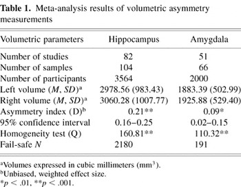
Among all studies that met initial inclusion criteria, 3 outliers were identified and removed from the hippocampus meta-analysis, resulting in 82 studies with 104 samples (N = 3,564 participants). In the amygdala meta-analysis 2 outliers were identified and removed, resulting in 51 studies with 66 samples (N = 2,000 participants). Table 1 presents the results from the hippocampal and amygdala volumetric asymmetry meta-analyses.
Meta-analysis results of volumetric asymmetry measurements

Hippocampal Asymmetry
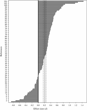
The overall weighted effect size (D) revealed a significant hippocampal volumetric asymmetry, suggesting that the right hippocampus is reliably larger than the left hippocampus in normal adults (p < .001). Hedges' g asymmetry estimates ranged from −0.90 to 1.53, with a mean value of g = 0.22. Unbiased asymmetry estimates ranged from −0.86 to 1.44, with a mean value of d = 0.22. There were 76 unbiased effect size estimates in the positive range, 26 unbiased effect size estimates in the negative range, and 2 unbiased effect size estimates equal to zero. Figure 1 displays the results of the 104 unbiased asymmetry estimates (d) for the hippocampus.

Effect size estimates of hippocampal asymmetry. Note. Positive values indicate larger right hippocampal volume. Solid line = overall weighted effect size (D); dashed lines = 95% confidence interval. Numbered references on the ordinate axis correspond to bracketed numbers at the end of each entry in the Appendix.
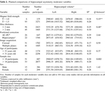
The overall homogeneity test was significant [Q(103) = 160.81, p < .001], suggesting that the heterogeneity in effect size estimates is likely attributable to the presence of moderator variables. There were significant between-category differences in asymmetry effect sizes for MRI magnet field strength (p < .01) and MRI slice thickness (p < .05). Studies that measured hippocampal volumes using a magnet field strength of 1.5 Tesla found a reliable hippocampal volumetric asymmetry, with the right hippocampus significantly larger than the left hippocampus [D = 0.20, C.I. (95%) = 0.15–0.25]. Alternatively, studies using a magnet field strength of 1.0 Tesla did not find a significant volumetric asymmetry [D = −0.18, C.I. (95%) = −0.42–0.06]. Imaging slice thickness also appeared to moderate the degree of hippocampal volumetric asymmetry. While there was no significant correlation between asymmetry effect size estimates and slice thickness per se (r = −.16, p = .13), a greater degree of right larger than left asymmetry was found in those studies that calculated hippocampal volumes using imaging slices less than or equal to 2 mm [D = 0.25, C.I. (95%) = 0.19–0.31] compared to those studies that used imaging slices of more than 2 mm [D = 0.10, C.I. (95%) = 0.01–0.19]. The results also suggested that head position corrections, plane of acquisition, age of participants, sample size, and sex of participants did not appear to significantly moderate the heterogeneity in effect size estimates. Indeed, the correlations between effect size estimates and age (r = .17), sample size (r = −.01), and sex (rpb = .32) were not statistically significant. The results of the planned comparisons are presented in Table 2.
Planned comparisons of hippocampal asymmetry moderator variables

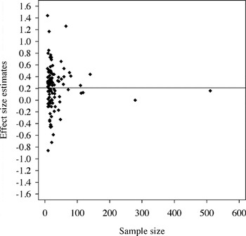
A funnel plot did not graphically reveal any publication bias, as adequate dispersion of effect size estimates was observed among studies with small samples (see Figure 2). In addition, we calculated the fail-safe number of studies with null effects required to bring the overall weighted effect size estimate to p = .05. The fail-safe N for the hippocampal asymmetry meta-analysis was 2,180. In other words, this number of null-effect, unpublished studies would have to exist in order to bring the asymmetry index to a minimal critical significance level.

Funnel plot of hippocampal asymmetry estimates. Note. Solid line corresponds to overall weighted effect size (D = 0.21).?bold?
Amygdala Asymmetry
In the amygdala meta-analysis, the overall weighted effect size (D) also revealed a significant volumetric asymmetry, suggesting that the right amygdala is reliably larger than the left amygdala in normal adults (p < .01). Hedges' g asymmetry estimates ranged from −0.83 to 1.2, with a mean value of g = 0.15. The unbiased asymmetry estimates ranged from −0.80 to 1.16, with a mean value of d = 0.15. There were 40 unbiased effect size estimates in the positive range, 23 unbiased effect size estimates in the negative range, and three unbiased effect size estimates equal to zero. Figure 3 displays the results of the 66 unbiased asymmetry estimates (d) for the amygdala.

Effect size estimates of amygdala asymmetry. Note. Positive values indicate larger right amygdala volume. Solid line = overall weighted effect size (D); dashed lines = 95% confidence interval. Numbered references on the ordinate axis correspond to bracketed numbers at the end of each entry in the Appendix.
The overall homogeneity test was significant [Q(65) = 110.32, p < .001], suggesting that in the amygdala meta-analysis the heterogeneity in effect size estimates is also likely attributable to the presence of moderator variables. There was a significant between-category difference in the asymmetry effect sizes for MRI magnet field strength (p < .05). Studies using a magnet field strength of 1.0 Tesla found a reliable volumetric asymmetry, with the right amygdala significantly larger than the left amygdala [D = 0.50, C.I. (95%) = 0.24–0.76]. Studies using a magnet field strength of 1.5 Tesla, however, did not find a significant degree of asymmetry [D = 0.05, C.I. (95%) = −0.01–0.11]. Imaging slice thickness, head position corrections, plane of acquisition, age of participants, sample size, and sex of participants did not appear to significantly moderate the heterogeneity in effect size estimates. As expected, the correlations between effect size estimates and slice thickness (r = −.04), age (r = −.05), sample size (r = −.11), and sex (rpb = .00) were not statistically significant. The results of these planned comparisons are presented in Table 3.
Planned comparisons of amygdala asymmetry moderator variables

The funnel plot for the amygdala meta-analysis did not reveal any publication bias (see Figure 4). The fail-safe N for the amygdala meta-analysis was 191. This number of null-effect, unpublished studies would have to exist in order to bring the asymmetry index to the minimal critical significance level of p = .05.

Funnel plot of amygdala asymmetry estimates. Note. Solid line corresponds to overall weighted effect size (D = 0.09).
DISCUSSION
Numerous studies over the past fifteen years have explored MRI-based hippocampal and amygdala volumetric measurements and investigated the relevance of differential volumetric values to symptom manifestation. A substantial number of these studies have focused on temporal lobe epilepsy, schizophrenia, mild cognitive impairment, and Alzheimer's disease, among other neurological and psychiatric conditions. In TLE, for example, the present consensus among investigators suggests that volumetric measurement of these structures correlates with neuropathological, neuropsychological, surgical, clinical, and prognostic factors. The aim of the present study was to complement and expand recent normative methodologies by using meta-analytic procedures to obtain reliable estimates of hippocampal and amygdala volumetric parameters in normal adults. As such, this investigation directly addressed three questions posed by Jack et al. (1995) within the context of hippocampal volumetric research: First, is there a normal relative, or interhemispheric volumetric asymmetry? Second, if such an asymmetry exists, which side is larger? And third, what are the normal absolute, or intrahemispheric volumes of the right and left hippocampus? These questions lay the framework for examining both hippocampal and amygdala volumetric measurements.
Hippocampal Volumetric Parameters
The results from the hippocampal meta-analysis suggest that a reliable parametric asymmetry exists in the hippocampus of normal adult individuals. The right hippocampus was found to be significantly larger than the left hippocampus. Although the magnitude of this asymmetry may be modest, it appears to be present across the adult age range, with no significant difference in hippocampal asymmetry between younger and older adults. There was also no significant volumetric difference between men and women, although the magnitude of hippocampal asymmetry was larger in the latter. At present it is not clear why there would be a greater degree of right larger than left hippocampal asymmetry in women. It should be noted, however, that the relatively small number of studies reporting volumes for men and women might have contributed to this effect, inasmuch as using fewer samples tends to provide a less accurate parametric estimate. The current results for volumetric asymmetry differences between men and women should therefore be interpreted cautiously. It would be useful in future MRI volumetric studies to report results separately for males and females, even if the two are eventually grouped for hypothesis testing.
Our findings suggest that magnet field strength is an important moderator of volumetric asymmetry in both the hippocampus and the amygdala. In hippocampal volumetric studies that used a magnet field strength of 1.5 Tesla, a reliable right greater than left asymmetry was obtained. Interestingly, studies that used a magnet field strength of 1.0 Tesla found a nonsignificant but comparable magnitude of volumetric asymmetry, albeit in the opposite direction. Under the assumption that an increase in magnet field strength generally contributes to greater image resolution capability, our results suggest that estimates obtained using a 1.5 Tesla magnet are more likely to yield reliable measurements of volumetric asymmetry.
Slice thickness also appears to be an important moderator of hippocampal volumetric asymmetry. The results from this meta-analysis suggest that measurements obtained using thinner imaging slices will correspond to a greater degree of hippocampal volumetric asymmetry. In a previous study by Laakso et al. (1997), no significant differences in hippocampal volumes were found when using slice thickness values of 1, 3, or 5 mm. Although the authors did not specifically address volumetric asymmetry, the mean left and right hippocampal volumes are reported for each of the 1, 3, and 5 mm slice thickness measurements. Interestingly, the mean left and right volumes are similar when a slice thickness of 5 mm was used (left = 3,040 mm3; right = 3,042 mm3). When a slice thickness of 3 mm was used, a left greater than right asymmetry was found (left = 3,086 mm3; right = 2,991 mm3). However, the opposite asymmetry pattern was found when a slice thickness of 1 mm was used (left = 2,939 mm3; right = 3,017 mm3). This observation is interesting because the authors used a small sample of 10 participants and only manipulated the thickness of the slices. Given the interest in exploring the relevance of slice thickness to reliable volume acquisition, the present results indicate that slice thickness does indeed play a moderating role in volume measurements of the hippocampus. There is generally a trade-off in using thinner slices since the overall measurement time may increase substantially. If thinner imaging slices do indeed provide a more reliable estimate of structural volumes, then these results would encourage the use of slice thickness less than or equal to 2 mm when a more precise measurement of hippocampal asymmetry is desired.
Finally, across all participants, the mean left hippocampal volume was 2,978.56 mm3 (SD = 983.43 mm3) while the mean right hippocampal volume was 3,060.28 mm3 (SD = 1,007.78 mm3). It should be clearly noted that this result does not reflect the hippocampal volumes for a given individual, but provides an estimate of the mean hippocampal volumes in the population of normal adults. As such, these values are appropriate estimates of the “absolute” left and right volumes of the hippocampus. When compared to hippocampal volume measurements reported in post-mortem investigations, the values obtained in this study are in concert with several previous reports (e.g., Bogerts et al., 1993), although some variability can be found across these studies. In a recent investigation comparing MRI-based hippocampal volumes obtained post-mortem to histology-based measurements from deceased normal elderly controls and Alzheimer's disease patients, Bobinski et al. (2000) found a very strong correlation (r = .97) between the two methods after controlling for tissue shrinkage. Although limited to post-mortem investigations, the authors concluded that MRI-based measurements can provide very accurate and anatomically valid estimates of hippocampal volume. It is thus expected that estimates of hippocampal volume and asymmetry obtained from a large series of post-mortem samples will be comparable to the values obtained in the present study.
Amygdala Volumetric Parameters
The results from the amygdala meta-analysis also suggest that a reliable parametric asymmetry exists in normal adult individuals. Although the magnitude was smaller than the asymmetry found for the hippocampus, the right amygdala was likewise found to be significantly larger than the left amygdala. This volumetric asymmetry was also present across the adult age range, with no significant difference between younger and older adults, and across sex, with no significant difference between men and women. When examining the potential moderating effect of sex composition, the absolute volumes for men and women were slightly larger for the right amygdala. However, the weighted effect size estimate for both was in the negative range (i.e., D = −0.05 in men and D = −0.09 in women). This discrepancy reflects the larger sample size in those studies that obtained a negative effect size estimate (d), thus leading to greater weighting in those calculations. Given the trivial differences in absolute amygdala volumes and the lack of a significant difference between men and women, this discrepancy was considered to be inconsequential.
As indicated previously, results from this study suggest that magnet field strength is an important moderating variable of volumetric asymmetry estimates. In those studies that used a magnet field strength of 1.5 Tesla, there was no significant difference between left and right amygdala volumes, while a significant right greater than left asymmetry was found in those studies using a magnet field strength of 1.0 Tesla. The overall weighted effect size estimate, which includes studies that used either a 1.0 or a 1.5 Tesla magnet, suggested that the amygdala was an asymmetrical structure (D = 0.09). This result, however, was in part moderated by the large magnitude of the asymmetry obtained in studies using a 1.0 Tesla magnet. In those particular studies the mean left amygdala volume was 1,766.60 mm3 while the mean right amygdala volume was 2,028.00 mm3, corresponding to an effect size estimate of D = 0.50. If once again we assume that an increase in magnet field strength generally contributes to greater image resolution capability, results obtained from the 1.5 Tesla studies would indicate that the amygdala is not an asymmetrical structure in normal adults. It remains open to consideration whether conclusions drawn from studies using a 1.0 versus a 1.5 Tesla MRI magnet should be comparable.
Slice thickness did not appear to be a significant moderator of volumetric asymmetry in the amygdala. It could be possible that the larger size of the hippocampus makes that structure more vulnerable to fluctuations in imaging parameters and methodological artifacts. Perhaps the greater anisotropic properties of the hippocampus in comparison to the amygdala additionally result in increased susceptibility to slice thickness parameters when reliably determining volumetric asymmetry. It is also conceivable that magnetic susceptibility artifact could have contributed to this difference.
Across all participants, the mean left amygdala volume was 1,883.39 mm3 (SD = 502.99 mm3) and the mean right amygdala volume was 1,925.88 mm3 (SD = 529.40 mm3). Once again, these values provide a response to Jack et al. (1995) and are estimates of the absolute left and right volumes of the amygdala. There is agreement when comparing these values to volumetric measurements obtained on several post-mortem investigations (e.g., Harding et al., 2002), although some variability exists across those studies in the reported amygdala volumes. Furthermore, the right greater than left asymmetry reported in this study is consistent with Murphy et al.'s (1987) detailed investigation of amygdala asymmetry in post-mortem human brains.
Additional Observations
There was no evidence of publication bias in either of the two meta-analyses, supporting the representativeness of the studies under review. For both asymmetry meta-analyses, a substantially large fail-safe N suggests that the obtained effect is very robust and a considerable number of studies with null effects would have to exist in order to reduce this finding to a minimal critical level. It was originally hypothesized that the sample size of each study might covary with estimates of volumetric asymmetry. However, our results indicate that sample size does not appear to be significantly related to asymmetry estimates.
Conclusion
Numerous studies during the past fifteen years have examined hippocampal and amygdala volumes in normal individuals. Results from these studies have often been at odds with each other and inconclusive regarding the magnitude of volume asymmetry in these structures. The present study provides a systematic analysis of hippocampal and amygdala volumetric asymmetry in normal adults using meta-analytic procedures. Results strongly suggest that both the hippocampus and the amygdala are asymmetrical structures, with larger volumes in the right hemisphere. A caveat, however, is in order. These results seem to be moderated by magnet field strength and, in the case of the hippocampus, slice thickness. Nevertheless, when only those hippocampal volumetric studies using a 1.5 Tesla magnet and slice thickness less than or equal to 2 mm are examined (N = 50), a comparable magnitude of asymmetry is obtained [D = 0.22, C.I. (95%) = 0.16–0.28]. Similarly, when only those amygdala volumetric studies using a 1.5 Tesla magnet and slice thickness less than or equal to 2 mm are examined (N = 34), the magnitude of the asymmetry is also comparable to the initial analyses [D = 0.09, C.I. (95%) = 0.02–0.17]. To the extent that greater confidence is placed on studies using a 1.5 Tesla magnet and slice thickness less than or equal to 2 mm, the results presented in Table 1 seem to be a robust indicator of hippocampal and amygdala asymmetry. Neither additional imaging parameters nor sample characteristics seemed to significantly moderate the observed results.
In conclusion, these results complement and expand previous imaging studies by providing additional normative information that may be useful to clinical investigators. It is hoped that these findings will stimulate further systematic analysis into the relationship between particular MRI imaging parameters, sample characteristics, and neuroanatomical volumetric estimates.
ACKNOWLEDGMENTS
Portions of this paper were initially presented at the November 2002 meeting of the Society for Neuroscience, Orlando, Florida.
APPENDIX1
Articles included in the meta-analysis:
Altshuler, L.L., Bartzokis, G., Grieder, T., Curran, J., & Mintz, J. (1998). Amygdala enlargement in bipolar disorder and hippocampal reduction in schizophrenia: An MRI study demonstrating neuroanatomic specificity. Archives of General Psychiatry, 55, 663–664. [H69; A52]
Anderson, J.E., Wible, C.G., McCarley, R.W., Jakab, M., Kasai, K., & Shenton, M.E. (2002). An MRI study of temporal lobe abnormalities and negative symptoms in chronic schizophrenia. Schizophrenia Research, 58, 123–134. [H9; A64]
Ashtari, M., Greenwald, B.S., Kramer-Ginsberg, E., Hu, J., Wu, H., Patel, M., Aupperle, P., & Pollack, S. (1999). Hippocampal/amygdala volumes in geriatric depression. Psychological Medicine, 29, 629–638. [H24; A39]
Barta, P.E., Pearlson, G.D., Powers, R.E., Richards, S.S., & Tune, L.E. (1990). Auditory hallucinations and smaller superior temporal gyral volume in schizophrenia. American Journal of Psychiatry, 147, 1457–1462. [A6]
Barta, P.E., Powers, R.E., Aylward, E.H., Chase, G.A., Harris, G.J., Rabins, P.V., Tune, L.E., & Pearlson, G.D. (1997). Quantitative MRI volume changes in late onset schizophrenia and Alzheimer's disease compared to normal controls. Psychiatry Research: Neuroimaging, 68, 65–75. [H22; A13]
Becker, T., Elmer, K., Mechela, B., Schneider, F., Taubert, S., Schroth, G., Grodd, W., Bartels, M., & Beckmann, H. (1990). MRI findings in medial temporal lobe structures in schizophrenia. European Neuropsychopharmacology, 1, 83–86. [H1]
Becker, T., Elmer, K., Schneider, F., Schneider, M., Grodd, W., Bartels, M., Heckers, S., & Beckmann, H. (1996). Confirmation of reduced temporal limbic structure volume on magnetic resonance imaging in male patients with schizophrenia. Psychiatry Research: Neuroimaging, 67, 135–143. [H5; A22]
Bernard, D., Walker, P.M., Baudouin-Poisson, N., Giroud, M., Fayolle, H., Dumas, R., Martin, D., Binnert, D., & Brunotte, F. (1996). Asymmetric metabolic profile in mesial temporal lobes: Localized H-1 MR spectroscopy in healthy right-handed and non-right handed subjects. Radiology, 199, 381–389. [H48, 60]
Bernasconi, A., Cendes, F., Lee, J., Reutens, D.C., & Gotman, J. (1999). EEG background delta activity in temporal lobe epilepsy: Correlation with volumetric and spectroscopic imaging. Epilepsia, 40, 1580–1586. [H47; A44]
Bernasconi, N., Bernasconi, A., Andermann, F., Dubeau, F., Feindel, W., & Reutens, D.C. (1999). Entorhinal cortex in temporal lobe epilepsy: A quantitative MRI study. Neurology, 52, 1870–1876. [H99; A55]
Bernasconi, N., Bernasconi, A., Caramanos, Z., Dubeau, F., Richardson, J., Andermann, F., Arnold, D.L. (2001). Entorhinal cortex atrophy in epilepsy patients exhibiting normal hippocampal volumes. Neurology, 56, 1335–1339. [H94; A47]
Bhatia, S., Brookheim, S.Y., Gaillard, W.D., & Theodore, W.H. (1993). Measurement of whole temporal lobe and hippocampus for MR volumetry: Normative data. Neurology, 43, 2006–2010. [H26, 32]
Bigler, E.D., Blatter, D.D., Anderson, C.V., Johnson, S.C., Gale, S.D., Hopkins, R.O., & Burnett, B. (1997). Hippocampal volume in normal aging and traumatic brain injury. American Journal of Neuroradiology, 18, 11–23. [H36, 46, 59, 70]
Bigler, E.D., Johnson, S.C., Anderson, C.V., Blatter, D.D., Gale, S.D., Russo, A.A., Ryser, D.K., Macnamara, S.E., Bailey, B.J., Hopkins, R.O., & Abildskov, T.J. (1996). Traumatic brain injury and memory: The role of hippocampal atrophy. Neuropsychology, 10, 333–342. [H45]
Bogerts, B., Ashtari, M., Degreef, G., Alvir, J.Ma.J., Bilder, R.M., & Lieberman, J.A. (1990). Reduced temporal limbic structure volumes on magnetic resonance images in first episode schizophrenia. Psychiatry Research: Neuroimaging, 35, 1–13. [H8, 16; A57, 61]
Bogerts, B., Lieberman, J.A., Ashtari, M., Bilder, R.M., Degreef, G., Lerner, G., Johns, C., & Masiar, S. (1993). Hippocampus-amygdala volumes and psychopathology in chronic schizophrenia. Biological Psychiatry, 33, 236–246. [H4; A59]
Breier, A., Buchanan, R.W., Elkashef, A., Munson, R.C., Kirkpatrick, B., & Gellad, F. (1992). Brain morphology and schizophrenia: A magnetic resonance imaging study of limbic, prefrontal cortex, and caudate structures. Archives of General Psychiatry, 49, 921–926. [H13; A26]
Bremner, J.D., Narayan, M., Anderson, E.R., Staib, L.H., Miller, H.L., & Charney, D.S. (2000). Hippocampal volume reduction in major depression. American Journal of Psychiatry, 157, 115–117. [H12; A20]
Bremner, J.D., Randall, P., Scott, T.M., Bronen, R.A., Seibyl, J.P., Southwick, S.M., Delaney, R.C., McCarthy, G., Charney, D.S., & Innis, R.B. (1995). MRI-based measurement of hippocampal volume in patients with combat-related posttraumatic stress disorder. American Journal of Psychiatry, 152, 973–981. [H62]
Bremner, J.D., Randall, P., Vermetten, E., Staib, L., Bronen, R.A., Mazure, C., Capelli, S., McCarthy, G., Innis, R.B., & Charney, D.S. (1997). Magnetic resonance imaging-based measurement of hippocampal volume in posttraumatic stress disorder related to childhood physical and sexual abuse—A preliminary report. Biological Psychiatry, 41, 23–32. [H6; A16]
Briellmann, R.S., Jackson, G.D., Kalnins, R., & Berkovic, S.F. (1998). Hemicranial volume deficits in patients with temporal lobe epilepsy with and without hippocampal sclerosis. Epilepsia, 39, 1174–1181. [H31]
Cendes, F., Andermann, F., Dubeau, F., Gloor, P., Evans, A., Jones-Gotman, M., Olivier, A., Andermann, E., Robitaille, Y., Lopes-Cendes, I., Peters, T., & Melanson, D. (1993). Early childhood prolonged febrile convulsions, atrophy and sclerosis of mesial structures, and temporal lobe epilepsy: An MRI volumetric study. Neurology, 43, 1083–1087. [H87; A45]
Chee, M.W.L., Low, S., Tan, J.S.P., Lim, W., & Wong, J. (1997). Hippocampal volumetry with magnetic resonance imaging: A cost-effective validated solution. Epilepsia, 38, 461–465. [H67]
Colchester, A., Kingsley, D., Lasserson, D., Kendall, B., Bello, F., Rush, C., Stevens, T.G., Goodman, G., Heilpern, G., Stanhope, N., & Kopelman, M.D. (2001). Structural MRI volumetric analysis in patients with organic amnesia, 1: Methods and comparative findings across diagnostic groups. Journal of Neurology, Neurosurgery, and Psychiatry, 71, 13–22. [H100]
Convit, A., McHugh, P., Wolf, O.T., de Leon, M.J., Bobinski, M., De Santi, S., Roche, A., & Tsui, W. (1999). MRI volume of the amygdala: A reliable method allowing separation from the hippocampal formation. Psychiatry Research: Neuroimaging, 90, 113–123. [A4]
Cook, M.J., Fish, D.R., Shorvon, S.D., Straughan, K., & Stevens, J.M. (1992). Hippocampal volumetric and morphometric studies in frontal and temporal lobe epilepsy. Brain, 115, 1001–1015. [H18]
Csernansky, J.G., Wang, L., Jones, D., Rastogi-Cruz, D., Posener, J.A., Heydebrand, G., Miller, J.P., & Miller, M.I. (2002). Hippocampal deformities in schizophrenia characterized by high dimensional brain mapping. American Journal of Psychiatry, 159, 2000–2006. [H103]
DeCarli, C., Hatta, J., Fazilat, S., Fazilat, S., Gaillard, W.D., & Theodore, W.H. (1998). Extratemporal atrophy in patients with complex partial seizures of left temporal origin. Annals of Neurology, 43, 41–45. [H88]
Deicken, R.F., Pegues, M., & Amend, D. (1999). Reduced hippocampal N-acetylaspartate without volume loss in schizophrenia. Schizophrenia Research, 37, 217–223. [H84]
Dickey, C.C., McCarley, R.W., Voglmaier, M.M., Niznikiewicz, M.A., Seidman, L.J., Hirayasu, Y., Fischer, I., The, E.K., Van Rhoads, R., Jakab, M., Kikinis, R., Jolesz, F.A., & Shenton, M.E. (1999). Schizotypal personality disorder and MRI abnormalities of temporal lobe gray matter. Biological Psychiatry, 45, 1393–1402. [H41; A49]
Driessen, M., Herrmann, J., Stahl, K., Zwaan, M., Meier, S., Hill, A., Osterheider, M., & Peterson, D. (2000). Magnetic resonance imaging volumes of the hippocampus and the amygdala in women with borderline personality disorder and early traumatization. Archives of General Psychiatry, 57, 1115–1122. [H29; A29]
Filipek, P.A., Richelme, C., Kennedy, D.N., & Caviness, V.S., Jr. (1994). The young adult human brain: An MRI-based morphometric analysis. Cerebral Cortex, 4, 344–360. [H11; A60]
Flaum, M., Swayze, V.W. II, O'Leary, D.S., Yuh, W.T.C., Ehrhardt, J.C., Arndt, S.V., & Andreasen, N.C. (1995). Effects of diagnosis, laterality, and gender on brain morphology in schizophrenia. American Journal of Psychiatry, 152, 704–714. [H14, 33]
Free, S.L., Bergin, P.S., Fish, D.R., Cook, M.J., Shorvon, S.D., & Stevens, J.M. (1995). Methods for normalization of hippocampal volumes measured with MR. American Journal of Neuroradiology, 16, 637–643. [H43, 63]
Fukuzako, H., Fukuzako, T., Hashiguchi, T., Hokazono, Y., Takeuchi, K., Hirakawa, K., Ueyama, K., Takigawa, M., Kajiya, Y., Nakajo, M., & Fujimoto, T. (1996). Reduction in hippocampal formation volume is caused mainly by its shortening in chronic schizophrenia: Assessment by MRI. Biological Psychiatry, 39, 938–945. [H89]
Fukuzako, H. Yamada, K., Kodama, S., Yonezawa, T., Fukuzako, T., Takenouchi, K., Kajiya, Y., Nakajo, M., & Takigawa, M. (1997). Hippocampal volume asymmetry and age at illness onset in males with schizophrenia. European Archives of Psychiatry and Clinical Neuroscience, 247, 248–251. [H82]
Gur, R.C., Gunning-Dixon, F., Bilker, W.B., & Gur, R.E. (2002). Sex differences in temporo-limbic and frontal brain volumes of healthy adults. Cerebral Cortex, 12, 998–1003. [H54, 92; A9, 12]
Gur, R.E., Turetsky, B.I., Cowell, P.E., Finkelman, C., Maany, V., Grossman, R.I., Arnold, S.E., Bilker, W.B., & Gur, R.C. (2000). Temporolimbic volume reductions in schizophrenia. Archives of General Psychiatry, 57, 769–775. [H71, 77; A11, 14]
Gurvits, T.V., Shenton, M.E., Hokama, H., Ohta, H., Lasko, N.B., Gilbertson, M.W., Orr, S.P., Kikinis, R., Jolesz, F.A., McCarley, R.W., & Pitman, R.K. (1996). Magnetic resonance imaging study of hippocampal volume in chronic, combat-related posttraumatic stress disorder. Biological Psychiatry, 40, 1091–1099. [H91; A63]
Hackert, V.H., den Heijer, T., Oudkerk, M., Koudstaal, P.J., Hofman, A., & Breteler, M.M.B. (2002). Hippocampal head size associated with verbal memory performance in nondemented elderly. Neuroimage, 17, 1365–1372. [H44]
Hasboun, D., Chantôme, M., Zouaoui, A., Sahel, M., Deladoeuille, M., Sourour, N., Duyme, M., Baulac, M., Marsault, C., & Dormont, D. (1996). MR determination of hippocampal volume: Comparison of three methods. American Journal of Neuroradiology, 17, 1091–1098. [H56]
Havermans, R., Honig, A., Vuurman, E.F.P.M., Krabbendam, L, Wilmink, J., Lamers, Th., Verheecke, C.J., Jolles, J., Romme, M.A.J., & Van Praag, H.M. (1999). A controlled study of temporal lobe structure volumes and P300 responses in schizophrenic patients with persistent auditory hallucinations. Schizophrenia Research, 38, 151–158. [A21]
Haznedar, M.M., Buchsbaum, M.S., Wei, T.C., Hof, P.R., Cartwright, C., Bienstock, C.A., & Hollander, E. (2000). Limbic circuitry in patients with autism spectrum disorders studied with positron emission tomography and magnetic resonance imaging. American Journal of Psychiatry, 157, 1994–2001. [H93; A19]
Hirayasu, Y., Shenton, M.E., Salisbury, D.F., Dickey, C.C., Fischer, I.A., Mazzoni, P., Kisler, T., Arakaki, H., Kwon, J.S., Anderson, J.E., Yurgelun-Todd, D., Tohen, M., & McCarley, R.W. (1998). Lower left temporal lobe MRI volumes in patients with first-episode schizophrenia compared with psychotic patients with first-episode affective disorder and normal subjects. American Journal of Psychiatry, 155, 1384–1391. [H7; A30]
Honeycutt, N.A., & Smith, C.D. (1995). Hippocampal volume measurements using magnetic resonance imaging in normal young adults. Journal of Neuroimaging, 5, 95–100. [H74]
Jack, C.R., Jr., Sharbrough, F.W., Cascino, G.D., Hirschorn, K.A., O'Brien, P.C., & Marsh, W.R. (1992). Magnetic resonance imaging-based hippocampal volumetry: Correlation with outcome after temporal lobectomy. Annals of Neurology, 31, 138–146. [H95]
Kälviäinen, R., Partanen, K., Äikiä, M., Mervaala, E., Vainio, P., Riekkinen, P.J., Sr., & Pitkänen, A. (1997a). MRI-based hippocampal volumetry and T2 relaxometry: Correlation to verbal memory performance in newly diagnosed epilepsy patients with left-sided temporal lobe focus. Neurology, 48, 286–287. [H79]
Kälviäinen, R., Salmenperä, T., Partanen, K., Vainio, P., Riekkinen, P., & Pitkänen, A. (1997b). MRI volumetry and T2 relaxometry of the amygdala in newly diagnosed and chronic temporal lobe epilepsy. Epilepsy Research, 28, 39–50. [A7]
Kälviäinen, R., Salmenperä, T., Partanen, K., Vainio, P., Riekkinen, P., & Pitkänen, A. (1998). Recurrent seizures may cause hippocampal damage in temporal lobe epilepsy. Neurology, 50, 1377–1382. [H81]
Kawasaki, Y., Maeda, Y., Urata, K., Higashima, M., Yamaguchi, N., Suzuki, M., Takashima, T., & Ide, Y. (1993). A quantitative magnetic resonance imaging study of patients with schizophrenia. European Archives of Psychiatry and Clinical Neuroscience, 242, 268–272. [H57; A53]
Kegeles, L.S., Shungu, D.C., Anjivel, S., Chan, S., Ellis, S.P., Xanthopoulos, E., Malaspina, D., Gorman, J.M., Mann, J.J., Laruelle, M., & Kaufmann, C.A. (2000). Hippocampal pathology in schizophrenia: Magnetic resonance imaging and spectroscopy studies. Psychiatry Research: Neuroimaging, 98, 163–175. [H21]
Laakso, M.P., Juottonen, K., Partanen, K., Vainio, P., & Soininen, H. (1997). MRI volumetry of the hippocampus: The effect of slice thickness on volume formation. Magnetic Resonance Imaging, 15, 263–265. [H40]
Laakso, M.P., Partanen, K., Lehtovirta, M., Hallikainen, M., Hänninen, T., Vainio, P., Riekkinen, P., Sr., & Soininen, H. (1995a). MRI of amygdala fails to diagnose early Alzheimer's disease. Neuroreport, 6, 2414–2418. [H98; A2]
Laakso, M.P., Soininen, H., Partanen, K., Helkala, E.L., Hartikainen, P., Vainio, P., Hallikainen, M., Hänninen, T., & Riekkinen, P.J., Sr. (1995b). Volumes of hippocampus, amygdala and frontal lobes in the MRI-based diagnosis of early Alzheimer's disease: Correlation with memory functions. Journal of Neural Transmission, 9, 73–86. [A1, 5, 8, 15]
Laakso, M.P., Soininen, H., Partanen, K., Lehtovirta, M., Hallikainen, M., Hänninen, T., Helkala, E.L., Vainio, P., & Riekkinen, P.J., Sr. (1998). MRI of the hippocampus in Alzheimer's disease: Sensitivity, specificity, and analysis of the incorrectly classified subjects. Neurobiology of Aging, 19, 23–31. [H75, 83]
Lehéricy, S., Baulac, M., Chiras, J., Piérot, L., Martin, N., Pillon, B., Deweer, B., Dubois, B., & Marsault, C. (1994). Amygdalohippocampal MR volume measurements in the early stages of Alzheimer disease. American Journal of Neuroradiology, 15, 927–937. [H104; A54]
Lemieux, L., Liu, R.S.N., & Duncan, J.S. (2000). Hippocampal and cerebellar volumetry in serially acquired MRI volume scans. Magnetic Resonance Imaging, 18, 1027–1033. [H17]
Mackay, C.E., Roberts, N., Mayes, A.R., Downes, J.J., Foster, J.K., & Mann, D. (1998). An exploratory study of the relationship between face recognition memory and the volume of medial temporal lobe structures in healthy young males. Behavioural Neurology, 11, 3–20. [H53; A40]
Mori, E., Yoneda, Y., Yamashita, H., Hirono, N., Ikeda, M., & Yamadori, A. (1997). Medial temporal structures relate to memory impairment in Alzheimer's disease: An MRI volumetric study. Journal of Neurology, Neurosurgery, and Psychiatry, 63, 214–221. [H101; A66]
Mu, Q., Xie, J., Wen, Z., Weng, Y., & Shuyun, Z. (1999). A quantitative MR study of the hippocampal formation, the amygdala, and the temporal horn of the lateral ventricle in healthy subjects 40 to 90 years of age. American Journal of Neuroradiology, 20, 207–211. [H28, 37, 38, 51; A24, 32, 33, 43]
Niemann, K., Hammers, A., Coenen, V.A., Thron, A., & Klosterkotter, J. (2000). Evidence of a smaller left hippocampus and left temporal horn in both patients with first episode schizophrenia and normal control subjects. Psychiatry Research, 99, 93–110. [H97; A25]
Pearlson, G.D., Barta, P.E., Powers, R.E., Menon, R.R., Richards, S.S., Aylward, E.H., Federman, E.B., Chase, G.A., Petty, R.G., & Tien, A.Y. (1997). Medial and superior temporal gyral volumes and cerebral asymmetry in schizophrenia versus bipolar disorder. Biological Psychiatry, 41, 1–14. [H65, 73; A3, 34]
Pearlson, G.D., Harris, G.J., Powers, R.E., Barta, P.E., Camargo, E.E., Chase, G.A., Noga, J.T., & Tune, L.E. (1992). Quantitative changes in mesial temporal volume, regional cerebral blood flow, and cognition in Alzheimer's disease. Archives of General Psychiatry, 49, 402–408. [H25; A10]
Pitkänen, A., Laakso, M., Kälviäinen, R., Partanen, K., Vainio, P., Lehtovirta, M., Riekkinen, P., Sr., & Soininen, H. (1996). Severity of hippocampal atrophy correlates with the prolongation of MRI T2 relaxation time in temporal lobe epilepsy but not in Alzheimer's disease. Neurology, 46, 1724–1730. [H85]
Pruessner, J.C., Collins, D.L., Pruessner, M., & Evans, A.C. (2001). Age and gender predict volume decline in the anterior and posterior hippocampus in early adulthood. Journal of Neuroscience, 21, 194–200. [H78; A51]
Pruessner, J.C., Li, L.M., Serles, W., Pruessner, M., Collins, D.L., Kabani, N., Lupien, S., & Evans, A.C. (2000). Volumetry of hippocampus and amygdala with high-resolution MRI and three-dimensional analysis software: Minimizing the discrepancies between laboratories. Cerebral Cortex, 10, 433–442. [H68; A18]
Quigg, M., Bertram, E.H., & Jackson, T. (1997). Longitudinal distribution of hippocampal atrophy in mesial temporal lobe epilepsy. Epilepsy Research, 27, 101–110. [H50]
Rajarethinam, R., DeQuardo, J.R., Miedler, J., Arndt, S., Kirbat, R., Brunberg, J.A., & Tandon, R. (2001). Hippocampus and amygdala in schizophrenia: Assessment of the relationship of neuroanatomy to psychopathology. Psychiatry Research: Neuroimaging 108, 79–87. [H96; A36]
Saukkonen, A., Kälviäinen, R., Partanen, K., Vainio, P., Riekkinen, P., & Pitkänen, A. (1994). Do seizures cause neuronal damage? A MRI study in newly diagnosed and chronic epilepsy. Neuroreport, 6, 219–223. [H55; A35]
Seidman, L.J., Faraone, S.V., Goldstein, J.M., Kremen, W.S., Horton, N.J., Makris, N., Toomey, R., Kennedy, D., Caviness, V.S., & Tsuang, M.T. (2002). Left hippocampal volume as a vulnerability indicator for schizophrenia. Archives of General Psychiatry, 59, 839–849. [H10]
Sheline, Y.I., Gado, M.H., & Price, J.L. (1998). Amygdala core nuclei volumes are decreased in recurrent major depression. Neuroreport, 9, 2023–2028. [A17]
Sheline, Y.I., Sanghavi, M., Mintun, M.A., & Gado, M.H. (1999). Depression duration but not age predicts hippocampal volume loss in medically healthy women with recurrent major depression. Journal of Neuroscience, 19, 5034–5043. [H23; A23]
Smith, C.D., Malcein, M., Meurer, K., Schmitt, F.A., Markesbery, W.R., & Pettigrew, L.C. (1999). MRI temporal lobe volume measures and neuropsychologic function in Alzheimer's disease. Journal of Neuroimaging, 9, 2–9. [H52, 66, 86, 102; A31, 38, 48, 56]
Steffens, D.C., Byrum, C.E., McQuoid, D.R., Greenberg, D.L., Payne, M.E., Blitchington, T.F., MacFall, J.R., & Krishnan, K.R.R. (2000). Hippocampal volume in geriatric depression. Biological Psychiatry, 48, 301–309. [H49, 76]
Stein, M.B., Koverola, C., Hanna, C., Torchia, M.G., & McClarty, B. (1997). Hippocampal volume in women victimized by childhood sexual abuse. Psychological Medicine, 27, 951–959. [H2]
Strakowski, S.M., DelBello, M.P., Sax, K.W., Zimmerman, M.E., Shear, P.K., Hawkins, J.M., & Larson, E.R. (1999). Brain magnetic resonance imaging of structural abnormalities in bipolar disorder. Archives of General Psychiatry, 56, 254–260. [H27; A42]
Sumich, A., Chitnis, X.A., Fannon, D.G., O'Ceallaigh, S., Doku, V.C., Falrowicz, A., Marshall, N., Matthew, V.M., Potter, M., & Sharma, T. (2002). Temporal lobe abnormalities in first-episode psychosis. American Journal of Psychiatry, 159, 1232–1234. [H64; A65]
Szabo, C.A., Xiong, J., Lancaster, J.L., Rainey, L., & Fox, P. (2001). Amygdalar and hippocampal volumetry in control participants: Differences regarding handedness. American Journal of Neuroradiology, 22, 1342–1345. [H19, 72; A41, 58]
Szeszko, P.R., Robinson, D., Alvir, J.Ma.J., Bilder, R.M., Lencz, T., Ashtari, M., Wu, H., & Bogerts, B. (1999). Orbital frontal and amygdala volume reductions in obsessive-compulsive disorder. Archives of General Psychiatry, 56, 913–919. [H3; A62]
Torres, I.J., Flashman, L.A., O'Leary, D.S., Swayze, V., II, & Andreasen, N.C. (1997). Lack of an association between delayed memory and hippocampal and temporal lobe size in patients with schizophrenia and healthy controls. Biological Psychiatry, 42, 1087–1096. [H30, 58]
Vakili, K., Pillay, S.S., Lafer, B., Fava, M., Renshaw, P.F., Bonello-Cintron, C.M., & Yurgelun-Todd, D.A. (2000). Hippocampal volume in primary unipolar major depression: A magnetic resonance imaging study. Biological Psychiatry, 47, 1087–1090. [H61]
Velakoulis, D., Pantelis, C., McGorry, P.D., Dudgeon, P., Brewer, W., Cook, M., Desmond, P., Bridle, N., Tierney, P., Murrie, V., Singh, B., & Copolov, D. (1999). Hippocampal volume in first-episode psychoses and chronic schizophrenia: A high-resolution magnetic resonance imaging study. Archives of General Psychiatry, 56, 133–140. [H80]
Villarreal, G., Hamilton, D.A., Petropoulos, H., Driscoll, I., Rowland, L.M., Griego, J.A., Kodituwakku, P.W., Hart, B.L., Escalona, R., & Brooks, W.M. (2002). Reduced hippocampal volume and total white matter volume in posttraumatic stress disorder. Biological Psychiatry, 52, 119–125. [H20]
von Gunten, A., Fox, N.C., Cipolotti, L., & Ron, M.A. (2000). A volumetric study of hippocampus and amygdala in depressed patients with subjective memory problems. Journal of Neuropsychiatry and Clinical Neuroscience, 12, 493–498. [H39; A46]
Vythilingam, M., Anderson, E.R., Goddard, A., Woods, S.W., Staib, L.H., Charney, D.S., & Bremner, J.D. (2000). Temporal lobe volume in panic disorder—A quantitative magnetic resonance imaging study. Psychiatry Research: Neuroimaging, 99, 75–82. [H34]
Watson, C., Andermann, F., Gloor, P., Jones-Gotman, M., Peters, T., Evans, A., Olivier, A., Melanson, D., & Leroux, G. (1992). Anatomic basis of amygdaloid and hippocampal volume measurement by magnetic resonance imaging. Neurology, 42, 1743–1750. [H90; A50]
Watson, C., Cendes, F., Fuerst, D., Dubeau, F., Williamson, B., Evans, A., & Andermann, F. (1997a). Specificity of volumetric magnetic resonance imaging in detecting hippocampal sclerosis. Archives of Neurology, 54, 67–73. [H35, 42; A27, 28]
Whitworth, A.B., Honeder, M., Kremser, C., Kemmler, G., Felber, S., Hausmann, A., Wanko, C., Wechdorn, H., Aichner, F., Stuppaeck, C.H., & Fleischhacker, W.W. (1998). Hippocampal volume reduction in male schizophrenic patients. Schizophrenia Research, 31, 73–81. [H15; A37]
References
REFERENCES
Allen, J.S.,
Damasio, H., &
Grabowski, T.J.
(2002).
Normal neuroanatomical variation in the human brain: An
MRI-volumetric study.
American Journal of Physical Anthropology,
118,
341–358.CrossRefGoogle Scholar
Bigler, E.D.,
Tate, D.F.,
Miller, M.J.,
Rice, S.A.,
Hessel, C.D.,
Earl, H.D.,
Tschanz, J.T.,
Plassman, B., &
Welsh-Bohmer, K.A.
(2002).
Dementia, asymmetry of temporal lobe structures, and Apolipoprotein
E genotype: Relationships to cerebral atrophy and neuropsychological
impairment.
Journal of the International Neuropsychological Society,
8,
925–933.Google Scholar
Bobinski, M.,
de Leon, M.J.,
Wegiel, J.,
Desanti, S.,
Convit, A.,
Saint Louis, L.A.,
Rusinek, H., &
Wisniewski, H.M.
(2000).
The histological validation of post mortem magnetic resonance
imaging-determined hippocampal volume in Alzheimer's disease.
Neuroscience,
95,
721–725.Google Scholar
Bogerts, B.,
Falkai, B.,
Schneider, T., &
Pfeiffer, U.
(1993).
The neuropathology of schizophrenia: Past and present.
Journal für Hirnforschung,
34,
193–205.Google Scholar
Breier, J.I.,
Leonard, C.M.,
Bauer, R.M.,
Roper, S.,
Lucas, T.H., &
Gilmore, R.L.
(1996).
Quantified volumes of temporal lobe structures in patients with
epilepsy.
Journal of Neuroimaging,
6,
108–114.CrossRefGoogle Scholar
Brierley, B.,
Shaw, P., &
David, A.S.
(2002).
The human amygdala: A systematic review and meta-analysis of
volumetric magnetic resonance imaging.
Brain Research Reviews,
39,
84–105.CrossRefGoogle Scholar
Castellanos, F.X.,
Giedd, J.N.,
Marsh, W.L.,
Hamburger, S.D.,
Vaituzis, A.C.,
Dickstein, D.P.,
Sarfatti, S.E.,
Vauss, Y.C.,
Snell, J.W.,
Lange, N.,
Kaysen, D.,
Krain, A.L.,
Ritchie, G.F.,
Rajapakse, J.C., &
Rapoport, J.L.
(1996).
Quantitative brain magnetic resonance imaging in attention-deficit
hyperactivity disorder.
Archives of General Psychiatry,
53,
607–616.CrossRefGoogle Scholar
Cooper, H. &
Hedges, L.V.
(1994).
Potentials and limitations of research synthesis. In
H. Cooper &
L.V. Hedges (Eds.),
Handbook of research synthesis (pp.
521–529).
New York:
Russell Sage Foundation.
Dickerson, B.C.,
Goncharova, I.,
Sullivan, M.P.,
Forchetti, C.,
Wilson, R.S.,
Bennett, D.A.,
Beckett, L.A., &
deToledo-Morrell, L.
(2001).
MRI-derived entorhinal and hippocampal atrophy in incipient and
very mild Alzheimer's disease.
Neurobiology of Aging,
22,
747–754.CrossRefGoogle Scholar
Dunlap, W.P.,
Cortina, J.M.,
Vaslow, J.B., &
Burke, M.J.
(1996).
Meta-analysis of experiments with matched groups or repeated measures
designs.
Psychological Methods,
1,
170–177.CrossRefGoogle Scholar
Durlak, J.A. &
Lipsey, M.W.
(1991).
A practitioner's guide to meta-analysis.
American Journal of Community Psychology,
19,
291–332.Google Scholar
Edland, S.D.,
Xu, Y.,
Plevak, M.,
O'Brien, P.,
Tangalos, E.G.,
Petersen, R.C., &
Jack, C.R., Jr.
(2002).
Total intracranial volume: Normative values and lack of association
with Alzheimer's disease.
Neurology,
59,
272–274.CrossRefGoogle Scholar
Galton, C.J.,
Patterson, K.,
Graham, K.,
Lambon-Ralph, M.A.,
Williams, G.,
Antoun, N.,
Sahakian, B.J., &
Hodges, J.R.
(2001).
Differing patterns of temporal atrophy in Alzheimer's disease
and semantic dementia.
Neurology,
57,
216–225.CrossRefGoogle Scholar
Ge, Y.,
Grossman, R.I.,
Babb, J.S.,
Rabin, M.L.,
Mannon, L.J., &
Kolson, D.L.
(2002).
Age-related total gray matter and white matter changes in normal
adult brain. Part II: Quantitative magnetization transfer ratio
histogram analysis.
American Journal of Neuroradiology,
23,
1334–1341.Google Scholar
Geroldi, C.,
Laakso, M.P.,
DeCarli, C.,
Beltramello, A.,
Bianchetti, A.,
Soininen, H.,
Trabucchi, M., &
Frisoni, G.B.
(2000).
Apolipoprotein E genotype and hippocampal asymmetry in
Alzheimer's disease: A volumetric MRI study.
Journal of Neurology, Neurosurgery, and Psychiatry,
68,
93–96.CrossRefGoogle Scholar
Giedd, J.N.,
Snell, J.W.,
Lange, N.,
Rajapakse, J.C.,
Casey, B.J.,
Kozuch, P.L.,
Vaituzis, A.C.,
Vauss, Y.C.,
Hamburger, S.D.,
Kaysen, D., &
Rapoport, J.L.
(1996).
Quantitative magnetic resonance imaging of human brain development:
Ages 4–18.
Cerebral Cortex,
6,
551–560.CrossRefGoogle Scholar
Gilmore, R.L.,
Childress, M.D.,
Leonard, C.,
Quisling, R.,
Roper, S.,
Eisenschenk, S., &
Mahoney, M.
(1995).
Hippocampal volumetrics differentiate patients with temporal lobe
epilepsy and extratemporal lobe epilepsy.
Archives of Neurology,
52,
819–824.CrossRefGoogle Scholar
Gloor, P.
(1997).
The temporal lobe and limbic system.
New York:
Oxford University Press.
Harding, A.J.,
Stimson, E.,
Henderson, J.M., &
Halliday, G.M.
(2002).
Clinical correlates of selective pathology in the amygdala of patients
with Parkinson's disease.
Brain,
125(Pt. 11),
2431–2445.CrossRefGoogle Scholar
Hedges, L.V. &
Olkin, I.
(1985).
Statistical methods for meta-analysis.
Orlando, FL:
Academic Press.
Hunter, J.E. &
Schmidt, F.L.
(1990).
Methods of meta-analysis: Correcting error and bias in research
findings.
Newbury Park, CA:
Sage Publications.
Jack, C.R., Jr.,
Petersen, R.C.,
Xu, Y.C.,
O'Brien, P.C.,
Smith, G.E.,
Ivnik, R.J.,
Boeve, B.F.,
Waring, S.C.,
Tangalos, E.G., &
Kokmen, E.
(1999).
Prediction of AD with MRI-based hippocampal volume in mild
cognitive impairment.
Neurology,
52,
1397–1403.CrossRefGoogle Scholar
Jack, C.R., Jr.,
Petersen, R.C.,
Xu, Y.C.,
Waring, S.C.,
O'Brien, P.C.,
Tangalos, E.G.,
Smith, G.E.,
Ivnik, R.J., &
Kokmen, E.
(1997).
Medial temporal atrophy on MRI in normal aging and very mild
Alzheimer's disease.
Neurology,
49,
786–794.CrossRefGoogle Scholar
Jack, C.R., Jr.,
Theodore, W.H.,
Cook, M., &
McCarthy, G.
(1995).
MRI-based hippocampal volumetrics: Data acquisition, normal ranges,
and optimal protocol.
Magnetic Resonance Imaging,
13,
1057–1064.CrossRefGoogle Scholar
Krasuski, J.S.,
Alexander, G.E.,
Horwitz, B.,
Daly, E.M.,
Murphy, D.G.M.,
Rapoport, S.I., &
Schapiro, M.B.
(1998).
Volumes of medial temporal lobe structure in patients with
Alzheimer's disease and mild cognitive impairment (and in healthy
controls).
Biological Psychiatry,
43,
60–68.CrossRefGoogle Scholar
Lawrie, S.M. &
Abukmeil, S.S.
(1998).
Brain abnormality in schizophrenia: A systematic and quantitative
review of volumetric magnetic resonance imaging studies.
British Journal of Psychiatry,
172,
110–20.CrossRefGoogle Scholar
Luft, A.R.,
Skalej, M.,
Welte, D.,
Kolb, R., &
Klose, U.
(1998).
Reliability and exactness of MRI-based volumetry: A phantom study.
Journal of Magnetic Resonance Imaging,
6,
700–704.Google Scholar
Murphy, G.M., Jr.,
Inger, P.,
Mark, K.,
Lin, J.,
Morrice, W.,
Gee, C.,
Gan, S., &
Korp, B.
(1987).
Volumetric asymmetry in the human amygdaloid complex.
Journal für Hirnforschung,
28,
281–289.Google Scholar
Nelson, M.D.,
Saykin, A.J.,
Flashman, L.A., &
Riordan, H.J.
(1998).
Hippocampal volume reduction in schizophrenia as assessed by
magnetic resonance imaging.
Archives of General Psychiatry,
55,
433–440.CrossRefGoogle Scholar
O'Brien, P.C. &
Dyck, P.J.
(1995).
Procedures for setting normal values.
Neurology,
45,
17–23.CrossRefGoogle Scholar
Orwin, R.G.
(1983).
A fail-safe N for effect size in meta-analysis.
Journal of Educational Statistics,
8,
157–159.CrossRefGoogle Scholar
Petersen, R.C.,
Doody, R.,
Kurz, A.,
Mohs, R.C.,
Morris, J.C.,
Rabins, P.V.,
Ritchie, K.,
Rossor, M.,
Thal, L., &
Winblad, B.
(2001).
Current concepts in mild cognitive impairment.
Archives of Neurology,
58,
1985–1992.CrossRefGoogle Scholar
Radhakrishnan, K.,
So, E.L.,
Silbert, P.L.,
Jack, C.R., Jr.,
Cascino, G.D.,
Sharbrough, F.W., &
O'Brien, P.C.
(1998).
Predictors of outcome of anterior temporal lobectomy for
intractable epilepsy: A multivariate study.
Neurology,
51,
465–471.CrossRefGoogle Scholar
Rosenthal, R.
(1991).
Meta-analysis: A review.
Psychosomatic Medicine,
53,
247–271.CrossRefGoogle Scholar
Rosenthal, R. &
DiMatteo, M.R.
(2001).
Meta-analysis: Recent developments in quantitative methods for
literature reviews.
Annual Review of Psychology,
52,
59–82.CrossRefGoogle Scholar
Salanova, V.,
Markand, O., &
Worth, R.
(1999).
Longitudinal follow-up in 145 patients with medically refractory
temporal lobe epilepsy treated surgically between 1984 and 1995.
Epilepsia,
40,
1417–1423.CrossRefGoogle Scholar
Shadish, W.R. &
Haddock, C.K.
(1994).
Combining estimates of effect size. In
H. Cooper &
L.V. Hedges (Eds.),
Handbook of research synthesis (pp.
261–281).
New York:
Russell Sage Foundation.
Wang, L.,
Joshi, S.C.,
Miller, M.I., &
Csernansky, J.G.
(2001).
Statistical analysis of hippocampal asymmetry in schizophrenia.
Neuroimage,
14,
531–545.CrossRefGoogle Scholar
Watson, C.,
Jack, C.R., Jr., &
Cendes, F.
(1997b).
Volumetric magnetic resonance imaging: Clinical applications and
contributions to the understanding of temporal lobe epilepsy.
Archives of Neurology,
54,
1521–1531.Google Scholar
Wright, I.C.,
Rabe-Hesketh, S.,
Woodruff, P.W.R.,
David, A.S.,
Murray, R.M., &
Bullmore, E.T.
(2000).
Meta-analysis of regional brain volumes in schizophrenia.
American Journal of Psychiatry,
157,
16–25.CrossRefGoogle Scholar
Meta-analysis results of volumetric asymmetry measurements
Effect size estimates of hippocampal asymmetry. Note. Positive values indicate larger right hippocampal volume. Solid line = overall weighted effect size (D); dashed lines = 95% confidence interval. Numbered references on the ordinate axis correspond to bracketed numbers at the end of each entry in the Appendix.
Planned comparisons of hippocampal asymmetry moderator variables
Funnel plot of hippocampal asymmetry estimates. Note. Solid line corresponds to overall weighted effect size (D = 0.21).?bold?
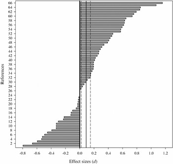
Effect size estimates of amygdala asymmetry. Note. Positive values indicate larger right amygdala volume. Solid line = overall weighted effect size (D); dashed lines = 95% confidence interval. Numbered references on the ordinate axis correspond to bracketed numbers at the end of each entry in the Appendix.
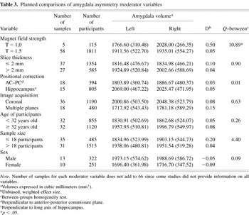
Planned comparisons of amygdala asymmetry moderator variables
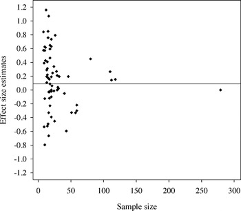
Funnel plot of amygdala asymmetry estimates. Note. Solid line corresponds to overall weighted effect size (D = 0.09).
 You have
Access
- 170
- Cited by