I. INTRODUCTION
Natural polysaccharides such as hyaluronic acid, chondroitin sulfate, dermatan sulfate, proteoglycans, etc. are involved in the biosynthesis of different fibrillar and non fibrillar collagen isotypes, which forms the basic component of a certain type of connective tissue.Reference Iordachescu, Campean and Stan1 These polysaccharides are difficult to extract and purify from animal tissues and consequently more expensive. That is why their replacement with vegetable and/or microbial polysaccharides or their derivatives (chitin, chitosan, cellulose, dextran, etc.) is studied.
Chemical derivatization of polysaccharides has a decisive effect on their macroscopic properties, such as solubility, stability, and viscosity characteristics. If the functionalization leads to polysaccharide derivatives bearing ionic or ionizable groups, they will behave as polyelectrolytes. The polyelectrolytes with phosphoric groups have become a hot topic due to their important potential use in the biomedical field. Thus, some of the sulfated polysaccharides, dideoxynucleoside analogs and derivatives of purines with phosphoryl groups have been investigated as potential drugs for AIDS treatment. The interactions of the protein sequences of HIV-1 or HIV-2 with curdlan sulfate have been studied and it has demonstrated its HIV inhibition ability, anticoagulating properties and antitumor activity.Reference Yoshida, Yasuda, Uryu, Nakashima, Yamamoto, Mimura and Kaneko2–Reference Jeon, Katsuraya, Inazu, Kaneko, Mimura and Uryu6
A recent application field is the use of natural polymers as crystallization modulators (inhibitors or promoters) in biomineralization processes that interfere in the formation of hard tissues. Thus, the effect of some polysaccharides with sulfate groups or of phosphorylated polysaccharides such as phosphorylated cellulose on the formation/growth of hydroxyapatite was investigated.Reference Shimabayashi, Hashimoto, Kawamura, Uno and Zahid7 The effect of natural polymers was attributed to the interfacial adsorption phenomena between organic and inorganic phases. The mineralization process and its estimation are of a great interest, as well as the investigation of polymer–phosphate interaction and the fabrication of polymer–hydroxyapatite biocomposites or, more generally, polymer–inorganic phosphate biocomposites.Reference Ficai, Andronescu, Trandafir, Ghitulica and Voicu8–Reference Ficai, Andronescu, Ficai, Voicu and Ficai11
Other applications of polysaccharides with phosphorus groups were reported in the pharmaceutical industry and medicine.Reference Granja, Barbosa, Pouysegu, De Jeso, Baquey and Ottenbrite12 New bacterial polysaccharides containing phosphate groups were tested as additives for preventing skin aging by acceleration of collagen production in fibroblasts.Reference Volkin, Tsai, Dabora, Gress, Burke, Linhardt and Middaugh13
Dextran is a family of neutral polysaccharides produced by bacterial enzymes, consisting of main chains from α-(1→6) linked D-glucose units with different ratios of linkages and branches, depending on the bacterial strains.Reference Heinze, Liebert, Heublein and Hornig14
Dextran is present in the extracellular matrix (ECM) and shows superior biocompatibility.Reference Robyt and Kroschwitz15, Reference Jing, Wu, Lin, Liu, Tang and Tian16 It is known that dextran promotes adhesion between protein and cells.Reference Volkin, Tsai, Dabora, Gress, Burke, Linhardt and Middaugh13
Several methods of phosphorylated dextran (PDex) synthesis are known. These methods use different phosphorylated agents such as polyphosphoric acid/tributyl amine (TBA)/dimethylformamide (DMF),Reference Duailibi, Duailibi, Young, Bartlett, Vacanti and Yelick17–Reference Suzuki, Suzuki and Matsumoto21 H3PO4/ fatty acids esters,Reference Suzuki, Suzuki and Mikami22 H3PO4/palmitic acid/tertiary amines,Reference Sacco, Klett-Zygmunt and Dellacherie18, Reference Makarova, Aptova, Litvak and Raldugina23 H3PO4/formamide,Reference Sato, Nishimura-Uemura, Shimosato, Kawai, Kitazawa and Saito24 H3PO4/urea (the reaction results in cross-linked dextran),Reference Suflet, Chitanu and Desbrières25 and H3PO3/urea.Reference Suflet, Chitanu and Popa26 These phosphorylated derivatives of dextran find applications in medicine and pharmaceutical industry as potential substitutes for erythrocytes, as antitumor agents or as inhibitors of virus development, anti tartar and anti caries agents in toothpastes, ion exchangers.Reference Sacco, Klett-Zygmunt, Vigneron and Dellacherie19–Reference Makarova, Aptova, Litvak and Raldugina23
Collagen can form a variety of homogeneous composites with other water-soluble materials like peptides, proteins, polysaccharides, and synthetic polymers. The obtaining methods involve chemical cross-linking, physical loading and coprecipitation. Some of our latest unpublished studies have focused on collagen/hydroxyapatite scaffolds with cytostatic drugsReference Iliescu, Andronescu, Voicu, Ficai and Covaliu27 for bone cancer applications and continuation with development of collagen- polysaccharide scaffolds.
The aim of this work is to prepare, to perform physical–chemical characterization and to test in vitro MG-63 cell lines on the biocomposites based on collagen–PDex with different substitution degrees in the forms of Na salt, mixed salts of Na and Zn, Mg, and Ca ions or in acid form. The 2D (membranes) and 3D (matrices) biocomposites were comparatively characterized to choose the proper scaffolds that can be used as drug delivery systems for bone cancer or as scaffolds for bone tissue engineering, being important both from theoretical and practical points of view.
II. MATERIALS AND METHODS
A. Materials
Monobasic dextran phosphate (PDex) with degree of substitution (DS) of 1.3 (PDex1.3) and 1.5 (PDex1.5) was prepared according to the method described in the literature.Reference Trandafir, Popescu, Albu, Iovu and Georgescu28 Zinc carbonate, magnesium oxide, and calcium hydroxide from Fluka, Switzerland, and natrium hydroxide, from Merck, Germany were used as received.
Type I collagen gel (Coll) at a concentration of 1.83% was obtained from calf hide by acid and alkaline treatments as previously described.Reference Siapi, Mavromoustakos, Trandafir, Albu and Budrugeac29, Reference Albu30
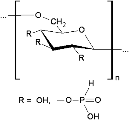
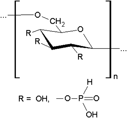
Mixed salts of PDex: monobasic dextran phosphate was obtained in Na salt form. The acid form of PDex (PDexH) was prepared by passing diluted solutions of Na salt of PDex1.3 through an ion exchange column filled with a sulfonic Dowex 50W × 8, 20–50 mesh resin. In aqueous solution of PDexH with a concentration of 5 × 10⁻³ equi./L was gradually added while mixing an aqueous solution of ZnCO3, MgO, or Ca(OH)2 respectively and then NaOH solution so that 2% of the phosphoric groups is neutralized by Zn2+, Mg2+ or Ca2+ ions and 98% of the acidic groups is neutralized by Na+ ions. The solid forms of mixed salt of PDex (PDexZn-Na, PDexMg-Na and PDexCa-Na) were obtained by freeze-drying. The schematic structure of PDex is presented in Fig. 1.
Schematic structure of phosphorylated dextran.Reference Suflet, Chitanu and Desbrières25
The biocomposites in the forms as matrices and membranes were prepared from 1.1% collagen gel (reported at 100 g of dry collagen) with pH = 7 in which 5% PDex was added.
Prior to this, PDex powder was dissolved into pure water. The biocomposite gels were plasticized with glycerin (2%), cross-linked with formaldehyde (0.2%) and then were conditioned by free-drying at 25 °C in Ventricel oven to obtain the membranes (MEM-PDex1.3Na; MEM-PDex1.5Na; MEM- PDexH; MEM-PDexZn-Na; MEM-PDexMg-Na; MEM-PDexCa-Na). The matrices were obtained from biocomposite gels as such, by freeze-drying with Delta 2–24 LSC lyophilizer, Martin Christ, Germany and named MAT-PDex1.3Na; MAT-PDex1.5Na; MAT-PDexH; MAT- PDexZn-Na; MAT- PDexMg-Na and MAT- PDexCa-Na.
B. Methods
1. Analysis and characterization of PDex samples
FTIR spectra were recorded on KBr pellet using a Bruker Vertex 70 spectrometer. NMR spectra were recorded in D2O using a Bruker Avance 400 MHz instrument. Potentiometric titration was performed using a Metrohm all-purpose 716 DMS Titrino apparatus equipped with a 6.0203.100 combined electrode. Monovalent bases LiOH and KOH were used for titration without or with added low molecular salt. Conductometric titrations were performed with same bases, with no added salt, using a 712 Metrohm conductometer with 6.0908.110 cell.
2. Physical–chemical characterization of biocomposite membranes and matrices
a. Thermal analysis
The DSC curves were recorded using DSC 204 F1 Phoenix apparatus manufactured by Netzsch—Germany. The following kinds of DSC measurements were performed: DSC analysis of the sample (1–5 mg) immersed in water (35 μL deionized water), hermetically sealed in an aluminium pan and stocked for 24 h. Each sample was heated from 25 to 110 °C, at a heating rate of 10 K/min. DSC analysis of the sample (1–5 mg) in nitrogen flow (purity is higher than 99.999%; 20 mL/min), in an open aluminum pan, at the heating rate of 10 K/min, and in the temperature range 25–280 °C.
Hydrophilic properties were emphasized by water absorption and water vapor absorption. To determine water absorption the following procedure was used: the samples were immersed in distilled water at 23 °C. At scheduled time intervals, samples were withdrawn, blotted with a tissue paper to remove the surface water and weighed. The water absorption was calculated using the following equation:

where, Wt denotes the weight of the swollen samples at immersion time t and Wd denotes the weight of the dry samples. The water vapor absorption was evaluated using standard methods for leather characterization: SR EN ISO17229: 2003 si SR EN ISO14268:2003.Reference Siapi, Mavromoustakos, Trandafir, Albu and Budrugeac29
3. “In vitro” biological testing for biocomposite matrices and membranes
“In vitro” biological testing for biocomposite matrices and membranes used human osteosarcoma MG 63 cell lines, which was grown in Dulbecco’s modified Eagle’s culture medium (DMEM) containing 1‰ glucose, 10% heat inactivated fetal bovine serum and antibiotics (100 U/L penicillin, 100 U/L streptomycin, 50 U/L neomycin). Collagen-dextran samples were sterilized for 24 h with 70% ethanol, and then conditioned in the same culture medium for 24 h and inoculated with osteosarcoma cell lines (50,000 cells/mL). The cells on samples were maintained in culture at 37 °C in incubators with 5% CO2 in air (v/v) and relative humidity over 95%. All experiments were done after 1 wk of culture. Chemicals used for cell cultures were obtained from Sigma (St. Louis, MO) and tissue culture plates from Costar (Cambridge, MA).
a. Hoechst staining
After 1 wk in culture, the cells on collagen biocomposites were washed in phosphate buffer (PB) solution, fixed in 2% paraformaldehyde (1 h) and then cryoprotected. After washing in PB (0.2 M Na2HPO4 and 0.2 M NaH2PO4) pH 7.2, the specimens were kept in a solution of 0.1 M PB with 5% sucrose overnight at 4 °C.
Then, the samples were immersed in PB containing 5% glycerin for 15 min, PB with 10% glycerin for 1 h, PB with 20% glycerin for 10 h and PB with 50% glycerin for 1 h at 4 °C. Specimens were frozen in liquid nitrogen and sectioned with a Leica CM 1800 cryotome; the thickness of the sections were 4–6 μm. The cryosections were washed with PB solution for 15 min, stained with Hoechst 258 (a specific DNA staining) for 15 min, washed in distilled water, mounted in glycerol and examined with a Nikon microscope equipped with epifluorescence and a filter G1-B; the micrographs were captured with a Sony DSC-S75 Digital Camera.
III. RESULTS AND DISCUSSION
A. Physical–chemical properties of PDex samples
The structure of PDex was confirmed by FTIR and NMR spectra. DS of 1.3 and 1.5 was calculated by potentiometric measurements.
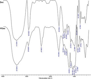
The FTIR spectra of phosphorylated samples are shown in Fig. 2, compared to the parent dextran. In the spectrum of dextran, the following characteristic bands are observed: a large band at 3400 cm-1 corresponding to OH groups; a band at 2924 cm-1 attributed to CH2 groups; characteristic bands at 1652 cm-1, 1462–1280 cm-1 for the glycosidic units and 1162–998 cm-1 for C—O—C from the glycosidic units and from β-(1→3)-glycosidic bonds. In the FTIR spectrum of PDex several new bands appeared: a band at 2396 cm-1 corresponding to the P—H bond, another one at 1228 cm-1 corresponding to the P = O bond, a shoulder at 1081–1057 cm-1 attributable to the P−OH bond, and a band at 859 cm-1 corresponding to the P—O—C bonds.Reference Purama, Goswami, Khan and Goyal31–Reference Budrugeac, Trandafir and Albu33
The FTIR spectra of dextran and phosphorylated dextran.

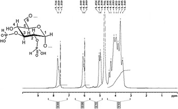
In the 1H NMR spectrum of PDex, presented in Fig. 3, two signals at 6.10 and 7.74 ppm are observed, which correspond to a doublet with a coupling constant of P–H bond of 640 Hz.
1H NMR analysis of phosphorylated dextran PDex.

DS of phosphorylated samples was evaluated by potentiometric and conductometric titration according to the methods already verified.Reference Trandafir, Popescu, Albu, Iovu and Georgescu28 DS of PDex samples was 1.3 and 1.5 depending on the reaction time. The EDX spectra of mixed salt of PDex samples confirm this degree of phosphorylation and the presence of Ca2+, Mg2+ and Zn2+ ions in a ratio of 2:98 toward Na+ ion in the mixed salts (PDexZn-Na, PDexMg-Na and PDexCa-Na).
B. Physical–chemical and biological properties of biocomposite membranes and matrices based on collagen and PDex
The fibrillar collagen gel forms inter macromolecular complexes with PDex salts by different types of interactions: electrostatic, hydrogen bonding or hydrophobic interactions. They depend on the DS and the nature of metal ions from PDex mixed salts or their absence from the acid form of PDex. The interactions in the gel phase and during process of drying influenced the thermal stability, the morphological properties and the biocompatibility of collagen-PDex matrices and membranes.
Thermal stability of skin connective tissue and collagen biomaterials (gels, matrices, membranes) based on this natural raw material is important for preparation of collagen-based biomaterials and their biomedical applications.Reference Siapi, Mavromoustakos, Trandafir, Albu and Budrugeac29
1. DSC analyses in nitrogen flow
It is well known that during gradual heating in inert atmosphere (nitrogen) and in the temperature range 20–260 °C of pure collagen matrices and membranes, two successive endothermic processes take place: dehydration occurs between 30 °C and ≈ 130 °C and is followed by crystalline zone melting that occurs in the temperature range 200–240 °C. Such experimental evidence, first detected by Okamoto and Saeki,Reference Okamoto and Saeki34 was recently observed by some of us for various collagen-based materials (parchments, vegetable-tanned leathers) and proposed as qualitative marker for damage assessing.Reference Badea, Miu, Budrugeac, Giurginca, Mašić, Badea and Della Gatta35, Reference Budrugeac and Miu36 Initially, this small endotherm was explained by a biphasic amorphous–crystalline structure of collagen according to which the crystalline triple helix is embedded into an amorphous matrixReference Budrugeac and Miu36 and thus ascribed to the melting of the crystalline fraction on the basis of literature XRD data.Reference Popescu, Budrugeac, Wortmann, Miu, Demco and Baias37 Very recent data from proton solid-state NMR on collagen-based materials (pure collagen, recently manufactured parchments and leathers, historical parchments and leathers) have led to a three-phase model of collagen-based materials consisting of mobile–amorphous phase, interphase and rigid crystalline phase.Reference Loke and Khor38 The minimum DSC temperatures and melting enthalpy can be correlated with cross-linking strength and cross-linking degree of biomaterial.
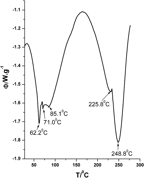
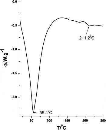
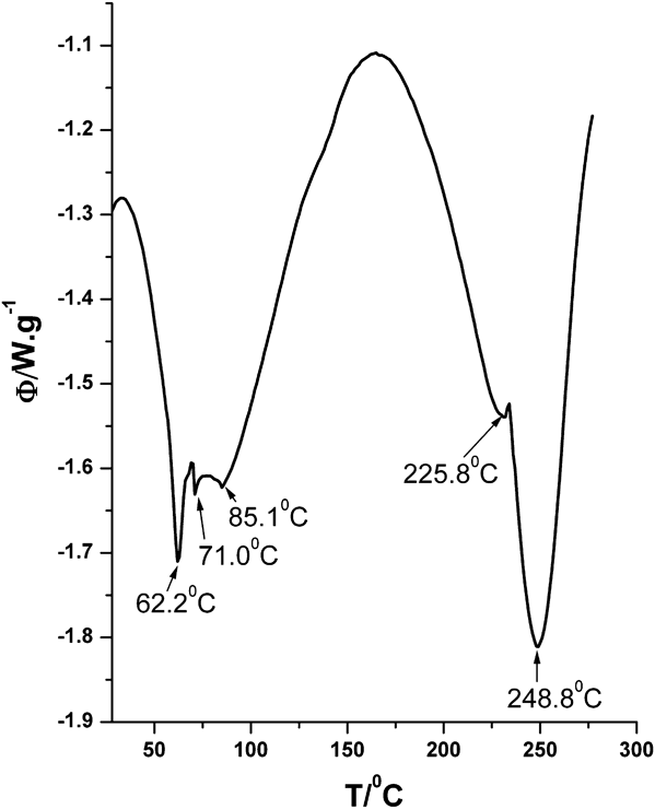
Some of the DSC curves obtained for the biocomposites matrices and membranes that are the subject matter of the present study exhibit complex shapes (Figs. 4–6). For example, DSC curve for MEM-PDex1.3 Na exhibits the splitting of the peak corresponding to water desorption and two endothermic peaks at temperatures higher than 200 °C. This is due to the complex processes resulting from the interactions between basic biomaterial (collagen) with metallic ions from PDex salts.
DSC curve in nitrogen flow of MEM-PDex1.3Na membrane.
DSC curve in nitrogen flow of MAT-PDex1.3Na matrix.

DSC curve in nitrogen flow of MAT-PDexMg-Na matrix.

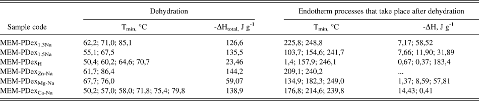
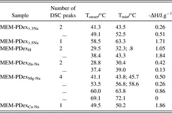
The results of DSC analysis in nitrogen flow for biocomposites revealed a dependence of thermal parameters on the DS of PDex and on the nature of metallic ions that form the mixed salt of PDex (Tables I and II). Thus, the DS influenced the thermal stability for both the biocomposites: the dehydration temperature decreased with the increasing of substitution degree of PDex. It is probably due to the phosphate groups from PDex weakening the bonds between water and polar groups of collagen as a result of the interaction between PDex and collagen.
Thermal parameters for biocomposite membranes, in nitrogen flow.
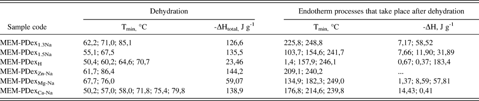
Tmin = temperature corresponding the minimum DSC; ΔH = enthalpy process.
Thermal parameters for biocomposite matrices, in nitrogen flow.

Tmin = temperature corresponding the minimum DSC; ΔH = enthalpy process.
An emphasis on separation of dehydration process/processes towards the melting crystalline zone was noticed from thermograms of all the studied matrices. Due to the specific drying process by lyophilization (freezing at −40 °C and ice sublimation in vapor phase at high vacuum 2 × 10⁻² Torr), there was only one species of bound water for MAT-PDex1.3Na, MAT-PDexH and MAT PDex1.5Na matrices, whereas for the matrices containing mixed salts of PDex a small shoulder appears at low temperature probably because of Zn, Mg and Ca metallic ions influence (Fig. 6).
2. DSC analyses of biocomposites immersed in water
Information about thermal stability of structural conformation of collagen from biocomposites was obtained by DSC analysis in water. In vitro or in vivo resorption of these biomaterials can be adjusted by cross-linking of collagen depending on the specific biomedical application (e.g., dressings have to be readily resorbable and tissues substituents/implants have to be resorbable between 1 and 6 mo). The cross-linking degree influenced the temperature of helix-coil conformational denaturation, also named shrinkage temperature (TS) for leather or membranes and matrices obtained from fibrillar collagen.
The collagen biomaterials must have a shrinkage/denaturation temperature over 40 °C to ensure a proper process of in vivo biointegration. For this reason, the native helicoidal structure of collagen has to be maintained. This structure was recognized by the connective tissue cells, which could then provide immediately their attachment and development, whereas the resorption of collagen scaffold took place at the same time with new tissue formation.
The pure collagen and some collagen-based materials (parchments, vegetable-tanned leathers) immersed in excessive water exhibit a single DSC endothermic peak in the temperature range 25–100 °C. This peak corresponds to the denaturation in water of material and its extrapolated onset temperature and the peak temperature reflect the hydrothermal stability of the investigated collagen. It was pointed out (ref. Reference Budrugeac and Miu36 and corresponding references given in this article) that the onset temperature of the DSC peak practically coincided with the value of the shrinkage temperature (Ts) of the material determined by other methods (Micro Hot Table, for example).
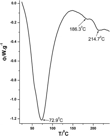
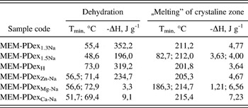
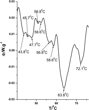
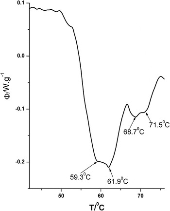
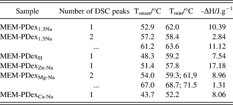
More complex DSC curves were obtained for the investigated biocomposite matrices and membranes, which are the subject matter of the present study (Figs. 7 and 8). This is due to the complex processes that result from the interactions between basic biomaterial (collagen) with metallic ions from PDex salts. Thermal parameters for biocomposite membranes and matrices immersed in water are listed in Tables III and IV.
DSC curve of MEM-PDexMg-Na membrane immersed in water.

DSC curve of MAT-PDexMg-Na matrix immersed in water.

Thermal parameters for biocomposite membranes immersed in water.

Tonset= extrapolated onset temperature; Tmin= temperature corresponding to minimum of DSC peak; ΔH = process enthalpy.
Thermal parameters for biocomposite matrices immersed in water.
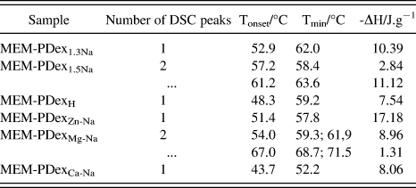
The MAT-PDex matrices showed TS higher than 52 °C and MEM-PDex membranes higher than 40 °C (Tables III and IV). The thermogram of MEM-PDexMg-Na membrane revealed many peaks with different intensities in the wide temperature range of 43.8–72 °C (Fig. 7). A similar situation can be seen for MAT-PDexMg-Na matrix (Fig. 8).
Most of the interactions that took place during interpolymeric complex collagen–PDex formation in aqueous medium are electrostatic and hydrogen bonds. Subsequent operations of cross-linking by aldehyde and the specifics of drying processes allowed the formation of covalent bonds, which were more hydrothermally resistant than new hydrogen bonds. Thus, the processes of autoassembling/restructuration of collagen macromolecule took place by free drying at 25 °C in the presence of PDex, which formed more orderly morphological structures but by sensitive bonds in aqueous medium. This fact was better noticed for membranes with mixed salts of PDex. In this case, the TS and the enthalpy values for collagen matrix without PDex were lower compared with the ones obtained for collagen matrices with PDex. The drying by lyophilization immediately after the formation of the biocomposite collagen–PDex gel did not allow such slow reassembling process, but cross-linking that took place increased the thermal stability of matrices in aqueous medium.
3. Hydrophilic characteristics of biocomposite matrices and membranes
Water can be regarded as an integral component of collagen structure and it was demonstrated that to maintain its fibrillar structure the presence of a certain amount of water was required. By removing this water, the arrangement of inter- and intramolecular bonds was changed with irreversible effect. Collagen, being a hydrophilic polymer with a specific supramolecular structure, in contact with physiological fluids absorbs a large amount of liquid. This ability was kept also in the case of matrices obtained from solubilized and lyophilized fibrillar collagen.
It is also known that both proteins and polysaccharides, particularly hyaluronic acid, plays an important role in controlling water content from connective tissues. In these tissues, there are two types of water: free and chemically bound. The latter no longer had the same properties as free water. Thus, in skin there is about 80% free water content and about 20% water bonded by polar groups and peptide bonds of type I fibrillar collagen; this water is kept in skin after a total drying.Reference Albu, Ghica, Ficai, Titorencu, Popa, Albu and Deselnicu39 Water absorption of dry skin was about 150% after 2 h and it was influenced by the density of the morphological structure, the thickness of the sample, and the temperature. The matrices and membranes absorbed water differently, depending on their microporous structure and the cross-linking degree. Thus, a collagen matrix can absorb about 2500–3000%water after 2 h at 23 °C and a collagen membrane absorbed 20 times less water than a matrix.Reference Chirita and Chirita40
Water absorption in collagen matrices and membranes took place immediately after their immersion in water. The first step of absorption, the diffusion one, was influenced by the hydrophilic properties of the surface and its microstructure (porosity). As we know, in the more compact structure of collagen membrane where the pores with medium diameter of 0.08 μm are predominant, the process of diffusion was slower and the subsequent process of swelling was restricted by membrane microstructure.Reference Siapi, Mavromoustakos, Trandafir, Albu and Budrugeac29 On the contrary, a collagen matrix with macro- and microporous structure accelerated these phases of absorption process. The medium pore diameter of matrices was 2–3 μm and the pore sizes and distribution was wide and depends mainly on parameters of lyophilization process.Reference Siapi, Mavromoustakos, Trandafir, Albu and Budrugeac29
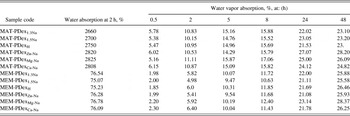
In this case, all MAT-PDex matrices absorbed after 2 h similar amount of water with reference collagen matrix and the values varied between 2700 and 2825% (Table V).
Water absorption and water vapor absorption for biocomposites.

The membranes MEM-PDex absorbed less water compared with reference one. The values varied between 75 and 77%. Even if the percentage of PDex was small (5% toward collagen), the parameters of free drying favored the process of autoassembling of macromolecular complex collagen-PDex, which led to a uniform and compact surface of the membrane. This hydrated surface of biocomposite membranes was proved to be an ideal scaffold for attachment and development of osteoblast cells, especially in the case of MEM-PDexMg-Na membrane.
Water vapor absorption depended on the initial water content and morphological structure of the samples, temperature and relative air humidity in which the analyses were performed. The values of water vapor absorption were about 15–20% after 24 h when this method is applied for the analysis of tanned animal hide which keep the native fibrillar structure of natural skin. A collagen membrane obtained from collagen gel extracted from hide, after cross-linking, plasticization and free drying presents a smaller value (18%) of water vapor absorption. Reference collagen matrix had a higher value for water vapor absorption (20–22%). These results provided information about micro- and nanoporous fibrillar structure when the water vapors are condensed. Collagen matrices and membranes kept at the fibrillar level the native microstructure of the skin connective tissue from which they are obtained. This fact is important for in vivo integration and biofunctionality of collagen implant.
Biocomposite matrices and membranes had higher values for water vapor absorption after 24 h compared with reference samples of collagen membrane and matrix. The values of 22–27% were obtained for matrix and 21–23% for membrane samples (Table V).
The differences were more evident in the first 8 h. Closer values could be observed for all samples after 48 h, especially in the case of matrices and membranes that contain mixed salts of PDex. The increasing of hydrophilic properties of collagen–PDex biocomposites due to the biomimetic structureReference Siapi, Mavromoustakos, Trandafir, Albu and Budrugeac29, Reference Albu, Ghica, Ficai, Titorencu, Popa, Albu and Deselnicu39 of the complex between collagen and Mg, Ca, Zn and Na salts of polysaccharide can explain the high degree of biocompatibility with osteoblast cells obtained by in vitro tests.
C. “In vitro” tests of biocomposite matrices and membranes
Interactions occurring in vivo at the interface between the biomaterial and the living tissue take place at different levels (nano, micro and macro). A medical implant must have well-defined physical–chemical properties and adequate surface characteristics (chemical reactivity, hydrophilic properties, topography) for a certain cellular response to be able to lead to a fast healing.Reference Roach, Eglin, Rohde and Perry41 The first reactions between ECM and biomaterial surface influences the subsequent stages: surface hydration with a layer of water molecule, protein absorption from ECM on this layer, cells attachment, their development and proliferation, new tissue formation.
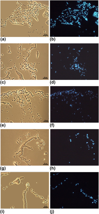
All collagen–PDex biocomposites were proved to be suitable biocompatible scaffolds for osteoblast cells colonization. Cells developed normally and proliferated both at the interface with solid samples and into the biocomposites. No cytotoxic effects were observed during 1 wk of experiment. The matrices were analyzed in section because the visualization of the surface colonization is difficult due to the high opacity and thickness of the matrices (5 mm) compared with the thickness of the membranes (0.3–0.5 mm). To check the distribution of osteoblast-like MG 63 on collagen biomaterials we performed Hoechst staining to label DNA (nucleus).
The morphological difference between the microstructure of matrices and membranes make colonization to be maximal on the membrane surface and in situ penetration was favored by macro- and microporous spongious structure of matrices (Fig. 9).
Osteoblast like cells cultured during 1 wk on 3D collagen stained with the blue-fluorescent Hoechst 258 nuclear counterstain are shown in this fluorescent micrograph (right panel), and the same field is shown in this phase-contrast image (left panel). (a), (b) MAT-PDex1.3Na; (c), (d) MAT-PDexH; (e), (f) MAT-PDexZn-Na; (g), (h) MAT-PDexMg-Na; (i), (j) MAT-PDexCa-Na.

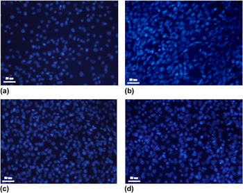
The fluorescent microscopy images show significant increase in cell number and proliferation on collagen membranes with anorganic components [Fig. 10(b)–10(d)].
Osteoblast like cells from human osteosarcoma cultured during 1 wk on surfaces of biocomposite membranes: Images obtained by fluorescent Hoechst staining. (a) MEM-PDexH; (b) MEM-PDexZn-Na; (c) MEM-PDexMg-Na; (d) MEM-PDexCa-Na.

IV. CONCLUSIONS
Biocompatible properties of interpolymeric complex collagen–PDex and its metallic ions (Mg, Ca, Zn, Na) salts are due to the nature of components and to the hydrophilic properties of specific microstructure of biocomposite membranes and matrices.
The DS of PDex as well as the nature of metallic ions from PDex mixed salts influences the thermal stability and hydrophilic properties of collagen–PDex biocomposites, especially in the case of membrane samples.
The existence of many species of bound water in the biocomposite membranes with different temperature of dehydration and lower enthalpy compared with the matrices reveals the importance of process of free drying at 25 °C in the autoassembling of collagen–PDex complex and in the formation of surface with a pattern recognized by osteoblast cells.
ACKNOWLEDGMENT
This research was financially supported by Project No. 71-147/2007 from The National Centre for Programme Management (CNMP) Bucharest, Romania.