Introduction
Trypanosoma cruzi, the causative agent of Chagas disease in humans, is an obligate intracellular protozoan parasite that invades a wide variety of cell types. In the vertebrate host, both trypomastigotes and amastigotes are exposed to various molecules involved in innate and adaptive immunity. To evade the defences of the vertebrate host, amastigotes and trypomastigotes shed different molecules from their surfaces, some of which have been implicated in the invasion of host cells or in the modulation of the immune response (Joiner et al., Reference Joiner, Dias da Silva, Rimoldi, Hammer, Sher and Kipnis1988; Affranchino et al., Reference Affranchino, Ibãnez, Luquetti, Rassi, Reyes, Macina, Aslund, Pettersson and Frasch1989; Gonçalves et al., Reference Gonçalves, Umezawa, Katzin, de Souza, Alves, Zingales and Colli1991; Norris and Schrimpf, Reference Norris and Schrimpf1994; Yokoyama-Yasunaka et al., Reference Yokoyama-Yasunaka, Pral, Oliveira, Alfieri and Stolf1994; Barros et al., Reference Barros, Silva, Versbick, Araguth, Tedesco, Procópio and Mortara1996; Norris, Reference Norris1996; Trocoli-Torrecilhas et al., Reference Trocoli-Torrecilhas, Tonelli, Pavanelli, da Silva, Schumacher, de Souza, E Silva, de Almeida Abrahamsohn, Colli and Manso Alves2009).
The shedding of ectodomains is an irreversible post-translational change in transmembrane proteins, which is driven by the action of endoproteases (shedases, secretases or convertases), releasing a large portion or even the entire extracellular portion (ectodomain) of proteins (Dello Sbarba and Rovida, Reference Dello Sbarba and Rovida2002). This phenomenon represents an important and efficient strategy for regulating the activity of different regulatory molecules such as receptor–ligand complexes, adhesion molecules and ectoenzymes (Werb and Yan, Reference Werb and Yan1998; Dello Sbarba and Rovida, Reference Dello Sbarba and Rovida2002). During Chagas disease progression, such shed membrane components can be detected in sera from infected animals and humans, explaining the immunopathological and morphological alterations observed in the kidney (Castro and Ribeiro dos Santos, Reference Castro and Ribeiro dos Santos1977).
Spontaneous and ligand-dependent capping and shedding have been described in vitro in T. cruzi trypomastigote and amastigote forms (Leon et al., Reference Leon, Villalta, Queiroz and Szarfman1979; Schmuñis et al., Reference Schmuñis, Szarfman, De Souza and Langembach1980; Szarfman et al., Reference Szarfman, Queiroz and De Souza1980; Gonçalves et al., Reference Gonçalves, Umezawa, Katzin, de Souza, Alves, Zingales and Colli1991; Barros et al., Reference Barros, Silva, Versbick, Araguth, Tedesco, Procópio and Mortara1996; Garcia-Silva et al., Reference Garcia-Silva, das Neves, Cabrera-Cabrera, Sanguinetti, Medeiros, Robello, Naya, Fernandez-Calero, Souto-Padron, de Souza and Cayota2014). Conventional multivalent ligands (lectins and antibodies against surface antigens) are known to induce small clusters (patches) of their receptors on the surface of lymphocytes, which subsequently form large polarized aggregates (caps/capping process) via an energy and temperature-dependent process (Butman et al., Reference Butman, Bourguignon and Bourguignon1980). Shedding is an energy, temperature and time-dependent phenomenon that involves plasma membrane vesiculation (Gonçalves et al., Reference Gonçalves, Umezawa, Katzin, de Souza, Alves, Zingales and Colli1991), proteases activities (Norris, Reference Norris1996) or solubilization of glycosylphosphatidylinositol (GPI)-membrane-anchored components such as Ssp4 (Andrews et al., Reference Andrews, Robbins, Ley, Hong and Nussenzweig1988). In contrast to what is observed in other cell types, vesicle shedding in T. cruzi is independent of the presence of serum (Taverna et al., Reference Taverna, Ghersi, Ginestra, Rigogliuso, Pecorella, Alaimo, Saladino, Dolo, Dell'Era, Pavan, Pizzolanti, Mignatti, Presta and Vittorelli2003).
The glycocalyx of T. cruzi cell surface is complex, varying according to the developmental stage of the parasite, exhibiting a high diversity of (GPI)-anchored glycoproteins, such as mucins, trans-sialidase (TS)/gp85 glycoproteins, mucin-associated surface proteins (MASPs) and trypomastigote small surface antigen. The protein-free GPIs (glycoinositolphospholipids) and other glycolipids are also important components of the T. cruzi glycocalyx (Fonseca et al., Reference Fonseca, da Costa, Chaves, Freire-de-Lima, Morrot, Mendonça-Previato, Previato and Freire-de-Lima2019).
Different molecules have been used to probe the cell membrane structure and dynamics of different cell types, including parasitic protozoans (Pinto da Silva et al., Reference Pinto da Silva, Martínez-Palomo and Gonzalez-Robles1975; Szarfman et al., Reference Szarfman, Queiroz and De Souza1980; Saraiva et al., Reference Saraiva, Vannier-Santos, Silva-Filho and de Souza1989; Barros et al., Reference Barros, Silva, Versbick, Araguth, Tedesco, Procópio and Mortara1996). Cationic molecules, such as cationized ferritin (CF), which have been used to identify the presence of negative charges on the cell surface at physiological pH (Danon et al., Reference Danon, Goldstein, Marikovsky and Skutelsky1972), bind to sialoglycoconjugates on the surfaces of all developmental forms of T. cruzi and Leishmania (Souto-Padrón and De Souza, Reference Souto-Padrón and De Souza1986; Saraiva et al., Reference Saraiva, Vannier-Santos, Silva-Filho and de Souza1989). CF also interferes with the interaction of these parasites with their host cells (Meirelles et al., Reference Meirelles, Souto-Padrón and De Souza1984; Saraiva et al., Reference Saraiva, Vannier-Santos, Silva-Filho and de Souza1989). Incubation of living L. amazonensis in the presence of CF induces capping and shedding of anionic sites, and these effects are more prominent in infective forms (Saraiva et al., Reference Saraiva, Vannier-Santos, Silva-Filho and de Souza1989).
Lectins have also been employed to characterize the differences in cell surface composition and physiology. The manner by which lectins bind and agglutinate distinct cell types or strains may be indicative of malignance or virulence. One of the best-studied lectins is Concanavalin A (Con A), which induces the capping and shedding of surface binding sites in parasites such as Leishmania (Saraiva et al., Reference Saraiva, Vannier-Santos, Silva-Filho and de Souza1989) and Entamoeba histolytica (Pinto da Silva et al., Reference Pinto da Silva, Martínez-Palomo and Gonzalez-Robles1975).
This study follows from our longstanding interest on the surface composition and dynamics in T. cruzi parasites, particularly in situations where modifications in the distribution of surface sites lead to changes in the parasite–host relationship. Here, we report the dynamics of anionic sites and Con A-binding sites in living tissue culture-derived trypomastigotes from two different strains and their influence on the adhesion and internalization of parasites to LLC-MK2 cells. Here, we discuss the ways trypomastigotes of the Y strain and the CL Brener clone modulate their own surface binding sites and the relationship these sites have with molecules involved in the innate immune system.
Materials and methods
Parasites
Tissue culture trypomastigotes (TCT) from T. cruzi Y strain and CL-Brener clone [belonging to the TcII and TcVI lineages, respectively, according to Zingales et al. (Reference Zingales, Andrade, Briones, Campbell, Chiari, Fernandes, Guhl, Lages-Silva, Macedo, Machado, Miles, Romanha, Sturm, Tibayrenc and Schijman2009)] were obtained from the supernatants of 5 to 6-day-old infected LLC-MK2 cells (American Type Culture Collection, Rockville, MD, USA), maintained in RPMI-1640 medium (Sigma Aldrich, St. Louis, MO, USA) supplemented with 2% fetal bovine serum (FBS), at 37 °C in a 5% humidified CO2 atmosphere as previously described (Adade et al., Reference Adade, Chagas and Souto-Padrón2012).
Binding of cationic compounds
Tissue culture supernatant of infected LLC-MK2 cells, containing trypomastigotes, were collected and centrifuged at 2000 × g for 10 min and washed in cold RPMI medium without serum. The purity of trypomastigote developmental forms derived from culture was over 90%, as assessed by direct counting in a Neubauer chamber, as well as by Giemsa staining. Subsequently, the parasites were resuspended (5 × 10⁶ cells sample−1) in cold PBS in the presence of 10 µg mL−1 CF or 1 mm poly-L-lysine-fluorescein isothiocyanate (FITC) (PLL), both from Sigma Aldrich, followed by incubation for 30 min at 4 °C (labelling at 4 °C – designated L4CF and L4PLL trypomastigotes, respectively). Labelling was stopped by centrifugation at 4 °C, 10 000 × g for 3 min. One sample was immediately fixed in 2% formaldehyde in PBS for the analysis described below, and the others were rinsed in cold RPMI medium without serum and without CF or poly-L-lysine for an additional period of 30 min at 37 °C in RPMI without serum (shedding at 37 °C – S37CF and S37PLL trypomastigotes). CF and poly-L-lysine labelled trypomastigotes were processed as described further below.
Binding of Con A
TCT were incubated in the presence of 10 µg mL−1 Con A, Alexa Fluor 488 conjugated with Con A (Life Technologies) and gold-labelled Con A (diluted 1:20) and obtained as described by Bourguignon et al. (Reference Bourguignon, De Souza and Souto-Padrón1998). Both Alexa Fluor and gold-labelled Con A were diluted in PBS and processed as described above and referred to the abbreviations L4ConA, and S37ConA. Con A-labelled parasites were processed for flow cytometry and fluorescence and electron microscopy as will be described below. Parasites pre-fixed in 2% formaldehyde in PBS for 10 min at room temperature were used as a control for the labelling with CF and poly-L-lysine-FITC.
We employed pre-fixed parasites, which are regularly used in immunofluorescence and ultrastructural studies, as controls for the localization of CF- and Con A-binding sites, based on the assumption that chemical fixation with glutaraldehyde or formaldehyde, under conditions that have been established for many years in different laboratories, is sufficient to maintain the distribution of surface sites in the absence of any stimuli (Souto-Padrón et al., Reference Souto-Padrón, Harth and de Souza1990; de Carvalho et al., Reference De Carvalho, Souto-Padrón and de Souza1991; Bourguignon et al., Reference Bourguignon, De Souza and Souto-Padrón1998).
Labelling of α-Gal surface sites
Control, S37CF and S37ConA trypomastigotes were fixed in PBS containing 3.7% formaldehyde for 10 min at 4 °C, then washed twice in PBS and incubated with 50 mm ammonium chloride for 30 min at room temperature. Non-specific sites were subsequently blocked with PBS containing 0.2% gelatin and 0.01% azide (PGN) for 30 min. Aliquots of the three populations of trypomastigotes were incubated for 1 h at room temperature with Chagasic anti-〈-galactosyl antibodies [Ch anti-〈-Gal – isolated from sera of patients with Chagas' disease, diluted 1:200 in PGN, and kindly provided by Dr Igor C. Almeida (University of Texas at El Paso)], as described previously (Almeida et al., Reference Almeida, Milani, Gorin and Travassos1991). After the incubation, the parasites were washed twice in PBS and incubated with Alexa-546-conjugated anti-human IgG antibody (diluted 1:20) for 1 h at room temperature. The samples were processed for Field Emission Scanning Electron Microscopy (FESEM) as described below.
Flow cytometric assays
Trypomastigotes previously labelled with poly-L-lysine-Con A-FITC conjugate were fixed in 2% formaldehyde in PBS, for 10 min, at 4 °C. Then, the parasites were rinsed and resuspended in PBS, pH 7.2, with 0.1% sodium azide (FACS buffer) and analysed in the same buffer in a FACScan flow cytometer (Becton Dickinson, Franklin Lakes, NJ, USA) equipped with a 15 mW argon laser emitting at 488 nm (FL1-Height) and the CellQuest software (Joseph Trotter, Scripps Research Institute, San Diego, CA, USA). The equipment was gauged to analyse 3 × 10⁴ cells. All results were analysed using the Windows Multiple Document Interface Flow Cytometry Application (WinMDI), version 2.8. All analyses were performed in at least three independent experiments.
Trypomastigote lysis assay
These experiments were carried out following previously described methods (Pereira-Chioccola et al., Reference Pereira-Chioccola, Acosta-Serrano, Almeida, Ferguson, Souto-Padrón, Rodrigues, Travassos and Schenkman2000). Briefly, trypomastigote suspensions (4 µL in RPMI medium without serum; 1 × 10⁷ parasites mL−1) were pre-treated with the same volume of either 10 µg mL−1 CF or Con A for 10 min at 37 °C. Next, 8 µL of Ch anti-α-Gal antibody at 20 µg mL−1 was added, and the mixture was incubated for an additional 30 min at 37 °C. The controls were incubated in RPMI without serum. We counted the numbers of motile and free (non-agglutinated) trypomastigotes with a haemocytometer. The results are expressed as the percentages (mean ± s.d.) of motile and free parasites relative to the control. These experiments were carried out in triplicate.
Cell adhesion and invasion assays
To investigate the effects of the cell surface modulation caused by CF and Con A on the adhesion and invasion of host cells, LLC-MK2 cells were seeded in 24-well plates containing glass coverslips, cultivated in RPMI medium supplemented with 10% FCS and then maintained at 37 °C in a 5% CO2 humidified atmosphere for 1 day. Subsequently, the cultures were washed with RPMI medium without FCS, to remove non-adherent cells, and cultivated in fresh RPMI containing 10% FCS. To analyse the parasite adhesion, host cells were then incubated at 4 °C, for 30 min, in the presence of S37CF and S37ConA trypomastigotes and control parasites at 10:1 parasite:cell ratio; then, the samples were rinsed in PBS and fixed. To analyse parasite internalization, LLC-MK2 cells previously incubated with trypomastigotes at 4 °C were washed in RPMI without serum, incubated for 24 h at 37 °C in a 5% humidified CO2 atmosphere, and then washed with PBS. All cells were then fixed in Bouin's solution, stained with Giemsa and mounted on glass slides with Permount. Parasite adhesion and infection were quantified using a Zeiss Axioplan 2 light microscope (Oberkochen, Germany) equipped with a Color View XS digital video camera. The numbers of adhered and internalized parasites per 100 cells were evaluated by counting a total of 400 cells in three different experiments.
Fluorescence microscopy
Trypomastigotes (Y and Cl-Brener stocks) were incubated with Con A-Alexa Fluor 488, rinsed in cold PBS, and fixed in 3.7% formaldehyde in PBS for 10 min at 4 °C. Parasites were spread onto poly-L-lysine (0.1% in PBS) coated coverslips and allowed to adhere at room temperature for 30 min. Coverslips were then washed twice, labelled with 1 g mL−1 4′,6-diamidino-2-phenylindole (Sigma Aldrich) in PBS for 5 min, and mounted with 0.2 M N-propyl galate in glycerol: PBS (9:1) solution. Images were acquired with using a Zeiss Axioplan 2 fluorescence microscope (Zeiss, Jena, Germany) coupled to a charge-coupled device colour video camera.
Electron microscopy
Samples to be analysed by scanning electron microscopy (SEM), transmission electron microscopy (TEM) and FESEM were processed as follows. Parasites to be observed via SEM were fixed in 2.5% glutaraldehyde in 0.1 M cacodylate buffer, pH 7.2, containing 3.7% sucrose and 5 mm CaCl2, for 1 h at room temperature. A 50 L sample of the parasite suspension was spread onto poly-L-lysine coated coverslips and allowed to adhere at room temperature for 30 min. After that, non-adherent parasites were removed by rinsing with PBS, and the coverslips were fixed in 1% OsO4, 0.8% potassium ferrocyanide and 5 mm CaCl2 in 0.1 M sodium cacodylate buffer for 20 min at room temperature. The coverslips were finally rinsed in PBS, dehydrated in graded ethanol, critical point dried in CO2, covered with a 20 nm gold layer and observed using a JEOL JSM 5310 scanning electron microscope.
For FESEM analysis, samples were fixed in 0.1% glutaraldehyde (grade I), 4% formaldehyde in 0.1 M cacodylate buffer, pH 7.2, containing 3.7% sucrose and 0.2% picric acid for 1 h at room temperature. After two washes in the same buffer, cells were spread onto poly-L-lysine-coated silicon chips and allowed to adhere at room temperature for 30 min. Then, the chips were washed twice in PBS and incubated for 30 min at room temperature in PBS containing 2% bovine serum albumin (BSA) (PBS-BSA). Then, the chips were incubated in the presence of Ch anti-α Gal antibody (Chagasic anti-α-galactosyl antibody) in PBS-BSA (1:400 dilution) for 30 min at room temperature, washed extensively with PBS-BSA and incubated in the presence of a gold-labelled anti-human IgG antibody (1:50 diluted, 15 nm in diameter) for 30 min at the same temperature. Following labelling, the chips were washed in PBS, dehydrated in a graded ethanol series, critical point dried in CO2, mounted on a stub and then sputter coated with a 10 nm carbon layer in a Gatan Model 681 apparatus. The samples were finally imaged using a JEOL JSM-6340-FESEM at 5 kV using the backscattered detection mode.
For TEM analysis, the parasites were fixed in 2.5% glutaraldehyde in 0.1 M cacodylate buffer, pH 7.2, containing 3.7% sucrose and 5 mm CaCl2 for 1 h at room temperature, washed twice in PBS and post-fixed in 1% OsO4, 0.8% potassium ferrocyanide and 5 mm CaCl2 in 0.1 M sodium cacodylate buffer for 1 h at room temperature. The cells were then rinsed once with 0.1 M cacodylate buffer, pH 7.2, containing 3.7% sucrose, dehydrated in a graded acetone series and embedded in PolyBed 812. Ultra-thin sections were stained with uranyl acetate (30 min) and lead citrate (5 min) and subsequently observed using FEI Morgani TEM operating at 80 Kv.
Statistical analysis
All data are presented as the mean of the results from at least three experiments [± standard deviations (s.d.)]. A P value < 0.05 indicates statistically significant differences between means.
Results
Cationized molecules induce capping and shedding in trypomastigotes
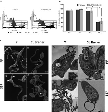
In order to analyse the dynamics of anionic sites on the surface of live T. cruzi, we used flow cytometry and TEM upon binding of polycationic molecules to the surface of strain Y and CL Brener trypomastigotes. In the flow cytometry assays, poly-L-lysine-FITC was used, and the fluorescence intensity and the percentage of labelled parasites in each population were analysed (Fig. 1A and B). We observed that the distribution of labelling in the two populations of fixed trypomastigotes was quite distinct. The distribution of the labelling intensity in the Y trypomastigotes was broad: parasites with several different degrees of labelling were observed, in addition to approximately 15% negatively labelled parasites (Fig. 1A). In comparison, the distribution of the labelling intensity in CL Brener trypomastigotes was more uniform and concentrated on the right side of the histogram, showing a higher percentage of labelled parasites (52.37%), and only 1.5% non-labelled parasites (Fig. 1A). The poly-L-lysine-FITC labelling of the parasites at 4 °C was quite different from that obtained for pre-fixed parasites, most likely due to the lack of binding site mobility within the plane of the membrane at 4 °C. In both types of samples, labelling performed at 4 °C resulted in two fluorescence intensity peaks. Approximately 95% of the trypomastigotes of both isolates were labelled, and the peaks corresponding to the highest labelling intensities accounted for 13 and 19% of the L4PLL Y and CL Brener trypomastigotes, respectively. The Y and CL Brener L4 trypomastigotes were unlabelled 0.55 and 7.7% of the time, respectively. However, incubation of S4PLL parasites for an additional 30 min at 37 °C (parasites S37PLL) completely changed the distribution of labelling among Y trypomastigotes, which showed a single fluorescence intensity peak (corresponding to 69% of the parasites) very close to the negative control (Fig. 1A). In contrast, the S37PLL parasites of the CL Brener clone showed a small percentage (8.5%) of highly labelled trypomastigotes (albeit with a marked reduction in the intensity of labelling as a whole – approximately 42% of the parasites were negative), which can be seen as a second peak of fluorescence intensity at the right end of the histogram (Fig. 1A). Analysis of the median fluorescence intensity in Y and CL Brener trypomastigotes showed a significant decrease in fluorescence in both those stocks at 37 °C (Fig. 1B).
Modulation of the surface of trypomastigotes by cationized molecules. (A) Kinetics of the poly-L-lysine-induced redistribution of negatively charged cell surface components in Y and CL Brener trypomastigotes. A representative histogram is shown for each experimental condition. A total of four experiments were conducted for each experimental condition. FL1-Height, Fluorescence gated for poly-L-lysine-FITC-labelled. Pre-fixed parasites, as described in section ‘Binding of Con A’, were used as a control. (B) Percentages of poly-L-lysine-labelled cells under the different experimental conditions. The results are expressed as the mean ± s.d. (n = 4). The pooled data are statistically significantly different (*P < 0.05); (C–F) pre-fixed (C, D) and S37CF (E, F) parasites were processed for scanning electron microscopy. Pre-fixed parasites present small shedding vesicles distributed along the cell body and flagella (small arrows in C and D). In S37CF parasites, the shedding vesicles are larger than those observed in C and D. These vesicles can be seen at the posterior edge of a Y trypomastigote, together with amorphous material (arrowheads in E), and associated with the flagella (F). The shedding vesicles in CL Brener S37CF trypomastigotes are up to five times larger than those found in PF trypomastigotes (arrow in F); (G–M) pre-fixed (G–J) and S37CF parasites (K–M) were processed for transmission electron microscopy. Pre-fixed parasites present a dense uniform layer of CF particles on the cell body and flagellar membranes. Membrane projections observed in Y strain trypomastigotes appear to be completely covered with CF (arrows in G). Small vesicles without CF labelling can also be seen budding from the flagellar membrane (arrow in H). Projections observed on the surface of CL Brener trypomastigotes either present CF particles (arrow in I) or not present these particles (arrowhead in J). These results show that Y and CL Brener parasites constitutively release portions of the membrane containing negative charges recognized by CF. CL Brener trypomastigotes also shed extended segments of the membrane, containing multiple layers that are not recognized by CF (arrows in J). Around S37CFY trypomastigotes, there is amorphous material labelled with CF particles that are being released from the posterior end of the parasite (small arrows in K), and several labelled shedding vesicles can be observed near the flagellum (arrows in L). A large vesicle weakly labelled with CF can be observed alongside a CL Brener trypomastigote (M). f, flagellum; fp, flagellar pocket; k, kinetoplast.

In ultrastructural analyses, CF was used to induce mobility of anionic sites on the cell surface of the parasites (Fig. 1C–M). Ferritin particles on the surface of the parasites cannot by analysed through conventional SEM. Nonetheless, CF contains an electron-dense iron core that can be easily visualized using TEM. Thus, after treatment with CF, significant differences in the number and size of shedding vesicles released by the parasites were observed (Fig. 1C–F). Importantly, CF does not exhibit toxicity toward T. cruzi, at the concentrations that are normally used (10 g mL−1). Pre-fixed parasites (which are frequently used to mimic the parasites in culture medium at 37 °C) displayed a smooth surface with small shedding vesicles (50 nm in diameter), which budded at the end of the flagellar pocket and the flagellum (Fig. 1C and D). The Y strain S3FCF parasites showed a greater number of larger vesicles, which were released from the same regions as in PF parasites (Fig. 1E and F). The vesicles shed from S37CF CL Brener parasites showed up to six times the size of those observed in previously fixed CL Brener trypomastigotes (Fig. 1E, F and M).
TEM analysis of the parasites that were pre-fixed and incubated with CF showed that trypomastigotes constitutively release vesicles containing negatively charged molecules from the cell body, the flagellum and the flagellar pocket (Fig. 1G–J). Trypomastigotes from clone CL Brener, in addition to presenting projections and membrane shedding vesicles that were intensely labelled with CF, exhibited some unlabelled projections released from the membrane of the flagellum (Fig. 1J). S37CF trypomastigotes of the Y strain showed a greater number of vesicle budding from the cell body and ferritin clusters being released from the posterior region of the body (Fig. 1K and L). S37CF trypomastigotes of the CL Brener clone also showed vesicles budding directly from the cell body and large vesicles that could be observed freely in the vicinity of parasites. Both types of vesicles were poorly labelled (Fig. 1M).
Dynamics of Con-A-binding sites on the surface of trypomastigote
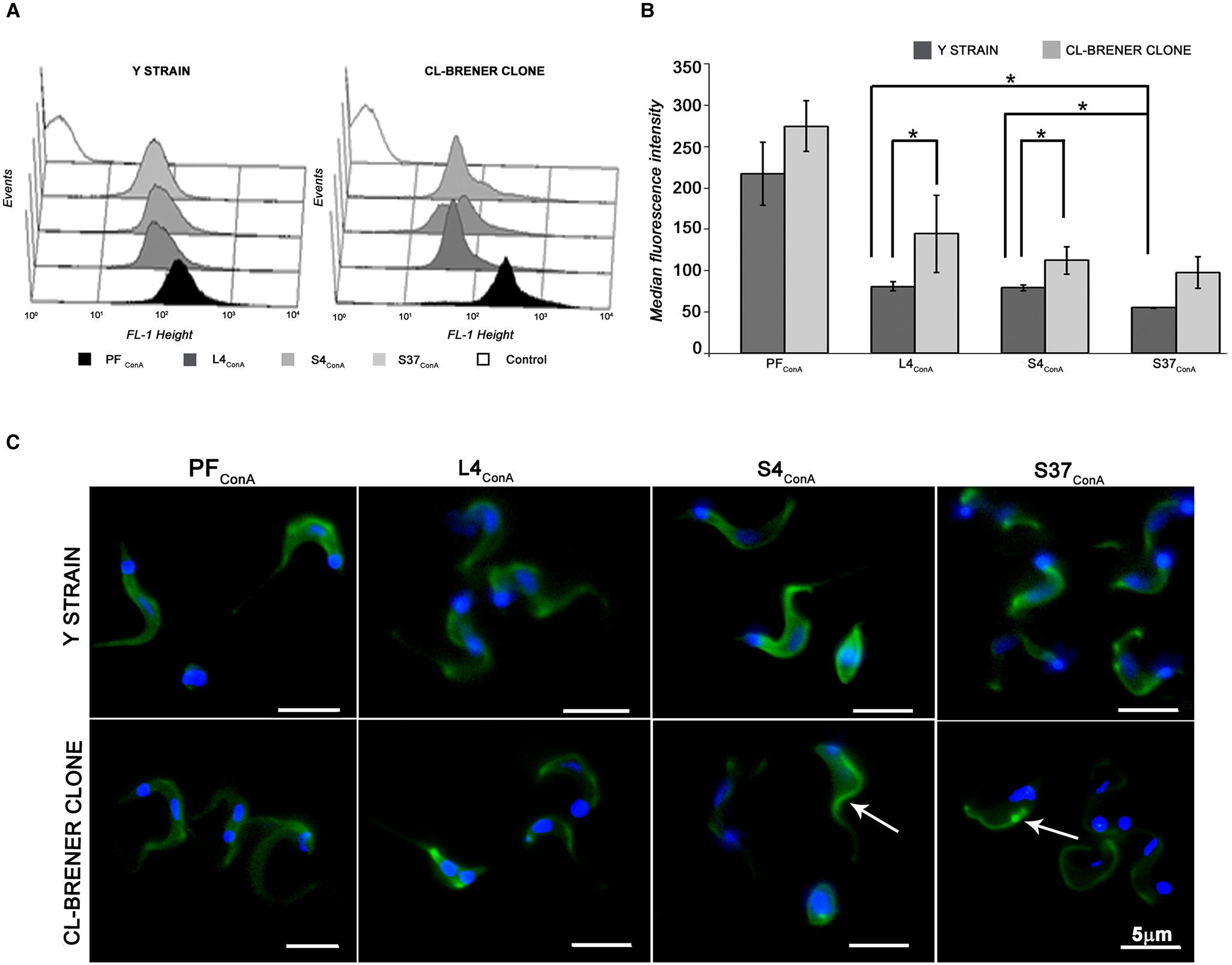
Analysis using flow cytometry showed that lectins bound more strongly to CL Brener L4ConA trypomastigotes, as compared to those of the Y strain (Fig. 2A). The distribution of labelling on the CL Brener parasites was also wider; approximately 20% of these parasites showed a median fluorescence intensity of 147.83. When the same analysis was applied to the L4ConA Y trypomastigotes, we observed that 34.6% of the parasites presented a median fluorescence intensity of 73.30. Following subsequent incubations at 4 and 37 °C, a significant reduction in the median fluorescence of Y trypomastigotes was shown (Fig. 2B), which may be observed in Fig. 2A as a uniform displacement of the labelling to the left end of the graph. CL Brener trypomastigotes showed a very different pattern. The reduction of the fluorescence intensity was not significant for S4ConA and S37ConA parasites (Fig. 2B), because approximately 20% of them continued to show a high median fluorescence intensity (141.54), very close to that observed in L4ConA parasites. As previously observed using poly-L-lysine-FITC, the PF parasites exhibited more intense labelling, as compared to those incubated at 4 °C.
Modulation of the surface of trypomastigotes by Concanavalin A. (A) The histograms indicate the different levels of fluorescence intensity observed in different samples. A total of four experiments were conducted for each experimental condition. Pre-fixed parasites, as described in the Material and methods section, were used as a control. FL1-Height, Fluorescence gated for Con A-FITC-labelled. (B) Comparative analysis of the median fluorescence intensity observed under different experimental conditions. The results are expressed as the mean ± s.d. (n = 4). The pooled data are statistically significantly different (*P < 0.05); (C) fluorescence microscopy of Y and CL Brener parasites subjected to the various experimental treatments, as described in the Methods section. Incubation of L4ConA parasites at 4 and 37 °C causes a decrease in the fluorescence intensity on the surface of Y trypomastigotes and concentration of the labelling in small regions of the flagellum. In S4ConA and S37ConA CL Brener trypomastigotes, the fluorescent labelling is progressively concentrated in the flagellum (white arrow in S4ConA) and the flagellar pocket (white arrow in S37ConA).

Fluorescence microscopy analysis (Fig. 2C) showed that the PF trypomastigotes of both types were homogeneously labelled. In the L4ConA parasites, even though the labelling was homogeneous on the cell body membrane, it was concentrated on the membrane of the flagellum. In S4ConA and S37ConA trypomastigotes from the Y strain, the labelling intensity was reduced, but the labelling pattern was homogeneous. The analysis of S4ConA and S37ConA CL Brener trypomastigotes showed that under these conditions, labelling was concentrated on the membrane of the flagellum or even in the flagellar pocket, while the labelling of the cell body was significantly reduced.
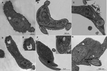
The TEM analysis of PF parasites of both types showed that these trypomastigotes were intensely labelled with the Con A–gold complex, presenting a continuous layer of gold particles on the cellular and flagellar membranes (Fig. 3). Of particular note, no labelling was observed in the flagellar pocket (Fig. 3E), confirming the results obtained by fluorescence microscopy. The labelling of S4ConA and S37ConA trypomastigotes of the Y strain was significantly less intense, as compared to PF parasites, although homogeneously distributed gold particles were observed on the cell body and flagellar membranes (Fig. 3B and C). A significant number of shedding vesicles containing gold particles was also observed (Fig. 3C). As observed using immunofluorescence, the Con A-binding sites were repositioned to the region of adhesion between the flagellum and cell body and within the flagellar pocket in S37ConA CL Brener parasites (Fig. 3F–H). This movement, observed by the presence of gold particles, was also observed in S4ConA parasites, indicating that even at 4 °C, Con A-binding sites show motility on the membrane plane (Fig. 3F and G).
Ultrastructural analysis of the modulation of the surface of trypomastigotes by Concanavalin A. Pre-fixed (A, D, E) and living parasites (B–C, F–H) were incubated in the presence of gold-labelled Con A and processed for transmission electron microscopy. Pre-fixed trypomastigotes from the Y strain (A) and CL Brener clone (D, E) present a continuous layer of particles on the cell and flagellar membranes. No gold particles were observed in the flagellar pocket (fp in E). In S4 and S37ConA Y trypomastigotes, the labelling is less intense than in PF parasites but is still distributed homogeneously on the cell surface (B, C). Labelled shedding vesicles are observed in the extracellular space (arrows in C). S4ConA CL Brener trypomastigotes present few gold particles distributed on the cell body, and some particles are concentrated at the flagellum-cell body attachment zone and in the flagellar pocket (arrows in F and G). Following additional incubation at 37 °C (I, J), gold-labelled vesicles are primarily observed in the flagellar pocket (arrows in H). f, flagellum; k, kinetoplast.

Pre-incubation with CF and Con A protects parasites from lysis with anti-α-Gal antibodies
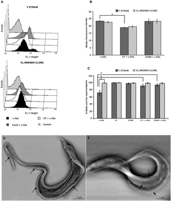
Flow cytometric and FESEM analysis of the binding of Ch anti-α-Gal antibodies on control trypomastigotes showed that these antibodies bind more strongly to Y strain than to CL Brener trypomastigotes (Fig. 4A and B, D and E). The histogram of the fluorescence intensities of Y parasites clearly shows two populations (Fig. 4A). The most intensely labelled population, which represents approximately 13% of the parasites, displayed a median fluorescent intensity that was six times higher than that observed in the second population. Control trypomastigotes from the CL Brener clone exhibited only a single population, in which the fluorescence intensity was similar to the less intensely labelled Y trypomastigotes. Incubation of both types of trypomastigotes with CF significantly reduced the binding of Ch anti-α-Gal antibodies, increasing the percentage of negative trypomastigotes and thereby eliminating the more intensely labelled peak of the Y parasites. On the other hand, incubation of the parasites with Con A did not alter the median labelling intensity relative to the control parasites (Fig. 4A and B). We next tested whether incubation of the parasites with CF and Con A was able to modify the susceptibility to lysis induced by Ch anti-α-Gal antibodies (Fig. 4C). The number of motile control Y trypomastigotes was reduced by 33% when they were previously incubated for 30 min in the presence of Ch anti-a-Gal, in contrast to what was observed for CL Brener trypomastigotes, in which only 2% reduction was observed. Incubation of both types of trypomastigotes in the presence of CF (10 µg mL−1) or Con A (10 µg mL−1) for 30 min did not promote lysis or agglutination of the parasites. Previous incubation of Y trypomastigotes with CF or Con A protected the parasites from the lysis induced by Ch anti-α-Gal antibodies. In contrast, these treatments did not alter the percentage of motile and free CL Brener trypomastigotes (Fig. 4C).
Modulation of α-Gal surface sites by CF and Con A. (A) Flow cytometric analysis showing the expression of α-Gal sites on the surface of Y and CL Brener trypomastigotes. Living parasites (S37) were incubated in the presence of 10 µg mL−1 CF or Con A and then fixed and incubated with Chagasic anti-α-Gal antibodies. S37 parasites incubated in the absence of CF and Con A were used as positive controls. Negative controls were prepared in the absence of Chagasic anti-α-Gal antibodies. The histograms represent the different fluorescence intensity levels in the different samples. FL1-Height, Fluorescence gated for Alexa-546-conjugated anti-human antibody. (B) Comparative analysis of the median of fluorescence intensity. The results are expressed as the mean ± s.d. (n = 3). Pooled data are statistically significantly different (*P < 0.05). (C) Percentage of living parasites following incubation with a fixed amount of Ch anti-α-Gal antibodies (20 µg mL−1), CF (10 µg mL−1) and Con A (10 µg mL−1) or after incubation with CF or Con A followed by Ch anti-α-Gal antibodies. The results are expressed as the mean ± s.d. (n = 3). The pooled data are statistically significantly different; (D, E) FESEM analysis of tissue-cultured trypomastigotes from the Y strain (D) and CL Brener clone (E) incubated in the presence of Ch anti-α-Gal antibodies (20 µg mL−1) and a secondary gold-labelled anti-human antibody (10 nm in diameter). Note the high labelling intensity in Y trypomastigotes. Arrows indicate the binding sites for α-Gal antibody.

The data show here that Y trypomastigotes presented a greater number of binding sites for α-Gal antibody, as compared to parasites of the CL Brener clone and that changes occurred on the surface of the parasites following incubation with Con A and CF, including blocking of negative charges and increased shedding of surface components. These changes caused a decrease in α-Gal residues or a rearrangement of surface molecules, hindering the action of Ch anti-α-Gal antibodies and therefore inhibiting the lysis of the parasite.
Pre-incubation with CF and Con A interferes with the adhesion and internalization of trypomastigotes
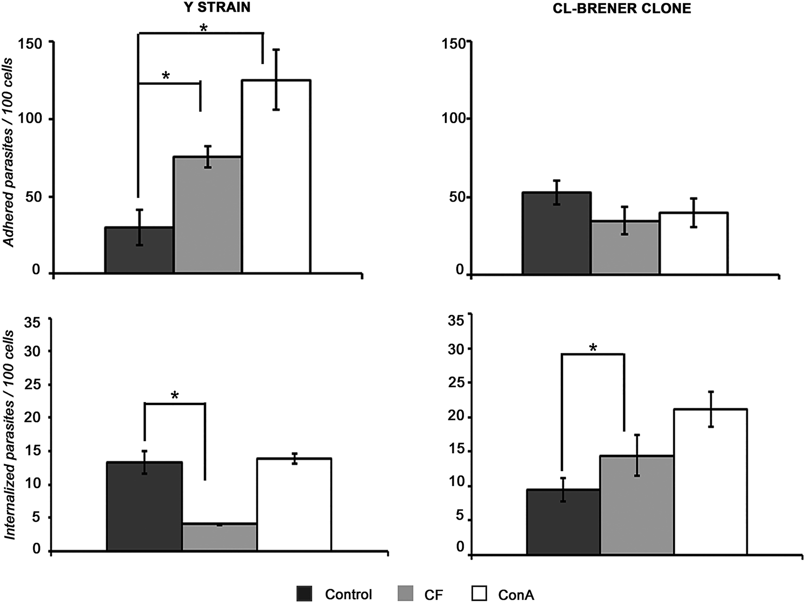
To determine whether the modifications induced by incubation with CF or Con A would interfere with the interaction of the parasites with host cells, S37, S37CF and S37ConA trypomastigotes from the Y strain were incubated in the presence of LLC-MK2 cells at 4 or 37 °C with host cells, which promoted and increase on the number of adhered parasites by approximately 2.5 and 4 times, respectively, as compared to the control. When the same sort of experiment was carried out using trypomastigotes from the CL Brener clone, no significant difference in the number of adherent parasites was observed (Fig. 5). Following adhesion, the internalization of the parasites was evaluated for an additional period of 24 h at 37 °C. We did not observe any increase in the number of internalized parasites in the Y strain, even though treatment with CF and Con A induced an increase in parasite adherence. Conversely, CF treatment promoted a 3-fold reduction in the number of intracellular parasites (Y strain). In trypomastigotes of the CL Brener clone, only the incubation with Con A promoted a significant increase in the internalization of the parasites.
Effects of CF and Con A on the adhesion and internalization of Y and CL Brener trypomastigotes. S37CF and ConA parasites of both types were incubated for 30 min at 4 °C (adhesion) and for an additional 1 h at 37 °C (internalization) in the presence of LLC-MK2 cells. The number of internalized parasites per 100 cells was quantified 24 h after the interaction. The results are expressed as the mean ± s.d. (n = 3). The pooled data are statistically significantly different (*P < 0.05).

Discussion
Capping and shedding in the presence of different ligands have previously been described in Trypanosoma and Leishmania (Schmuñis et al., Reference Schmuñis, Szarfman, Langembach and de Souza1978, Reference Schmuñis, Szarfman, De Souza and Langembach1980; Szarfman et al., Reference Szarfman, Queiroz and De Souza1980; Frevert and Reinwald, Reference Frevert and Reinwald1988; Saraiva et al., Reference Saraiva, Vannier-Santos, Silva-Filho and de Souza1989). In species of the genus Leishmania, capping and shedding were observed in amastigotes and promastigotes, in the presence of CF or antibodies (Doyle et al., Reference Doyle, Behin, Mauel and Rowe1972; Saraiva et al., Reference Saraiva, Vannier-Santos, Silva-Filho and de Souza1989). In T. cruzi, capping and shedding induced by immunoglobulins from Chagasic patients or infected mice, as well as lectins and cationic molecules, such as poly-L-lysine, have been described in vitro in trypomastigotes and amastigotes (Kloetzel and Deane, Reference Kloetzel and Deane1977; Schmuñis et al., Reference Schmuñis, Szarfman, Langembach and de Souza1978, Reference Schmuñis, Szarfman, De Souza and Langembach1980; Leon et al., Reference Leon, Villalta, Queiroz and Szarfman1979; Szarfman et al., Reference Szarfman, Queiroz and De Souza1980; Gonçalves et al., Reference Gonçalves, Umezawa, Katzin, de Souza, Alves, Zingales and Colli1991; Barros et al., Reference Barros, Silva, Versbick, Araguth, Tedesco, Procópio and Mortara1996).
Trypomastigotes from different strains release different amounts of proteins with similar molecular weights, most often incorporated into shedding vesicles that are recognized by specific receptors in diverse cell types, thus influencing the infection process and modulating the immune response (Gonçalves et al., Reference Gonçalves, Umezawa, Katzin, de Souza, Alves, Zingales and Colli1991; Acosta-Serrano et al., Reference Acosta-Serrano, Almeida, Freitas-Junior, Yoshida and Schenkman2001; Aparício et al., Reference Aparício, Scharfstein and Lima2004; Trocoli-Torrecilhas et al., Reference Trocoli-Torrecilhas, Tonelli, Pavanelli, da Silva, Schumacher, de Souza, E Silva, de Almeida Abrahamsohn, Colli and Manso Alves2009; Bayer-Santos et al., Reference Bayer-Santos, Aguilar-Bonavides, Rodrigues, Cordero, Marques, Varela-Ramirez, Choi, Yoshida, da Silveira and Almeida2013; Garcia-Silva et al., Reference Garcia-Silva, das Neves, Cabrera-Cabrera, Sanguinetti, Medeiros, Robello, Naya, Fernandez-Calero, Souto-Padron, de Souza and Cayota2014). The currently available data constitute a complex puzzle, largely due to the use of different strains and host cells.
In the current study, we investigated whether the differences between strains regarding infectivity, tropism and resistance to lysis by complement are caused by the manner in which the surface of each strain is modulated during the interaction process or during the recognition of the cells by the immune system, rather than simply the composition of the cell surface.
The choice of cationic molecules such as poly-L-lysine, CF and Con A was made not only due to the importance of negative surface charges and mannose residues in T. cruzi cell biology (Krettli et al., Reference Krettli, Weiss-Carrington and Nussenzweig1979; Kipnis et al., Reference Kipnis, David, Alper, Sher and da Silva1981; Meirelles et al., Reference Meirelles, Martinez-Palomo, Souto-Padrón and De Souza1983, Reference Meirelles, Souto-Padrón and De Souza1984; Souto-Padrón, Reference Souto-Padrón2002; Fonseca et al., Reference Fonseca, da Costa, Chaves, Freire-de-Lima, Morrot, Mendonça-Previato, Previato and Freire-de-Lima2019), but also to allow comparison with the action of antimicrobial cationic peptides, defensins and mannose-binding lectins (MBLs), which are essential components of the innate immune system. The role of defensins in controlling T. cruzi populations has been demonstrated in both in vivo and in vitro studies (Madison et al., Reference Madison, Kleshchenko, Nde, Simmons, Lima and Villalta2007; Waniek et al., Reference Waniek, Jansen and Araújo2011), and the recognition of mannose sites on the parasite surface has been proven to be important in the regulation of host immune responses, parasite–host cell interactions, immunotherapeutic diagnostic and vaccine development strategies (Kahn et al., Reference Kahn, Wleklinski, Aruffo, Farr, Coder and Kahn1995; Cestari et al., Reference Cestari, Krarup, Sim, Inal and Ramirez2009; Giorgi and de Lederkremer, Reference Giorgi and de Lederkremer2011; Calvet et al., Reference Calvet, Melo, Garzoni, Oliveira, Neto, Meirelles and Pereira2012; Rothfuchs et al., Reference Rothfuchs, Roffê, Gibson, Cheever, Ezekowitz, Takahashi, Steindel, Sher and Báfica2012; Campo et al., Reference Campo, Riul, Carvalho and Baruffi2014; Serna et al., Reference Serna, Lara, Rodrigues, Marques, Almeida and Maldonado2014; Portillo et al., Reference Portillo, Zepeda, Iniguez, Olivas, Karimi, Moreira, Marques, Michael, Maldonado and Almeida2019).
For our analyses, we chose to use Y and CL strains, with the latter being represented by the CL Brener clone, in view of previous studies revealing they present variable tissue tropisms, as well as induce different responses from the host immune system (Brener, Reference Brener1965; Melo e Brener, Reference Melo and Brener1978; Krettli et al., Reference Krettli, Weiss-Carrington and Nussenzweig1979). Here, we showed that the Y and CL Brener populations were heterogeneous when labelled with fluorescent poly-L-lysine. Indeed, individuals within the same stock showed great variability in fluorescence intensity, displaying the differences in the number of negative charges on the cell surface. Further studies are needed to better analyse the surface of the subpopulations shown by flow cytometry. Such analyses were not performed because our laboratory ceased its activity in 2018. These data are consistent with previous findings by our group regarding the electrophoretic mobility (EPM) of Y and CL trypomastigotes (Meirelles et al., Reference Meirelles, Souto-Padrón and De Souza1984; Souto-Padrón et al., Reference Souto-Padrón, De Carvalho, Chiari and de Souza1984; Souto-Padrón, Reference Souto-Padrón2002). We showed that there are mobility and shedding of surface anionic sites even at 4 °C, as previously reported (Giannini and D'Alessandro, Reference Giannini and D'Alessandro1978; Gonçalves et al., Reference Gonçalves, Umezawa, Katzin, de Souza, Alves, Zingales and Colli1991).
Previous results from our group, regarding the behaviour of surface charges on parasites living in the presence of CF, also demonstrated that Y and LC stocks display similar reduction (approximately 50%) of EPM after incubation at 37 °C (Meirelles et al., Reference Meirelles, Souto-Padrón and De Souza1984). We showed that living parasites actively release the negative surface charges bound to CF in the form of membrane vesicles and CF aggregates, as previously shown in L. amazonensis (Saraiva et al., Reference Saraiva, Vannier-Santos, Silva-Filho and de Souza1989). It was clear that Y and CL Brener behaved differently after binding to cationic molecules.
The Y strain presents a greater capacity for shedding than the CL Brener clone (Schmuñis et al., Reference Schmuñis, Szarfman, De Souza and Langembach1980), discarding the negative surface charges bound by both poly-L-lysine and CF. The increased mobility observed on the surface of the Y strain could facilitate the formation of pores in the plasma membrane, lysis by defensins and loss of negative charges, which is one of the predominant factors involved in the lysis of trypomastigotes by complement (Krettli et al., Reference Krettli, Weiss-Carrington and Nussenzweig1979; Kipnis et al., Reference Kipnis, David, Alper, Sher and da Silva1981) and by anti-〈 Gal antibodies (Pereira-Chiocolla et al., Reference Pereira-Chioccola, Acosta-Serrano, Almeida, Ferguson, Souto-Padrón, Rodrigues, Travassos and Schenkman2000).
In this study, we demonstrated that the kinetics of the Con A-binding site observed in the Y and CL Brener parasites were very different from those observed using cationic molecules. Increased recognition of the CL strain by cardiomyocytes (tropism), and resistance to the complement system are two important aspects of T. cruzi cell biology that can be correlated with the recorded behaviour of Con A-binding sites. With respect to tropism, previous studies have shown that different strains display contrasting behaviours towards diverse cell types. For T. cruzi, the Y strain represents a model of macrophage tropic strain, while the CL strain is myotropic (Melo and Brener, Reference Melo and Brener1978; Araújo-Jorge et al., Reference Araújo-Jorge, Barbosa, Moreira, De Souza and Meirelles1986). Additionally, mannose receptors in cardiomyocytes are involved in the invasion of these cells (Soeiro et al., Reference Soeiro, Paiva, Barbosa, Meirelles and Araújo-Jorge1999). We demonstrate the release of labelled vesicles to the extracellular medium in Y strain parasites. It is worth mentioning that trypomastigotes may constitutively shed vesicles (Bayer-Santos et al., Reference Bayer-Santos, Aguilar-Bonavides, Rodrigues, Cordero, Marques, Varela-Ramirez, Choi, Yoshida, da Silveira and Almeida2013), and membrane vesicles containing mannose may bind to the surface of cardiomyocytes soon thereafter; also, the parasites recognize mannose receptors on the host cell membrane, which may trigger the budding of the vesicles, as observed for Con A. Both events negatively modulate the binding of the trypomastigotes to the host cells. We believe that in the case of CL Brener parasites, which lose a much smaller percentage of mannose residues and show a redistribution of their localization, the remaining sites, despite being localized to the flagellum-cell body adhesion region or flagellar pocket, are still sufficient to ensure the binding of parasites to the host cell.
The glycoproteome of tissue-derived trypomastigotes of T. cruzi (Y strain) shows that these parasites express almost exclusively glycoproteins of the gp85/trans-sialidase family (gp85/TS), MASP, mucins and surface protease GP63 (Alves et al., Reference Alves, Kawahara, Viner, Colli, Mattos, Thaysen-Andersen, Larsen and Palmisano2017). The most abundant surface glycoproteins belong to the mucin family, with members containing up to 60% of their molecular mass composed of O-glycans (Almeida et al., Reference Almeida, Ferguson, Schenkman and Travassos1994a; Acosta-Serrano et al., Reference Acosta-Serrano, Almeida, Freitas-Junior, Yoshida and Schenkman2001). Regarding resistance to complement, our data lead us to hypothesize that the effects on the mobility of Con A-binding sites in vivo may be triggered by MBLs, which may actively bind to the parasite and regulate T. cruzi infection in mice, in a parasite strain-dependent manner (Kahn et al., Reference Kahn, Wleklinski, Aruffo, Farr, Coder and Kahn1995, Reference Kahn, Wleklinski, Ezekowitz, Coder, Aruffo and Farr1996; Alves and Colli, Reference Alves and Colli2007; Cestari et al., Reference Cestari, Krarup, Sim, Inal and Ramirez2009; Rothfuchs et al., Reference Rothfuchs, Roffê, Gibson, Cheever, Ezekowitz, Takahashi, Steindel, Sher and Báfica2012). We believe that the pattern of distribution of mannose residues observed in Y trypomastigotes should facilitate their recognition by MBLs, the function of the complement system, as well as the internalization of the parasites by macrophages. In contrast, the recruitment of Con A-binding sites in CL Brener parasites should prevent these processes. The greater resistance of CL trypomastigotes to lysis by complement in the presence of antibodies, as compared to that of Y parasites, has been related to differences in the distribution of the antibodies bound to the parasite surface, and to more effective shedding of the antibodies (Krettli and Brener, Reference Krettli and Brener1976; Krettli et al., Reference Krettli, Weiss-Carrington and Nussenzweig1979).
Revisiting the T. cruzi proteome, Brunoro et al. (Reference Brunoro, Caminha, Ferreira, da Leprevost, Carvalho, Perales, Valente and Menna-Barreto2015) showed that bloodstream trypomastigotes have 2214 proteins exclusively present in these forms, 3215 proteins common to metacyclic forms (De Godoy et al., Reference De Godoy, Marchini, Pavoni, de Rampazzo, Probst, Goldenberg and Krieger2012; Bayer-Santos et al., Reference Bayer-Santos, Aguilar-Bonavides, Rodrigues, Cordero, Marques, Varela-Ramirez, Choi, Yoshida, da Silveira and Almeida2013), but only nine proteins common to cultured-derived trypomastigotes (Atwood et al., Reference Atwood, Weatherly, Minning, Bundy, Cavola, Opperdoes, Orlando and Tarleton2005). Therefore, we decided to also analyse the influence of binding of cationic proteins and lectins in the recognition of trypomastigotes by anti-α-Gal antibodies. These antibodies are present in large quantities in the serum of chronic Chagasic patients, and they bind to trypomastigote mucins with a high affinity and cause the lysis of trypomastigotes in a complement-independent manner (Gazzinelli et al., Reference Gazzinelli, Pereira, Romanha, Gazzinelli and Brener1991; Almeida et al., Reference Almeida, Krautz, Krettli and Travassos1993, Reference Almeida, Ferguson, Schenkman and Travassos1994a, Reference Almeida, Ferguson, Schenkman and Travassos1994b).
Lysis by anti-α-Gal antibodies is closely related to the degree of sialylation of the surface of trypomastigotes; in the absence of sialic acid, the aggregation of mucins by the antibodies causes irreversible damage to the surface of the parasites and eventually their lysis (Pereira-Chioccola et al., Reference Pereira-Chioccola, Acosta-Serrano, Almeida, Ferguson, Souto-Padrón, Rodrigues, Travassos and Schenkman2000). Here, we showed that the Y strain trypomastigotes bind a greater quantity of anti-α-Gal antibodies than those from the CL Brener clone. We subsequently found that treatment with CF and Con A caused a decrease in the binding of anti-α-Gal antibodies to the parasites and, consequently, increased survival of the parasites, especially in the Y strain. This effect is most likely related to the shedding and blocking of binding sites for anti-α-Gal antibodies in the presence of CF and Con A, respectively. Previous data from our group have shown that treatment of TCT with Con A did not significantly reduce the surface charge of the parasites of the Y strain (Souto-Padrón and De Souza, Reference Souto-Padrón and De Souza1986). The less intense binding of anti-α-Gal to the surface of the CL Brener trypomastigotes is an additional feature that may explain their better survival in the bloodstream, as compared to the Y strain, along with other previously described characteristics (Krettli and Brener, Reference Krettli and Brener1976).
Finally, we showed that the applied treatments differentially modulate the adhesion and internalization of Y and CL Brener parasites to non-professional phagocytic cells. The current data regarding the effects of CF conflict with previously published results (Meirelles et al., Reference Meirelles, Martinez-Palomo, Souto-Padrón and De Souza1983, Reference Meirelles, Souto-Padrón and De Souza1984), but we believe that the differences are related to the concentration of CF used in earlier studies. Nevertheless, in agreement with the findings of other authors, in the present study, we show that Con A did not interfere with the uptake of trypomastigotes of the Y strain, but did stimulate the internalization of the CL Brener parasites (Meirelles et al., Reference Meirelles, Martinez-Palomo, Souto-Padrón and De Souza1983). Our results corroborate the experiments elsewhere, which demonstrate the close relationship between galactose, mucins and trans-sialidases, essential for the trypomastigote forms to interact and invade host cells (Alves and Colli, Reference Alves and Colli2007; Giorgi and de Lederkremer, Reference Giorgi and de Lederkremer2011; Alves et al., Reference Alves, Kawahara, Viner, Colli, Mattos, Thaysen-Andersen, Larsen and Palmisano2017; Fonseca et al., Reference Fonseca, da Costa, Chaves, Freire-de-Lima, Morrot, Mendonça-Previato, Previato and Freire-de-Lima2019).
Conclusions
This study strengthens the idea that a ligand can differentially modulate the cell surface of T. cruzi trypomastigotes belonging to different strains, resulting in variable immune system activities and recognition by host cells. The existence of reticulotropic and myotropic strains, a concept previously introduced by Mello and Brener (Reference Melo and Brener1978), and the great variability among parasites of the same strain or clone impose an additional complexity to the understanding of the control of infection both in vitro and in vivo.
Acknowledgements
We are grateful to Dr Rodrigo Coelho Ventura Pinto for the image acquisition by FESEM, Tarcisio Corrêa and Paulo Coleto Miguel for valuable technical assistance and Unidade de Microscopia Multiusuário Souto-Padrón & Lins (UniMicro) for all microscopy support.
Financial support
We thank Conselho Nacional de Desenvolvimento Científico e Tecnológico (CNPq), Fundação de Amparo a Pesquisa do Estado do Rio de Janeiro (FAPERJ-E-26/201.553/2018), and Coordenação de Aperfeiçoamento de Pessoal de Nível Superior (CAPES) for financial support and scholarships.
Conflict of interest
None.
Ethical standards
Not applicable.