Implications
Many studies are reported the importance of providing the adequate nutrients to pregnancy cows, for the better progeny performance. However, studies evaluating the capacity of tropical grass to supply nutrients necessary for foetal development are scarce. These studies are essential due to the grass have many qualitative and quantitative variations during the gestation period. This study aimed to establish whether the grass can optimize the foetal development and evaluated the effects of supplementary on pregnancy cows and their progeny. Our results showed that even the grass present a good nutritional status, the supplementation improve the muscle development and adipogenesis in the progeny.
Introduction
Development and growth of foetal skeletal muscle in cattle are processes regulated by interactions between undifferentiated mesenchymal cells in the embryo phase, and also nutrients supplied from the mother, which may be supplied from diet or by mobilization from body reserves (energy and protein source) (Jennings et al., Reference Jennings, Gonda, Underwood, Wertz and Blair2016). Undifferentiated mesenchymal cells have the ability to create myocytes, adipocytes and fibroblasts (Du et al., Reference Du, Huang, Das, Yang, Duarte, Dodson and Zhu2013). These cells can present hypertrophy or hyperplasia, processes that are regulated by nutrition during gestation. For example, hyperplasia in myocytes is stimulated by nutrient supply in the early-mid gestation. On the other hand, myocyte and adipocyte hypertrophy, adipogenesis and fibrogenesis, are stimulated by nutrient supply during late gestation (Picard et al., Reference Picard, Lefaucheur, Berri and Duclos2002; Bonnet et al., Reference Bonnet, Cassar, Chilliard and Picard2010; Du et al., Reference Du, Tong, Zhao, Underwood, Zhu, Ford and Nathanielsz2010).
Consequently, the nutritional manipulation of the feed of pregnant cows has been used to improve the foetal development and performance of their progeny, a process known as ‘foetal programming’ (Du et al., Reference Du, Zhao, Yan, Huang, Nicodemus, Yue, McCormick and Zhu2011). Considering the fact that there is no increase in the number of muscle fibres in cattle after birth (Russell and Oteruelo, Reference Russell and Oteruelo1981; Picard et al., Reference Picard, Lefaucheur, Berri and Duclos2002), muscle tissue development programming during the foetal phase is a priority, since it can lead to an increase in the number of muscle fibres and muscle mass, improving the carcass yield (Zhu et al., Reference Zhu, Ford, Means, Hess, Nathanielsz and Du2004). Moreover, maternal nutrition may also change energy metabolism of skeletal muscle, where fatty acids may become the major energy source instead of carbohydrate (Ozanne et al., Reference Ozanne, Olsen, Hansen, Tingey, Nave, Wang, Hartil, Petry, Buckley and Mosthaf2003; Selak et al., Reference Selak, Storey, Peterside and Simmons2003; Aragão et al., Reference Aragão, Guzman, Perez, Manhaes and Bolanos2014).
In production systems for growing beef cattle, qualitative and quantitative pasture variations are one of the major concerns for a short cycle of animal production. In this case, supplementation system becomes a viable tool to enhance animal performance (Paulino et al., Reference Paulino, Detmann and Valadares2008). Nevertheless, the ability of pastures to provide required nutrients and the existence of additional gains in foetal muscle development when grazing cows are supplemented during gestation is not completely understood. In this sense, the present work aimed to determine the gestation period at which supplementation of grazing cows exerts higher influence on performance and carcass composition as well as on the messenger RNA (mRNA) expression of skeletal muscle development and energy metabolism markers of the progeny during the suckling phase.
Material and methods
Location and weather conditions
This study was approved by the Brazilian Ethics Committee on Animal Use (CEUAP/UFV – process no. 26/2014), according to ethical principles of animal experimentation established by the National Council of Animal Experimentation Control (CONCEA). The experiment was performed at the Beef Cattle Farm of Animal Science Department of Universidade Federal de Viçosa, Viçosa-MG, Brazil. Weather conditions during the experiment were: 284 mm of rain and an average temperature of 22.6°C (first trimester of pregnancy); 94 mm of rain and average temperature 18.6°C (second trimester of pregnancy); 61 mm of rain and average temperature 18.7°C (last trimester of pregnancy); and 983 mm of rain and average temperature 22.6°C (rearing phase of progeny).
Animals, experimental design and supplements
In the present study, 27 pregnant Nellore cows were used with a 490±12.82 kg average initial weight. Cows were artificially inseminated at a fixed time using semen from the same bull. At 60 days of gestation, foetal sexing was performed to obtain homogeneous treatment. Animals were allocated to 6-ha paddocks per treatment, evenly covered with Urochloa decumbens grass, equipped with a drinker and feeders.
The study was performed as a completely randomized design, with three treatments and nine replications as follows: UNS, unsupplemented during gestation; MID, supplemented from 30 to 180 days of gestation; LATE, supplemented from 181 to 281 days of gestation. The supplementation periods were chosen based on the theoretical development of skeletal muscle during gestation as suggested by Du et al. (Reference Du, Tong, Zhao, Underwood, Zhu, Ford and Nathanielsz2010). We hypothesized that the supplementation periods tested would be the most effective period to modulate the skeletal muscle development, because the mid-gestation is when most of myogenesis process occurs (Bonnet et al., Reference Bonnet, Cassar, Chilliard and Picard2010; Du et al., Reference Du, Tong, Zhao, Underwood, Zhu, Ford and Nathanielsz2010) while late gestation is when intramuscular adipogenesis/fibrogenesis would initiate (Du et al., Reference Du, Tong, Zhao, Underwood, Zhu, Ford and Nathanielsz2010). We also hypothesized that maternal supplementation in different stages of gestation would also change the transcriptional metabolic switch from carbohydrate to lipid oxidation, which would contribute for less intramuscular fat deposition in the offspring at slaughter.
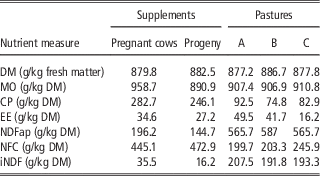
Composition of supplements and pastures are shown in Table 1. Percentage composition of supplements was the ground corn grain (26.25%), wheat meal (26.25%) and soya bean meal (47.5%) for cows. A total of 150 kg of supplement per animal were provided during the total experiment period for each treatment (1 and 1.5 kg/day to cows in the MID and LATE groups, respectively), accompanied by a mineral mixture offered ad libitum (Table 1). The control group, the UNS cows, received only a mineral mixture ad libitum during gestation.
Nutrient content of dietary supplements and pastures at times A, B, C offered to cows during gestation and your progeny

A=manual grazing simulation, early-mid gestation; B=manual grazing simulation, late gestation; C=manual grazing simulation, growing period; DM=dry matter; MO=matter organic; EE=ether extract; NDFap=NDF corrected to ash and protein; NFC=non-fibrous carbohydrates; iNDF=indigestible NDF.
Mineral mix – CaHPO4=50.00%; NaCl=47.15%; ZnSO4=1.50%; Cu2SO4=0.75%; CoSO4=0.05%; KIO3=0.05% and MnSO4=0.05%.
After birth, all progeny were kept at the same conditions and supplemented for 8 months until weaning, with the same supplement in equal amounts. Percentage composition of supplements was the ground corn grain (30%), wheat meal (30%), soya bean meal (35%) and molasses (5%). The supplement was offered in an amount of 6 g/kg of the live weight, and the mineral mixture was offered ad libitum. The experiment was carried out for 16 months, corresponding to the last 8 months of gestation (after pregnancy diagnosis, 30 days after insemination) and 8 months of growth of the progeny.
Experimental procedures and sampling
Cows were weighed after 14 h of fasting at the beginning (30 days of gestation) and at the end of the experiment (immediately after birth of calves) to determine their average daily gain (ADG). Calves were weighed immediately after birth and at the end of the suckling period (after 14 h of fasting) and every 30 days calves were weighed (without fasting) for performance monitoring and adjustment of the supplement offered.
In order to minimize possible paddock effects, all animals were rotated every 7 days; all groups stayed the same period in each paddock. Pastures samples were collected via manual grazing simulation, every 15 days, dried in an forced-air circulation oven (Ar SL – 102; SOLAB®, Piracicaba, São Paulo, Brasil) at 55°C to 60°C for 72 h and then grounded with 1 and 2 mm knife mill type Willye (TE-680, SOLAB®).
To evaluate the nutritional characteristics of diet, a digestibility trial was performed for 9 days at both 100 and 230 days of gestation as well as 120 days post calving. The three-marker method was used as follows: chromic oxide (Cr2O3) was used estimate the faecal excretion of animals, which was wrapped in paper cartridges in an amount of 20 g/animal per day for cows and 10 g/animal per day for calves and administered with a metal probe via the oesophagus at 10 h (Detmann et al., Reference Detmann, Paulino, Zervoudakis, Valadares Filho, Lana and Queiroz2001a). Titanium dioxide (TiO2) was used to estimate the individual supplement intake, provided via supplement at the proportion of 10 g/kg of supplement (Titgemeyer et al., Reference Titgemeyer, Armendariz, Bindel, Greenwood and Loest2001). Indigestible NDF (iNDF) was used to estimate the pasture dry matter (DM) intake (Detmann et al., Reference Detmann, Paulino, Zervoudakis, Valadares Filho, Euclydes, Lana and Queiroz2001b). The first 5 days of each trial were used for animal adaptation to TiO2 and Cr2O3. Faecal samples were collected immediately after defecation or directly from the rectum of the animals in the last 4 days of the digestibility period (one sample for day) at the times 1800, 1400, 1000 and 0600 h. After collection, faeces were dried in a forced-air circulation oven (Ar SL – 102; SOLAB®) at 60°C for 72 h and were grounded with 1 and 2 mm knife mill type Willye (TE-680; SOLAB®). A composite sample was prepared for each animal and stored for subsequent analysis.
Chemical analysis
Dry matter content, CP, ether extract (EE), NDF corrected to ash and protein (NDFap) and iNDF were quantified in supplement, pasture and faecal samples processed as mentioned above. Measurements of DM, CP and EE were performed according to Association of Official Analytical Chemists (Association of Official Analytical Chemists and Helrich, Reference Helrich1990); NDFap, according to Mertens (Reference Mertens2002), using thermostable α-amylase, without using sodium sulphite; insoluble NDF containing neutral detergent insoluble nitrogen (NDIN) was quantified by following the recommendations of Van Soest and Robertson (Reference Van Soest and Robertson1985), with ash corrections; iNDF, according to Valente et al., (Reference Valente, Detmann, Queiroz, Valadares, Gomes and Figueiras2011), obtained after in situ incubation in F57 Ankom® bags (Ankom, Macedon, NY, USA) for 288 h. In faecal samples, Cr2O3 was determined by atomic absorption spectrophotometry (Williams et al., Reference Williams, David and Iismaa1962) and TiO2 by colorimetric determination (Titgemeyer et al., Reference Titgemeyer, Armendariz, Bindel, Greenwood and Loest2001).
Faecal DM excretion was estimated based on the amount of indicator ingested and its concentration in the faeces (Lopes et al., Reference Lopes, Paulino, Detmann, de Campos, Valente, Barros, Cardenas, Almeida, Martins and Silva2014):
 $${\rm Faecal}\,{\rm DM}\,({\rm kg/day})\,{\equals}\,{\rm AOI}/{\rm ICF}$$
where AOI is the amount of indicator ingested (g) and ICF the indicator concentration in faeces (g/kg of faeces).
Estimates of individual supplement intake were obtained as follows:
 $${\rm ISI}\,{\equals}\,\left[ {\left( {{\rm FE}{\times}{\rm ICaF}} \right)/{\rm IOG}} \right]{\times}{\rm SOG}$$
where ISI is the individual supplement intake (kg/day); FE the faecal excretion (kg/day); ICaF the indicator concentration in animal faeces (kg/kg); IOG the indicator present in the supplement offered to each group (kg/day); and SOG the supplement amount offered to the group of animals or treatment (kg/day).
Estimation of voluntary DM intake of forage (DMIF) was performed using iNDF, as the internal marker, as follows:
 $${\rm DMIF}\,({\rm kg/day})\,{\equals}\,\left( {{\rm FE}{\times}{\rm FIC}} \right)/{\rm FOIC}$$
where FIC is the indicator concentration in the faeces (kg/kg); FOIC the indicator concentration in forage (kg/kg); and FE the faecal extraction (kg/day).
Total DM intake was obtained by the sum of cows concentrate and forage consumption.
To estimate the DM, CP and EE of milk consumed by calves, three milk collections were made with a mechanical milking machine (Milkban MB 300-1) at 30, 75 and 135 days of the suckling phase. Before milking, oxytocin (Forte UCB, 2.5 IU, 0.5 ml/animal) was applied to facilitate the milk descent. For calves, the milk DM intake was also considered in addition to forage and concentrate to quantify total DM intake.
Histological analyses
At 30 days after birth, biopsies of skeletal muscle tissue from calves were collected to obtain samples of longissimus lumborum (LL) muscle (Arrigoni et al., Reference Arrigoni, Júnior, Dias, Ludovico, Cervieri, Silveira, Oliveira and Chardulo2004). A 2 cm skeletal muscle samples were immediately fixed in 4% paraformaldehyde (pH=7.03) and incubated at room temperature for 24 h. After incubation, samples were dehydrated in an increasing concentration series of ethanol solutions (70%, 80%, 90% and 100%) for 2 h, diaphonized in xylene for 1 h and embedded in paraffin. Sections of 4 μm were stained with haematoxylin–eosin (Timm, Reference Timm2005). A total of 30 images (magnification=10×, scale bar=20 µm, image’s resolution=1600×1200 pixels) were obtained from each animal using an Olympus BX50 microscope with attached camera CMOS 1.3 MP BioCAM (Takachiho, Miyazaki, Japan). Muscle cell number and size were analysed with ImageJ software (Schindelin et al., Reference Schindelin, Rueden, Hiner and Eliceiri2015).
Gene expression
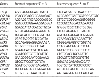
Total RNA was extracted from 500 mg of muscle tissue biopsies (n=9) with Trizol® reagent (Invitrogen, Carlsbad, CA, USA) according to the manufacturer instructions. To remove contamination by genomic DNA, RNA samples were treated with DNase-I Amplification Grade (Invitrogen). Complementary DNA (cDNA) synthesis was performed from 1 µg of total RNA using the GoScript Reverse Transcription System (Promega, Madison, WI, USA). The mRNA expression levels of markers for fibrogenesis (transforming growth factor β (TGF-β), collagen type I, α 1 (COL1A1), collagen type III, α 3 (COL3A3)), adipogenesis (peroxysome proliferator-activated receptor γ (PPARγ), zinc finger protein 423 (ZFP423), CCAAT enhancer binding protein α (C/EBPα)), muscle hypertrophy (fibroblast growth factor 2, receptor 1 (FGF2R1), FGF2) and energy metabolism (PPARα, medium-chain acyl-CoA dehydrogenase (MCAD), uncoupling protein 3 (UCP3), protein kinase AMP-activated catalytic subunit α 2 (PRKAA2), hydroxyacyl-CoA dehydrogenase (HADH), myosin heavy chain 7 (MYH7), pyruvate dehydrogenase kinase 4 (PDK4), peroxisome proliferator-activated receptor γ coactivator 1 α (PGC1α), carnitine palmitoyltransferase 1 (CPT1)) were determined by the SYBR Green RT-PCR kit from Bio-Rad using a 7300 Real-Time PCR System (Applied Biosystems, Foster City, CA, USA). We have not evaluated the mRNA expression for myogenesis markers because formation of skeletal muscle fibres in bovine occurs prenatally (Bonnet et al., Reference Bonnet, Cassar, Chilliard and Picard2010; Du et al., Reference Du, Tong, Zhao, Underwood, Zhu, Ford and Nathanielsz2010). Thus, we have only evaluated the effects of maternal nutrition on myogenesis through histological analysis. The endogenous gene used was 18S and the sequence primers are shown in Table 2. Polymerase chain reaction reactions were performed at 95°C for 3 min and subjected to 40 cycles of 95°C for 10 s, and 60°C for 30 s, followed by melting curve (0.01°C/s). Results are expressed by ΔΔCt method (Bustin, Reference Bustin2002; Nolan et al., Reference Nolan, Hands and Bustin2006; Duarte et al., Reference Duarte, Bueno, Silva, Campos, Gionbelli, Guimarães, Silva, Lopes, Hausman and Dodson2013; Martins et al., Reference Martins, Sanglard, Silva, Chizzotti, Rennó, Serão, Silva, Guimarães, Ladeira, Dodson, Du and Duarte2015).
Primer sequence for amplifying mRNA transcripts of genes of interest in longissimus muscle tissue of the progeny of cows supplemented during pregnancy

TGFβ=transforming growth factor, β; COL1A1=collagen type I, α 1; FGF2R1=fibroblast growth factor 2, receptor 1; COL3A3=collagen type III, α 3; FGF2=fibroblast growth factor 2; PPARα, PPARγ=peroxysome proliferator-activated receptor, α/γ; MCAD=medium-chain acyl-CoA dehydrogenase; UCP3=uncoupling protein 3; HADH=hydroxyacyl-CoA dehydrogenase; MYH7=myosin heavy chain 7; PDK4=pyruvate dehydrogenase kinase 4; PGC1α=peroxisome proliferator-activated receptor γ coactivator 1 α; CPT1=carnitine palmitoyltransferase 1; ZFP423=zinc finger protein 423; C/EBPα=CCAAT enhancer binding protein, α.
Evaluation of carcass traits by ultrasonography
In the final measurement in the suckling phase, carcass traits of the calves were evaluated by ultrasound (model: SSD 500v, with linear probe of 18 cm; Aloka). Carcass images were obtained from the right side of the animal where images for ribeye area measurement was taken from transversal section of the LL muscle (between the T12 and T13 thoracic vertebrae); subcutaneous fat thickness, from an average of two measurements (the first from the same place where the image for ribeye area was taken and the second at the pelvic region, between the ischium and pubis. Images were analysed in the BioSoft Toolbox® II for Beef software (Biotronics Inc., Ames, IA, USA).
Statistical analysis
The PROC MIXED procedure of SAS software (version 9.0) was used for all statistical analyses. For all statistical procedures we adopted α=0.10 of probability for type I error, due the high incidence of type-II error in this kind of study. Means were submitted to ANOVA and regression, and UNS, MID and LATE periods of supplementation were compared. Sex of the foetus was tested as fixed effect and removed from the model as it was not significant. Initial BW was used as a co-variable when significant and removed from the model when not significant. The variables on the effect of different supplementation (treatments) on the measured parameters were analysed in a completely randomized design.
Results
Performance
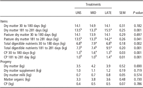
A greater intake of total digestible nutrients (TDN) and protein (CP) was observed in cows from MID group (P=0.002) in the 30- to 180-day period of gestation, when compared with those from LATE and UNS treatments (Table 3). Cows from LATE group presented greater consumption of DM, pasture DM, TDN and CP (P<0.1), at the 181- to 281-day period of gestation (Table 3). Cow supplementation did not affect the consumption (P>0.1) in calves during the suckling phase (Table 3).
Dry matter and nutrient intake of dietary treatments offered to cows during gestation and their progeny

UNS=cows unsupplemented during the gestation; MID=cows supplemented during 30 to 180 days of gestation; LATE=cows supplemented during 181 to 281 days of gestation.
a,bValues within a row with different superscript letters differ significantly at P<0.1.
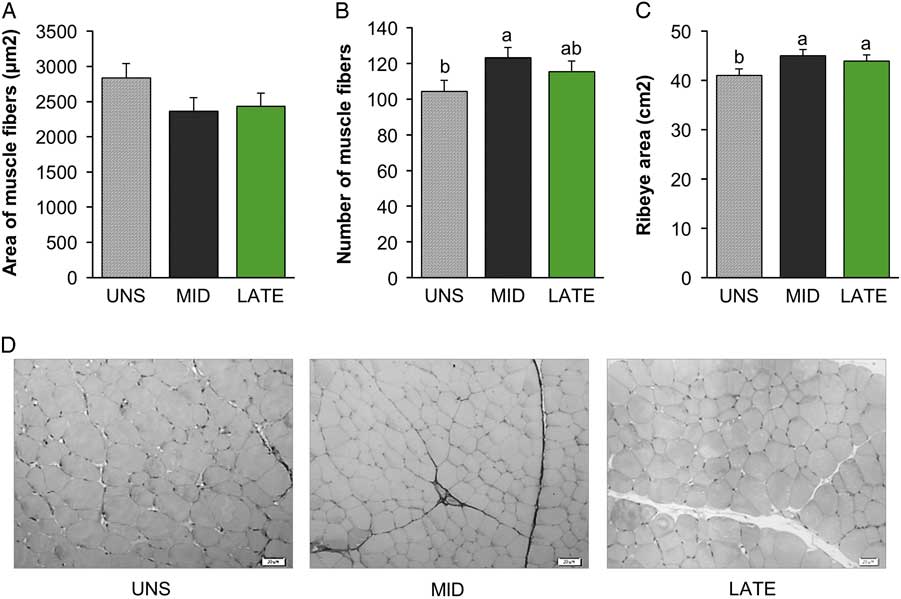
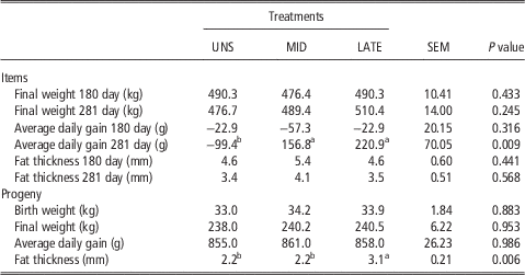
In cows, no difference were observed in the daily weight gain (P=0.316) among MID, LATE and UNS, during the 30- to 180-day period of gestation. The ADG was lower in the UNS group, at 181- to 281-day period of gestation (P=0.009). Supplementation during gestation did not influence the birth weight (P=0.883), final weight (P=0.953), ADG (P=0.986) and muscle fibre size (P=0.208) in calves. Higher values of subcutaneous fat thickness were observed in the progeny of LATE, also, higher values of ribeye area were observed in calves born from cows of LATE and MID groups, respectively (Table 4 and Figure 1A and C). Muscle fibre number was greater in calves born from cows of MID compared with those born from cows of UNS group but was not different compared with LATE group (Figure 1B).
Effect of maternal nutrition during gestation on characteristics of histological muscle of progeny of cows supplemented or unsupplemented during the gestation. UNS cows (unsupplemented during the gestation), MID cows (supplemented during 30 to 180 days of gestation) and LATE cows (supplemented during 181 to 281 days of gestation). Samples were collected per biopsies of longissimus lumborum muscle. Bars represent means±SEM (n=9). a,bSignificant differences between the groups (P<0.1). There was no significant variation by sex effect. Cell number per reading area (10×).

Performance and body condition of cows during gestation and their progeny

UNS=cows unsupplemented during the gestation; MID=cows supplemented during 30 to 180 days of gestation; LATE=cows supplemented during 181 to 281 days of gestation.
a,bValues within a row with different superscript letters differ significantly at P<0.1.
Messenger RNA expression
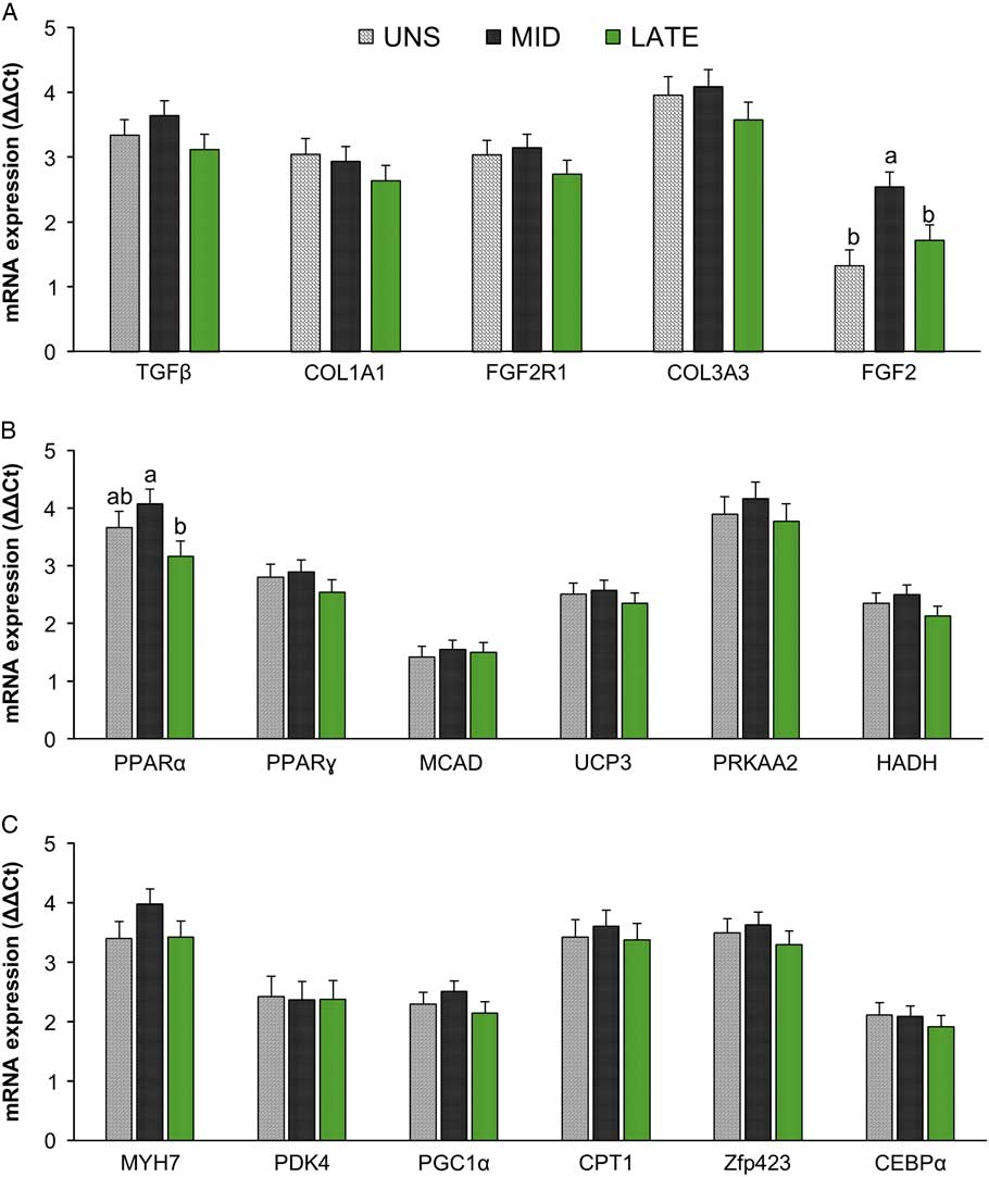
The progeny of MID group presented greater mRNA expression of FGF2 (P=0.003) and PPARα (P=0.073) genes. The mRNA expression of PPARα was decreased in skeletal muscle of calves from LATE group (Figure 2). Biomarkers associated with fibrogenic (TGFβ, FGF2R1, COL1A1, COL3A3), adipogenic (ZFP423, PPARγ, C/EBPα) and β oxidation (MCAD, UCP3, PRKAA2, HADH, MYH7, PDK4, PGC1α, CPT1) processes did not differ among the treatments (P>0.10) (Figure 2).
Real-time PCR analysis of the influence of maternal nutrition during gestation on characteristics muscle of progeny of cows supplemented or unsupplemented during the gestation. UNS cows (unsupplemented during gestation), MID cows (supplemented 30 to 180 days of gestation) and LATE cows (supplemented 181 to 281 days of gestation). Samples were collected per biopsies of longissimus lumborum muscle. Bars represent means±SEM (n=9). a,bSignificant differences between the groups (P<0.1). There was no significant variation by sex effect. TGFβ=transforming growth factor β; COL1A1=collagen type I, α 1; FGF2R1=fibroblast growth factor 2, receptor 1; COL3A3=collagen type III, α 3; PPARα=peroxysome proliferator-activated receptor α; MCAD=medium-chain acyl-CoA dehydrogenase; UCP3=uncoupling protein 3; HADH=hydroxyacyl-CoA dehydrogenase; MYH7=myosin heavy chain 7; PDK4=pyruvate dehydrogenase kinase 4; PGC1α=peroxisome proliferator-activated receptor γ coactivator 1 α; CPT1=carnitine palmitoyltransferase 1; ZFP423=zinc finger protein 423; C/EBPα=CCAAT enhancer binding protein α.

Discussion
It is well known that maternal nutrition during gestation affects the foetuses skeletal muscle development and thus impacts the postnatal development of the progeny (Du et al., Reference Du, Tong, Zhao, Underwood, Zhu, Ford and Nathanielsz2010). Among several ways that maternal nutrition may affects foetuses skeletal muscle development is by changing the mesenchymal stem cell commitment into different cell types that composes skeletal muscle (Du et al., Reference Du, Huang, Das, Yang, Duarte, Dodson and Zhu2013; Duarte et al., Reference Duarte, Paulino, Das, Wei, Serao, Fu, Harris, Dodson and Du2014). Among these types of cells are myocytes, adipocytes and fibroblasts, which are all derived from the same pool of mesenchymal stem cells and are the cells that contribute not only on muscle mass but also on marbling and collagen deposition. Thus, in the present study we investigated not only if maternal supplementation would affect the animal performance but also if it would change the number of muscle fibres and the transcription profile of adipogenesis, fibrogenesis and energy metabolism markers in skeletal muscle of the offspring.
With regard to myogenesis, our data revealed that maternal supplementation during mid-gestation increased the number of myofibres in skeletal muscle of the offspring when compared with calves born from dams that were not supplemented during gestation. However, the number of myofibres in skeletal muscle of calves born from dams supplemented at late gestation did not differ from those born from non-supplemented cows. As reported by Bonnet et al. (Reference Bonnet, Cassar, Chilliard and Picard2010) the total number of muscle fibres is set by the end of the second trimester of gestation. Therefore, a greater availability of nutrients due to maternal supplementation at mid-gestation may led to a greater number of myofibres in skeletal muscle of calves. On the other hand, it has been suggested that during the last trimester of gestation is when foetal skeletal muscle mass increase mainly due to muscle fibres hypertrophy (Du et al., Reference Du, Tong, Zhao, Underwood, Zhu, Ford and Nathanielsz2010). Thus, our results indicate that supplementation at late gestation may not substantially contributed to increase myogenesis in foetal skeletal muscle, which may explain the similarity of number of myofibers in skeletal muscle of calves from LATE and UNS groups. Collectively, these data indicates that supplementation at mid-gestation may be more effective to increase the commitment of mesenchymal stem cells into myogenesis as well the proliferation of myogenic cells, allowing the formation of more secondary muscle fibres, leading to a greater number of myofibres at birth.
Our data revealed that supplemental feed for grazing beef cows during gestation did not influence the progeny birth weight, weaning weight and the ADG. However, despite the lack of differences in BW gain, maternal supplementation during gestation did change the carcass composition of the calves. A greater ribeye area was observed in calves from cows that were supplemented at both mid and late gestation compared with those born from non-supplemented cows. This difference in ribeye area may be a consequence of an increase in both hyperplasia of skeletal muscle myofibers of calves born from supplemented dams. It should be mentioned that no differences were observed in nutrient intake by calves between treatments, which strongly suggest that differences observed in calves were likely due to changes occurred in skeletal muscle development during gestation.
As hyperplasia of skeletal muscle cells does not occur after birth, we evaluated the mRNA expression of FGF2 as a marker for skeletal muscle hypertrophy. It has been shown that FGF2 is up-regulated during skeletal muscle hypertrophy being predominantly localized to the myofibers during skeletal muscle hypertrophy (Mitchell et al., Reference Mitchell, Steenstrup and Hannon1999). The main role of FGF2 during postnatal skeletal muscle cell development is to stimulate myoblast (satellite cells) proliferation while inhibits its differentiation (Allen and Rankin, Reference Allen and Rankin1990; Mitchell et al., Reference Mitchell, Steenstrup and Hannon1999). We observed a greater mRNA expression of FGF2 in skeletal muscle of calves born from dams that were supplemented at mid-gestation compared with the other treatments. Previous study has demonstrated that proliferation capacity of skeletal muscle satellite cells in lambs’ offspring is affected by maternal nutrition during gestation (Raja et al., Reference Raja, Hoffman, Govoni, Zinn and Reed2016). Collectively, our results suggest that maternal supplementation during mid-gestation leads not only to an increased number of myofibres but also stimulate the proliferation capacity of satellite cells by increasing FGF2 expression, which may contribute to an increase in skeletal muscle deposition later in life.
It has been shown that maternal overnutrition increases the mRNA expression of adipogenesis markers in foetal skeletal muscle of beef cattle (Duarte et al., Reference Duarte, Paulino, Das, Wei, Serao, Fu, Harris, Dodson and Du2014) and fibrogenesis in skeletal muscle of ovine offspring (Huang et al., Reference Huang, Zhao, Yan, Zhu, Long, McCormick, Ford, Nathanielsz and Du2012). Therefore, we hypothesized that maternal supplementation at late gestation would shift the commitment the mesenchymal stem cells towards to adipogenesis and fibrogenesis as both adipocytes and fibroblasts share common immediate common progenitor cells (Uezumi et al., Reference Uezumi, Ito, Morikawa, Shimizu, Yoneda, Segawa, Yamaguchi, Ogawa, Matev, Miyagoe, Takeda, Tsujikawa, Tsuchida, Yamamoto and Fukada2011). However, no differences were observed for mRNA expression of any of adipogenic and fibrogenic markers evaluated in this study. Although the number of intramuscular adipocytes and the amount of connective tissue was not evaluated, the mRNA expression data indicates that maternal supplementation did not affect the transcriptional level of the main markers of adipogenesis and fibrogenesis. It is noteworthy that although cows were supplemented, it does not characterize overnutrition of the dams as was described by previous studies. Thus, our data suggests that to enhance intramuscular fat deposition in skeletal muscle of the offspring through maternal nutrition during gestation it would be necessary a higher energy or global nutrient intake by the dams during pregnancy, which warranty further studies using grazing beef cows as a model.
Energy metabolism of skeletal muscle of the offspring is also affected by maternal nutrition during gestation (Zhu et al., Reference Zhu, Ford, Nathanielsz and Du2006). These adaptive changes that occurs in skeletal muscle of the offspring due to maternal nutrition includes decreased glucose transport and ATP production (Selak et al., Reference Selak, Storey, Peterside and Simmons2003) and impairment of transcriptional metabolic flexibility of skeletal muscle, which means the decrease of skeletal muscle capacity to change the source for ATP synthesis. Thus, in certain cases maternal nutrition may programme skeletal muscle of the offspring for a preferable use of fatty acid instead the use of carbohydrates as a source to produce ATP, even when high levels of glucose are available for the skeletal muscle (Aragão et al., Reference Aragão, Guzman, Perez, Manhaes and Bolanos2014). Consequently, this scenario would lead to a lower marbling deposition in beef animals even when they are fed high-energy diets.
In the present study we did not observe differences in mRNA expression of energy metabolism markers in the skeletal muscle of the offspring, with exception of PPARα.
The PPARα gene regulates the mitochondrial biosynthesis of muscle through adipose triglyceride lipase expression. This enzyme is responsible for fatty acid oxidation at the mitochondrial level, providing energy primarily (in the ATP form) for muscle metabolism (Biswas et al., Reference Biswas, Ghosh, Kumar and Chakrabarti2016). Biosynthesis of muscle fibres requires a major input of energy for metabolism and hyperplasic growing of muscle. In this context, the PPARα expression observed in this study can be associated to increase of muscle fibre number. Thus, the greater expression of PPARα in skeletal muscle of calves born from damns supplemented at mid-gestation may be due to the greater number of myofibres presented by these animals. As a biological role of intramuscular fat is to provide readily carbon source for oxidation by muscle cells (Duarte et al., Reference Duarte, Gionbelli, Paulino, Serão, Nascimento, Botelho, Martins, Filho, Dodson, Guimarães and Du2017), the greater number of myofibres would decrease the lipid storage by intramuscular adipocytes in these animals, leading to a decrease in intramuscular fat deposition. The lower mRNA expression of PPARα in skeletal muscle may also explain the greater subcutaneous fat deposition in calves from the LATE group, as this might suggest a lower energy demand by the skeletal muscle, meaning more of the energy is partitioned into fat depots.
Additionally, it must be noted that the main focus of the present study was the skeletal muscle development and the cellularity of other fat depots was not investigated. Thus, as hypothesized for intramuscular fat depot, maternal supplementation at late gestation may also increase adipogenesis in subcutaneous fat depot, leading to a greater carcass back fat in the offspring, which need further investigation.
Conclusion
Supplementation of grazing beef cows during gestation promotes additional gain of skeletal muscle fibres number and ribeye area of the progeny but does not seem to improve intramuscular adipogenesis and/or fibrogenesis, which might require a greater energy or global nutrient intake. Calves born from damns supplemented during mid-gestation had increased mRNA expression of FGF2, which may indicate a higher hypertrophy ability of skeletal muscle of the offspring. On the other hand, calves born from dams supplemented at late gestation had greater subcutaneous fat deposition. Further studies are needed to investigate not only when but also the level of maternal supplementation at different stages of gestation to develop a precise maternal plane of nutrition to improve performance of the offspring.
Acknowledgements
This research was funded by Fundação de Amparo à Pesquisa do Estado de Minas Gerais (FAPEMIG, grant no. PPM-00366-15), Instituto Nacional de Ciência e Tecnologia – Ciência Animal (INCT-CA).