Non-technical Summary
Terebratulides, a major group of brachiopods in today’s oceans, are divided into two main subgroups distinguished by the shape of the loop, an internal structure supporting the lophophore. The fossil family Angustothyrididae is particularly intriguing because it mixes features from both subgroups, potentially representing a link in their evolution. Our study of Triassic fossils from Hungary and China, previously classified under the genus Angustothyris, reveals that they actually belong to several different taxa, allowing establishment of two new genera and one new species. Furthermore, morphological evidence from fossils conflicts with the evolutionary timeline suggested by genetic studies of living terebratulides. This mismatch indicates that the current understanding of how these brachiopods evolved and are classified might need a significant revision.
Introduction
The order Terebratulida Waagen, Reference Waagen1883 represents the most diverse group of brachiopods occurring during the Mesozoic–Cenozoic (Curry and Brunton, Reference Curry, Brunton and Selden2007; Carlson, Reference Carlson2016; Guo et al., Reference Guo, Benton, Stubbs and Chen2024). A fundamental division within this order separates it into two major suborders: the Terebratulidina Waagen, Reference Waagen1883, typically characterized by a short loop, and the Terebratellidina Muir-Wood, Reference Muir-Wood1955, distinguished by a long loop. Understanding the evolutionary transition between these groups is a central question in brachiopod paleontology. Fossil evidence and morphological comparisons have long suggested that the Terebratellidina evolved from terebratulidine ancestors during the Permian–Triassic transition, with the family Angustothyrididae Dagys, Reference Dagys1972 identified as a likely candidate for this evolutionary link (Lee et al., Reference Lee, MacKinnon, Smirnova, Baker, Jin, Sun and Kaesler2006). This hypothesis is supported by the cardinalia and loop ontogeny of the family, which displays a combination of features characteristic of both suborders.
Although the importance of the Angustothyrididae in the phylogeny and evolution of terebratulides has been recognized for decades (Dagys, Reference Dagys1972, Reference Dagys1974; Smirnova, Reference Smirnova1984, Reference Smirnova2008; Lee et al., Reference Lee, MacKinnon, Smirnova, Baker, Jin, Sun and Kaesler2006), the taxonomy of its type genus, Angustothyris Dagys, Reference Dagys1972, remains problematic and poorly resolved. This genus has been widely reported from the Middle Triassic at numerous localities across both the western and eastern parts of the Tethys Ocean, with nearly all occurrences assigned to a single species, Angustothyris angustaeformis (Böckh, Reference Böckh1872). However, the external morphology of these specimens is highly variable, raising doubts about the monophyly of the genus. More importantly, the internal structures of Angustothyris angustaeformis from its type locality, which are essential for terebratulide classification, are inadequately documented. Preliminary studies suggested that the internal morphology of specimens assigned to Angustothyris differs significantly (Dagys, Reference Dagys1972; Pálfy, Reference Pálfy2003; Guo et al., Reference Guo, Chen and Harper2019). This taxonomic ambiguity directly undermines the reliability of using Angustothyris as a stable reference point for evolutionary studies, casting doubt on the evolutionary scenarios that it is meant to illustrate.
To address this critical taxonomic issue, we conducted a comprehensive re-examination of ‘Angustothyris’ specimens from key Middle Triassic localities in Hungary and southwestern China. This study incorporates material from the type locality (Köveskál, Hungary) of Waldheimia angustaeformis Böckh, Reference Böckh1872, the species subsequently designated as the type species of Angustothyris by Dagys (Reference Dagys1972), thereby providing an essential baseline for comparison. Our primary objectives were to: (1) clarify the diagnostic characteristics and taxonomic composition of Angustothyris; (2) describe new taxa revealed by our analysis of internal and external morphologies; and (3) discuss the implications of our revised taxonomy for understanding the evolution and phylogenetic relationships of the Terebratulida.
Materials and methods
The specimens analyzed in this study originate from three key regions: southwestern China, Hungary, and northwestern Italy, all within the Tethyan realm.
Specimens of Angustothyris qingyanensis Guo, Chen, and Harper, Reference Guo, Chen and Harper2019 were collected from the Leidapo Member of the Qingyan Formation at the Leidapo and Wachangpo localities near Qingyan, Guizhou, southwestern China. The fossil-bearing strata, dated to the late middle to early late Anisian (late Pelsonian–early Illyrian; Stiller and Bucher Reference Stiller and Bucher2008), are renowned for hosting one of the world’s most diverse Anisian brachiopod faunas (Guo et al., Reference Guo, Chen and Harper2019, Reference Guo, Chen, Huang, Chen, Qiu and Guo2022). Detailed descriptions of the strata and fossil localities are available in previous studies (Stiller and Bucher, Reference Stiller and Bucher2008; Guo et al., Reference Guo, Chen and Harper2019).
Four specimens from the Balaton Uplands in western Hungary, previously assigned to Angustothyris angustaeformis by Pálfy (Reference Pálfy2003), were re-examined. These were originally collected from the Felsőörs Formation of Pelsonian (Anisian) age, with two specimens from the type locality at Köveskál and two from the nearby locality at Aszófő (for stratigraphic details, see Vörös et al., Reference Vörös, Budai, Kovács, Piros and Szabó2003).
We also re-examined the published serial sections of specimens assigned to Angustothyris by Torti and Angiolini (Reference Torti and Angiolini1997), including Angustothyris angustaeformis, Angustothyris sp. aff. A. raxana (Bittner, Reference Bittner1892), and Angustothyris sp. aff. A. ladina (Bittner, Reference Bittner1890). These specimens were collected from the lower Ladinian Esino Limestone in Val Parina, Lombardy, northwestern Italy.
Brachiopod specimens were photographed after coating with ammonium chloride to enhance surface detail. Serial sections were made to investigate their internal characters. All sections were oriented perpendicular to the commissure plane, starting at the posterior ends of the ventral or dorsal umbones. The resulting sections were photographed under a microscope and their outlines traced using drawing software.
Repositories and institutional abbreviations
BGEG, State Key Laboratory of Biogeological and Environmental Geology (now State Key Laboratory of Geomicrobiology and Environmental Changes), China University of Geosciences (Wuhan), China; MTM, Department of Paleontology and Geology, Hungarian Natural History Museum, Hungary; SZTFH, Department of Collections, Geological Survey, Supervisory Authority for Regulatory Affairs, Hungary.
Systematic paleontology
Order Terebratulida Waagen, Reference Waagen1883
Suborder Terebratulidina Waagen, Reference Waagen1883
Superfamily Dielasmatoidea Schuchert, Reference Schuchert and von Zittel1913
Family Angustothyrididae Dagys, Reference Dagys1972
Revised diagnosis
Small to large, biconvex or planoconvex, smooth or plicate anteriorly; foramen mesothyrid or permesothyrid; dental plates absent; hinge plates present; adult loop deltiform or teloform; transverse band derived from secondary elements.
Remarks
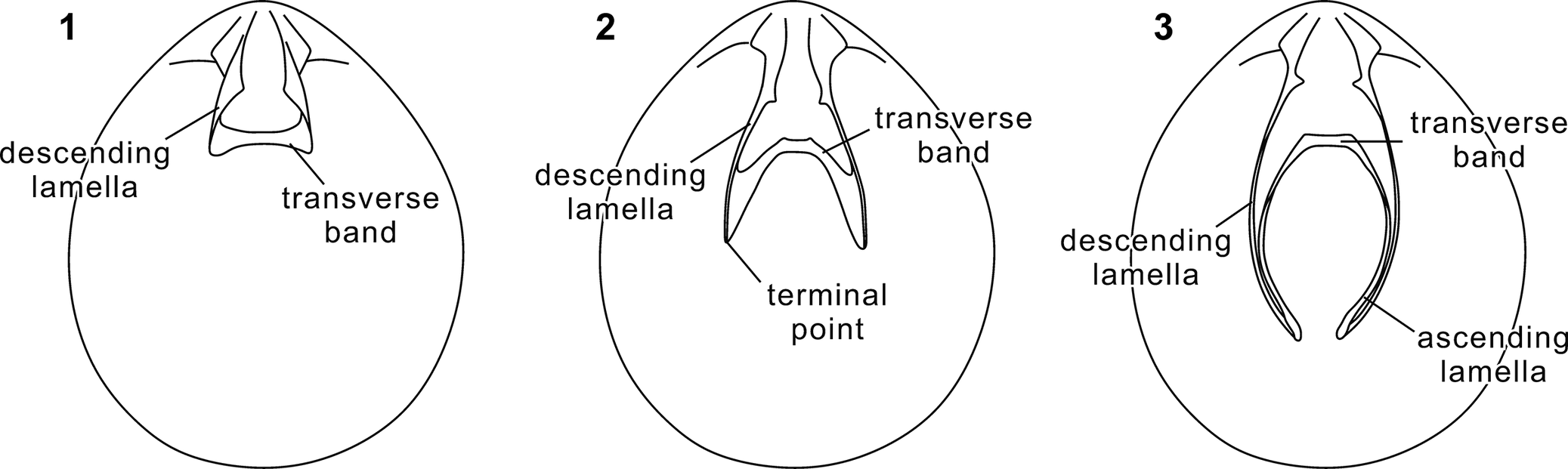
As reflected in the original family diagnosis (Dagys, Reference Dagys1972; also Lee et al., Reference Lee, MacKinnon, Smirnova, Baker, Jin, Sun and Kaesler2006), all genera previously assigned to this family are characterized by deltiform loops (or earlier phases) or long-flanged deltiform loops (Fig. 1) and lack clearly differentiated ascending lamellae in adult shells. The present study reveals the presence of a long teloform loop in Qianothyris new genus (see below; Fig. 1), resembling those of members of the superfamily Zeillerioidea Allan, Reference Allan1940. Owing to its close morphological affinity with other angustothyridid genera, particularly Angustothyris, Qianothyris n. gen. is herein regarded as a member of the Angustothyrididae, and the diagnosis of the family is accordingly expanded.
Three representative loop morphologies of terebratulides. (1) Deltiform loop, typical of Paleozoic–Cenozoic short-looped terebratulides. The transverse band connects directly to the descending lamellae, and ascending lamellae are absent. (2) Long-flanged deltiform loop, typical of Mesozoic Loboidothyridoidea Makridin, Reference Makridin1964 and also present in Angustothyris Dagys, Reference Dagys1972. This morphology differs from typical deltiform loop in having anteriorly extended terminal points beyond the transverse band (i.e., long flanges). Separate ascending lamellae are not developed. (3) Teloform loop, typical of Paleozoic–Cenozoic long-looped terebratulides. The transverse band laterally connects with ascending lamellae, which are clearly differentiated from the descending lamellae.

Figure 1. Long description
A three-panel diagram showing anatomical variations in brachiopod loops within oval shell outlines.
Panel 1 shows a deltiform loop. At the top hinge, two descending lamellae extend downward and connect directly to a horizontal transverse band. The structure is short and lacks additional branches.
Panel 2 shows a long-flanged deltiform loop. Similar to the first, descending lamellae extend from the hinge to a transverse band. However, the structure continues past the band, ending in two sharp terminal points that extend further toward the bottom of the shell.
Panel 3 shows a teloform loop. Long descending lamellae curve downward along the sides of the shell. They curve back upward to form ascending lamellae, which then connect to a transverse band located higher up, creating a complex, elongated circular loop structure.
The loop morphology of Waldheimia angustaeformis (formerly the type species of Angustothyris, here re-assigned to Balatonithyris new genus) remains uncertain, but its long loop with slender descending lamellae suggests a teloform phase. Whether other taxa possess a teloform loop is difficult to determine, because loop structures have not been documented for most occurrences. Moreover, current knowledge of loop ontogeny within this family remains fragmentary. Dagys (Reference Dagys1972) restricted the family to taxa with complex loop development, yet such development is documented for only a few taxa. Rhaetina Waagen, Reference Waagen1882 appears to exhibit a simple loop development (Dagys, Reference Dagys1958, Reference Dagys1963), but was nevertheless included in this family by Lee et al. (Reference Lee, MacKinnon, Smirnova, Baker, Jin, Sun and Kaesler2006). Given the lack of more comprehensive studies, we refrain from revising the assignment of Rhaetina and related taxa here, and instead follow Lee et al. (Reference Lee, MacKinnon, Smirnova, Baker, Jin, Sun and Kaesler2006). A deeper understanding of the evolutionary history of this family will require further investigation, particularly across the Permian to Lower Triassic interval, for which the fossil record is sparse.
Genus Angustothyris Dagys, Reference Dagys1972
Type species
Angustothyris dagysi Guo, Chen, and Harper, Reference Guo, Chen and Harper2019 from the Anisian of northwestern Caucasus, fixed as the type species by Guo et al. (Reference Guo, Chen and Harper2019) under Article 70.3.2 of the International Code of Zoological Nomenclature (ICZN, 1999).
Diagnosis
Shell small, ventribiconvex, unisulcate; beak erect, ridges rounded, foramen mesothyrid; pedicle collar distinct, dental plates rudimentary or absent; cardinal process absent, inner socket ridges very high, outer hinge plates absent, inner hinge plates fused with median septum to form septalium; loop long-flanged deltiform, approximately half of dorsal valve length.
Occurrence
Anisian of northwestern Caucasus and Aszófő, Balaton Uplands, Hungary.
Remarks
The genus Angustothyris was established by Dagys (Reference Dagys1972), who designated Waldheimia angustaeformis Böckh, Reference Böckh1872 as its type species. The type locality of W. angustaeformis is Köveskál in the Balaton Uplands of Hungary (Böckh, Reference Böckh1872, Reference Böckh1873; Pálfy, Reference Pálfy2003). However, the specimens studied by Dagys (Reference Dagys1972) to erect Angustothyris were from the Anisian of northwestern Caucasus (Dagys, Reference Dagys1972). Subsequent re-examination of the Hungarian material by Pálfy (Reference Pálfy2003) revealed that the internal morphology of W. angustaeformis from Köveskál differs markedly from the Caucasian specimens, a finding confirmed by the present study. Consequently, the Caucasian specimens on which Dagys (Reference Dagys1972) erected the genus Angustothyris do not belong to the true W. angustaeformis originally described from Hungary.
Because Dagys (Reference Dagys1972) (1) examined the Caucasian material that does not belong to true Waldheimia angustaeformis, (2) believed that it represented W. angustaeformis, (3) explicitly designated that species as the type species, and (4) based the generic diagnosis on that material, the genus was actually established with a misidentified type species in the sense of Article 70.3 of the ICZN (1999). Insisting on strict adherence to the original type-species designation would therefore threaten the nomenclatural stability of Angustothyris (see questions raised by Pálfy, Reference Pálfy2003). Moreover, the misidentification was unintentional and does not represent a deliberate action. This case falls under Article 70.3 of the ICZN (1999), according to which, if an author discovers that a type species was misidentified, the author can select and thereby fix as type species, one that will in his or her judgment best serve stability and universality, either: Article 70.3.1, the nominal species previously cited as type species, or Article 70.3.2, the taxonomic species actually involved in the misidentification. To align with Dagys’s (Reference Dagys1972) original intent and to preserve the diagnosis that has been in use for decades, Guo et al. (Reference Guo, Chen and Harper2019) erected a new species Angustothyris dagysi for the Caucasian material and fixed it as the type species of Angustothyris under Article 70.3.2 (ICZN, 1999). Waldheimia angustaeformis from Köveskál, Hungary, is here assigned to a new genus, Balatonithyris n. gen. (see below).
Dagys (Reference Dagys1972) tentatively assigned to Angustothyris the following species: Terebratula aulacothyroidea Bittner, Reference Bittner1890, Aulacothyris incurvata Bittner, Reference Bittner1903, and Terebratula suspecta Bittner, Reference Bittner1903. Terebratula aulacothyroidea was later re-assigned to Sulcatothyris Dagys, Reference Dagys1974 by Dagys (Reference Dagys1974). Aulacothyris incurvata, although externally similar, possesses dental plates (Bittner, Reference Bittner1903). Terebratula suspecta possesses a median septum extending to the midlength of the dorsal valve and a short deltiform loop (Bittner, Reference Bittner1903), excluding it from Angustothyris. Given that many previously described specimens of ‘Angustothyris’ lack internal characters, their generic assignment remains uncertain. The only species exhibiting morphology comparable to that of the type species is Angustothyris aszofoensis new species from the Balaton Uplands in Hungary.
Angustothyris aszofoensis new species
Angustothyris aszofoensis n. sp. from the Anisian Felsőörs Formation of Aszófő, Hungary: (1–4) MTM M.92.234-1, dorsal, ventral, lateral, and anterior views, respectively; (5–8) MTM M.92.234-2, dorsal, ventral, lateral, and posterior views, respectively.
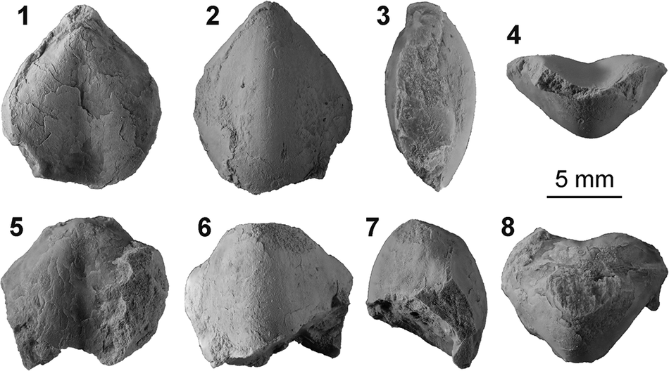
Figure 2. Long description
A multi-panel photo displaying two fossil specimens of the brachiopod Angustothyris aszofoensis.
The top row contains four views of specimen M T M M dot 92 dot 234-1:
1. Dorsal view showing a teardrop-shaped shell with a pointed beak.
2. Ventral view showing the opposite side of the shell.
3. Lateral view showing the side profile and the curvature of the valves.
4. Anterior view showing the front opening where the two valves meet.
The bottom row contains four views of specimen M T M M dot 92 dot 234-2:
5. Dorsal view showing a slightly more weathered or fragmented shell surface.
6. Ventral view.
7. Lateral view.
8. Posterior view showing the hinge line and beak area from the back.
A scale bar labeled 5 mm is positioned to the right of the first row, indicating the physical size of the specimens. The fossils are light gray with visible surface textures and some minor chipping at the edges.
Transverse serial sections of Angustothyris aszofoensis n. sp. (1) Sections based on MTM M.92.234-1 (12.74 mm long, 10.80 mm wide, 5.59 mm thick). Numbers above sections indicate distance (in mm) to the posterior end of ventral umbo. (2) Sections based on an incomplete specimen, MTM M.92.234-2. Numbers indicate distance (in mm) to the posterior end of dorsal umbo.
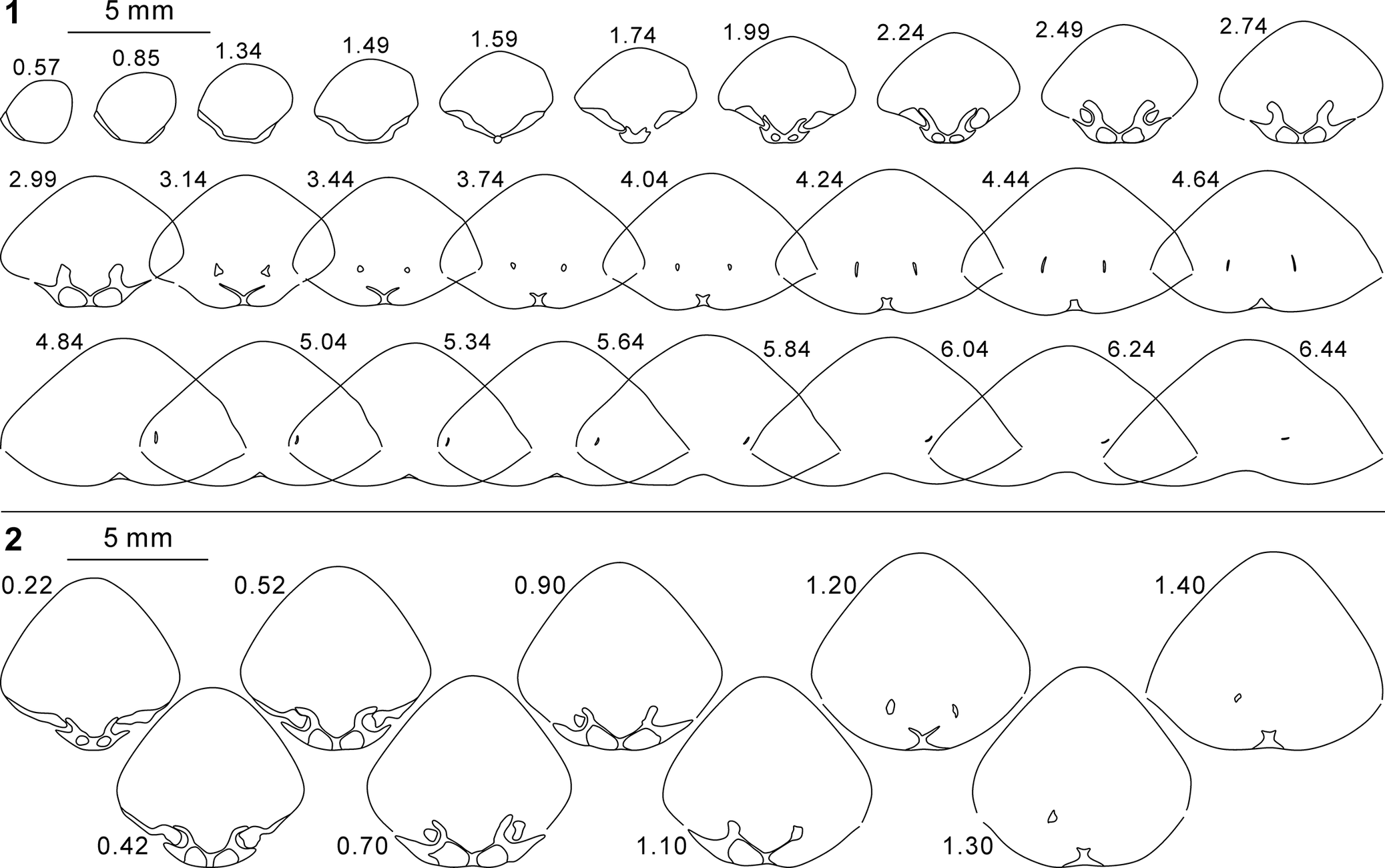
Figure 3. Long description
The image consists of two panels of line drawings showing the internal anatomy of brachiopod specimens through serial cross-sections.
Panel 1 contains 29 sections arranged in three rows. A 5 mm scale bar is located at the top left. The sections begin at 0.57 mm and progress to 6.44 mm.
* The first row shows the initial development of the hinge and dental plates from 0.57 to 2.74 mm.
* The second row, from 2.99 to 4.64 mm, shows the expansion of the brachidium and the appearance of small internal loops.
* The third row, from 4.84 to 6.44 mm, shows the gradual thinning and disappearance of the internal structures as the section moves toward the anterior.
Panel 2 contains 9 larger sections arranged in a staggered two-row format. A 5 mm scale bar is at the top left.
* The top row includes sections at 0.22, 0.52, 0.90, 1.20, and 1.40 mm.
* The bottom row includes sections at 0.42, 0.70, 1.10, and 1.30 mm.
These sections provide a more detailed view of the posterior end of the dorsal umbo, highlighting the complex hinge plates and the initial formation of the crura.
Reference Pálfy2003 Angustothyris? angustaeformis (Böckh, Reference Böckh1872); Pálfy, pl. Br-I, fig. 33 (non fig. 32).
Holotype
MTM M.92.433 from the Felsőörs Formation, Aszófő, Balaton Uplands, Hungary (Pálfy, Reference Pálfy2003, pl. Br-I, fig. 33).
Materials
Two specimens (MTM M.92.234-1, M.92.234-2) from the Felsőörs Formation of Aszófő, Balaton Uplands, Hungary.
Diagnosis
Dorsal valve weakly convex; anterior commissure strongly sulcate; dorsal sulcus wide and deep.
Occurrence
Anisian of Aszófő, Balaton Uplands, Hungary.
Description
Shell small, subrhombic in outline, longer than wide, greatest width slightly anterior to midlength; ventribiconvex or nearly planoconvex; anterior commissure strongly unisulcate; lateral commissure curved toward dorsal valve.
Ventral valve moderately convex, greatest convexity at midline of valve, forming blunt ridge, lateral flanks flat; beak erect, with rounded ridges extending to posterolateral commissure of shell; foramen possibly mesothyrid; deltidial plates conjunct. Dorsal valve weakly convex; sulcus starting from umbo, prominent and deep in umbonal area, becoming wider and deeper anteriorly; lateral flanks slightly convex. Shell smooth.
Pedicle collar not observed due to breakage of ventral beak; dental plates absent. Cardinal process not observed; inner socket ridges very high; outer hinge plates absent; inner hinge plates wide, inclined medianly, fused with median septum to form V-shaped septalium; median septum low, extending to one-third of valve length; crural bases merged with inner socket ridges, connected to or not connected to inner hinge plates; crural process low; loop long, reaching half of valve length, with slender descending lamellae, phase unknown.
Etymology
Named after the type locality, Aszófő, Hungary.
Remarks
Although the specimens from Aszófő were assigned to Angustothyris angustaeformis by Pálfy (Reference Pálfy2003), our re-examination indicates a distinct internal morphology characterized by the presence of a septalium and a median septum. Externally, these specimens can be further distinguished from ‘Angustothyris’ angustaeformis from Köveskál by their flatter dorsal valve and a more pronounced sulcus.
In having a well-developed septalium, a median septum, and very high inner socket ridges, this species is closely allied to the Angustothyris dagysi from the northwestern Caucasus (Dagys, Reference Dagys1972, Reference Dagys1974). The new species differs from Angustothyris dagysi in its external profile, possessing a slightly convex to nearly flat dorsal valve and a wider, deeper dorsal sulcus. The loop of this species is not completely known; its descending lamellae are slenderer than those of the type species, and a teloform loop cannot be ruled out.
Genus Balatonithyris new genus
Type species
Waldheimia angustaeformis Böckh, Reference Böckh1872 from the Pelsonian (Anisian) Felsőörs Formation of Köveskál, Balaton Uplands, Hungary, by original designation.
Diagnosis
Shell small to medium-sized, ventribiconvex, smooth, unisulcate; beak erect, with rounded ridges, foramen mesothyrid; pedicle collar present; cardinal process absent; outer hinge plates absent, inner hinge plates connected to valve floor without forming septalium; median septum absent; crural processes low; loop longer than half of valve length, with slender descending lamellae.
Occurrence
Anisian of Köveskál, Balaton Uplands, Hungary.
Etymology
From Balaton, referring to the type locality in the Balaton Uplands, combined with -thyris (Greek, opening), a suffix commonly used in terebratulide generic names.
Remarks
The species Waldheimia angustaeformis was selected as the type species of Angustothyris by Dagys (Reference Dagys1972). As discussed above, the type species of Angustothyris has been changed to Angustothyris dagysi from the northwestern Caucasus (Guo et al. Reference Guo, Chen and Harper2019). Consequently, a new genus is here proposed to accommodate the true Waldheimia angustaeformis species from Köveskál, Hungary.
Balatonithyris n. gen. can be readily distinguished from Angustothyris in lacking a well-developed septalium and a median septum in the dorsal valve. This new genus closely resembles several Late Triassic angustothyridid genera in the weak development of the dorsal median septum. Rhaetina is easily distinguished from Balatonithyris n. gen. by its sulciplicate or uniplicate anterior commissure. Sanqiaothyris Yang and Xu, Reference Yang and Xu1966 is characterized by a rectimarginate anterior commissure and the complete absence of a sulcus and fold. The closest genus to Balatonithyris n. gen. is Rhaetinopsis Yang and Xu, Reference Yang and Xu1966, a Late Triassic genus reported from Guizhou, Yunnan, and Qinghai in China, and shares with Balatonithyris n. gen. an erect ventral umbo, a mesothyrid foramen, and a unisulcate anterior commissure. However, Rhaetinopsis possesses a deltiform loop shorter than half the valve length (Yang and Xu, Reference Yang and Xu1966; Jin et al., Reference Jin, Ye, Xu and Sun1979); although the loop type of Balatonithyris n. gen. is unknown, its loop length (greater than half the valve length) and slender descending lamellae suggest a teloform phase in this genus. In addition, the inner hinge plates of Sanqiaothyris and Rhaetinopsis typically form a sessile to very low septalium, whereas in Balatonithyris n. gen. they remain separated. We note that both Rhaetinopsis and Sanqiaothyris were regarded as junior synonyms of Rhaetina by Lee et al. (Reference Lee, MacKinnon, Smirnova, Baker, Jin, Sun and Kaesler2006). However, because species of Rhaetina typically have a plicate anterior commissure and never show a sulcate one in adults (Dagys, Reference Dagys1963; Pearson, Reference Pearson1977), we consider them separate genera. Key differences are summarized in Table 1.
Morphological comparisons of some allied angustothyridid genera. Diagnostic characters are summarized as follows: Balatonithyris n. gen., unisulcate without a septalium; Angustothyris Dagys, Reference Dagys1972, unisulcate with a well-developed septalium and a long-flanged deltiform loop; Qianothyris n. gen., unisulcate with a well-developed septalium and a long teloform loop; Rhaetinopsis Yang and Xu, Reference Yang and Xu1966, unisulcate with a poorly developed septalium and a short deltiform loop; Sanqiaothyris Yang and Xu, Reference Yang and Xu1966, rectimarginate with a poorly developed septalium and a deltiform loop; Rhaetina Waagen, Reference Waagen1882, sulci- or uniplicate with a poorly developed septalium and a short deltiform loop

Table 1. Long description
The table compares Balatonithyris n. gen., Angustothyris, Qianothyris n. gen., Rhaetinopsis, Sanqiaothyris, and Rhaetina.
* Ventral umbo: Erect for most, except Qianothyris which is inclined and Rhaetina which is suberect to erect.
* Beak ridges: Prominent in most, absent in Qianothyris, and variable in Rhaetina.
* Foramen: Labiate in Qianothyris; nonlabiate in all others.
* Anterior commissure: Unisulcate in most; rectimarginate in Sanqiaothyris; sulci- or uniplicate in Rhaetina.
* Cardinal process: Absent in Balatonithyris and Angustothyris; present in the other four genera.
* Inner socket ridges: High in Angustothyris; low in all others.
* Outer hinge plates: Absent in Balatonithyris and Angustothyris; present in Qianothyris, Rhaetinopsis, and Sanqiaothyris; variable in Rhaetina.
* Median septum: High in Angustothyris and Qianothyris; absent in Balatonithyris; absent or low in the remaining three.
* Septalium: High in Angustothyris and Qianothyris; absent in Balatonithyris; sessile to low or absent in the others.
* Loop type: Unknown for Balatonithyris; long-flanged deltiform for Angustothyris; teloform for Qianothyris; deltiform for the remaining three.
* Loop length: Greater than 1/2 valve length for Balatonithyris; approximately 1/2 for Angustothyris; approximately 2/3 for Qianothyris; less than 1/2 for Rhaetinopsis and Rhaetina; unknown for Sanqiaothyris.
Beyond the angustothyridids, some Triassic taxa in the suborder Terebratellidina and other families of the Terebratulidina (e.g., the zeilleriids Silesiathyris Brügge, Reference Brügge1977, Aulacothyroides Dagys, Reference Dagys1965, and Qilianoconcha Jin, Sun, and Ye in Jin et al., Reference Jin, Ye, Xu and Sun1979, emended by Guo et al., Reference Guo, Chen, Huang, Chen, Qiu and Guo2022; the dielasmatids Dareithyris Siblík, Reference Siblík1991 and Sulcatinella Dagys, Reference Dagys1974) are externally comparable to Balatonithyris n. gen., but can be distinguished by the presence of dental plates.
The type species Waldheimia angustaeformis has been reported from numerous places worldwide. However, because its internal morphology was poorly understood and owing to the misdiagnosis by Dagys (Reference Dagys1972, Reference Dagys1974), most of these records represent misidentifications. At present, the genus is considered monotypic and restricted to the type locality at Köveskál, Hungary.
Balatonithyris angustaeformis (Böckh, Reference Böckh1872)
Balatonithyris angustaeformis (Böckh, Reference Böckh1872) from the Anisian Felsőörs Formation of Köveskál, Hungary: (1–5) holotype, SZTFH T. 64, dorsal, ventral, lateral, anterior, and posterior views, respectively; (6–9) MTM M.92.366-1, dorsal, ventral, lateral, and anterior views, respectively; (10–13) MTM M.92.366-2, dorsal, ventral, lateral, and anterior views, respectively.

Figure 4. Long description
A multi-panel photo displaying three fossilized brachiopod specimens.
Specimen S Z T F H T dot 64 (holotype) is shown in panels 1 through 5 with a smooth, tan-colored shell.
* 1. Dorsal view showing a rounded triangular outline.
* 2. Ventral view.
* 3. Lateral view showing the biconvex profile.
* 4. Anterior view showing a sulcate frontal commissure.
* 5. Posterior view showing the hinge line.
Specimen M T M M dot 92 dot 366-1 is shown in panels 6 through 9 with a matte gray surface texture.
* 6. Dorsal view.
* 7. Ventral view.
* 8. Lateral view.
* 9. Anterior view.
Specimen M T M M dot 92 dot 366-2 is shown in panels 10 through 13, also in matte gray.
* 10. Dorsal view.
* 11. Ventral view.
* 12. Lateral view.
* 13. Anterior view.
A horizontal scale bar labeled 5 mm is positioned above panel 13 to indicate the physical dimensions of the fossils.
Transverse serial sections of Balatonithyris angustaeformis (Böckh, Reference Böckh1872): (1) sections based on MTM M.92.366-1 (16.34 mm long, 13.71 mm wide, 9.40 mm thick); (2) sections based on MTM M.92.366-2 (17.72 mm long, > 12.87 mm wide, 9.92 mm thick). Numbers above sections indicate distance (in mm) to the posterior end of ventral umbo.

Figure 5. Long description
The diagram presents two sets of line drawings representing serial cross-sections of brachiopod shells. A 5 mm scale bar is provided at the top left of each panel.
Panel 1 contains 48 sections from specimen M T M M dot 92 dot 366-1. The sections begin at 0.28 mm and end at 10.14 mm.
* The first row (0.28 to 2.64 mm) shows the initial appearance of the ventral and dorsal valves, with the development of the hinge teeth and dental sockets.
* The second row (2.84 to 4.24 mm) illustrates the formation of the brachidium and crural bases.
* Subsequent rows (4.44 to 10.14 mm) show the gradual widening of the shell cavity and the thinning of the internal skeletal structures as they move toward the anterior margin.
Panel 2 contains 28 sections from specimen M T M M dot 92 dot 366-2. The sections begin at 0.47 mm and end at 10.92 mm.
* The first row (0.47 to 4.52 mm) shows a similar progression to Panel 1 but with a more rapid increase in shell height and more robust internal hinge structures.
* The second row (4.72 to 7.82 mm) displays the internal loop structures and the separation of the valves.
* The final row (8.22 to 10.92 mm) shows the large, open internal chamber of the shell as the internal supports terminate.
Reference Böckh1872 Waldheimia angustaeformis Böckh, p. 160, pl. 11, fig. 20.
Reference Böckh1873 Waldheimia angustaeformis; Böckh, p. 172, pl. 11, fig. 20.
?Reference Bittner1890 Waldheimia angustaeformis; Bittner, p. 8, pl. 36, figs. 37–40.
?Reference Salomon1895 Waldheimia angustaeformis; Salomon, p. 104, pl. 3, figs. 29–31.
?Reference Arthaber1915 Waldheimia angustaeformis; Arthaber, p. 194, pl. 18, fig. 10.
non Reference Wang, Yang and Wang1955 Rhaetina angustaeformis; Wang, p. 170, pl. 102, figs. 9, 10, 13, 14.
non Reference Wang, Jin and Fang1964 Rhaetina angustaeformis; Wang et al., p. 680, pl. 136, figs. 1, 2, 6, 7.
non Reference Yang and Xu1966 Rhaetina angustaeformis; Yang and Xu, p. 77, pl. 12, figs. 3–8, text-figs. 75–78.
?Reference Siblík1972 ‘Zeilleria’ angustaeformis; Siblík, p. 195, pl. 62, fig. 5.
non Reference Dagys1972 Angustothyris angustaeformis; Dagys, figs. 11–14, 25.
non Reference Dagys1974 Angustothyris angustaeformis; Dagys, text-fig. 131, pl. 48, fig. 1.
nonReference Liao and Sun1974 Rhaetina angustaeformis; Liao and Sun, p. 351, pl. 184, figs. 18–21.
nonReference Feng and Jiang1978 Rhaetina angustaeformis; Feng and Jiang, p. 303, pl. 108, fig. 2.
?Reference Kochanová and Pevný1982 Angustothyris angustaeformis; Kochanová and Pevný, p. 29, pl. 5, fig. 12, pl. 6, fig. 3.
non Reference Popiel-Barczyk and Senkowiczowa1983 Angustothyris angustaeformis; Popiel-Barczyk and Senkowiczowa, figs. 3–8, pls. 1–4.
non Reference Usnarska-Talerzak1988 Angustothyris angustaeformis; Usnarska-Talerzak, fig. 27.
non Reference Usnarska-Talerzak1990 Angustothyris angustaeformis; Usnarska-Talerzak, p. 681, fig. 3.
nonReference Torti and Angiolini1997 Angustothyris angustaeformis; Torti and Angiolini, p. 168, pl. 1, figs. 35–36, pl. 2, fig. 5.
Reference Pálfy2003 Angustothyris? angustaeformis; Pálfy, p. 152, fig. Br-10, pl. Br-I, fig. 32 (non fig. 33).
non Reference Chen, Chen and Tong2010 Rhaetina angustaeformis; Chen et al., figs. 5.1–5.4, 6.1, 6.2.
non Reference Chen, Tong, Song, Luo, Huang and Xiang2015 Rhaetina angustaeformis; Chen et al., fig. 13.5.
?Reference Gaetani2016 Angustothyris angustaeformis; Gaetani, p. 72, pl. 1, fig. 8.
Holotype
SZTFH T. 64 from the Anisian Felsőörs Formation, Köveskál, Balaton Uplands, Hungary (Böckh, Reference Böckh1872, pl. 11, fig. 20a–d; refigured herein as Fig. 4.1–4.5).
Occurrence
Anisian of Köveskál, Balaton Uplands, Hungary.
Description
Shell small to medium-sized, subpentagonal, subrhombic or drop-shaped in outline, longer than wide, greatest width at or slightly anterior to midlength of shell; ventribiconvex; anterior commissure moderately unisulcate; lateral commissure curved to variable extent.
Ventral valve moderately to strongly convex in anterior and lateral profiles, with greatest convexity along midline, forming blunt ridge, lateral flanks nearly flat; beak erect, with prominent and rounded ridges extending to one-third to one-half of valve length; foramen circular, nonlabiate, mesothyrid; deltidial plates conjunct. Dorsal valve moderately convex; sulcus weakly to moderately deep, starting from dorsal umbo, widening and deepening anteriorly. Shell surface smooth.
Ventral interior with distinct pedicle collar; dental plates absent. Cardinal process not observed; inner socket ridges moderately high; outer hinge plates absent, inner hinge plates wide and flat, inclined medially and connected to valve floor without forming septalium; median septum absent; crural bases merged with inner socket ridges; crural processes low; loop longer than one-half of valve length, possibly reaching two-thirds of valve length, with slender descending lamellae.
Materials
Two topotypes (MTM M.92.366-1, M.92.366-2) from the Anisian Felsőörs Formation of Köveskál, Balaton Uplands, Hungary.
Remarks
Of the two topotypes investigated, one (Fig. 4.6–4.9) closely matches the subpentagonal to subrhombic outline of the holotype (Fig. 4.1–4.5), whereas the other exhibits a more drop-shaped outline with less curved lateral commissures (Fig. 4.10–4.13). Despite this variation, both specimens share identical internal structures consistent with the observations of Pálfy (Reference Pálfy2003), confirming the absence of a median septum and septalium as a stable diagnostic feature for B. angustaeformis. The loops in the two investigated specimens of B. angustaeformis are incomplete, preventing a definitive morphological characterization. However, we can confirm that the loops are relatively long (exceeding half the valve length) and possess slender descending lamellae, features that are indicative of a teloform stage.
The observed intraspecific morphological variation, coupled with the high degree of external similarity among allied genera, emphasizes that taxonomic assignments based solely on external morphology are unreliable. Specimens previously assigned to Balatonithyris angustaeformis with unknown internal characters must therefore be treated with caution and their classification remains provisional pending further investigation. The only specimens that could be correctly assigned to Balatonithyris angustaeformis outside of the type locality are the ones reported by Bittner (Reference Bittner1890, pl. 36, figs. 37–40) from the Anisian of northeastern Italy, which exhibit the strongest external resemblance to the holotype and a long and possibly teloform loop. Nevertheless, given the lack of information on their dorsal septal apparatus, a confident assignment of these specimens to Balatonithyris angustaeformis cannot be made.
Genus Qianothyris new genus
Type species
Angustothyris qingyanensis Guo, Chen, and Harper, Reference Guo, Chen and Harper2019 from the Anisian Qingyan Formation of Qingyan, Guizhou, southwestern China, by original designation.
Occurrence
Anisian of Guizhou, southwestern China, and possibly lower Ladinian of northwestern Italy.
Diagnosis
Shell small to medium-sized, ventribiconvex, unisulcate; beak slightly inclined, without prominent ridges, foramen mesothyrid, labiate; pedicle collar present, dental plates absent; cardinal process distinct, inner socket ridges low, outer hinge plates present, inner hinge plates fused with median septum to form septalium; loop teloform with slender transverse band and ascending lamellae, approximately two-thirds of valve length.
Etymology
Qian (in Chinese pinyin), short name of the Guizhou Province in China, where the type species was discovered, combined with the suffix -thyris (Greek, opening), commonly used in terebratulide generic names.
Remarks
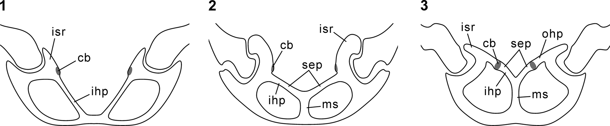
Qianothyris n. gen. is distinguished from related genera by a suite of external and internal characteristics. Externally, it differs from Angustothyris and Balatonithyris n. gen. by its slightly curved ventral umbo, labiate foramen, and the absence of defined beak ridges. The primary internal distinction is the combination of a septalium and a long teloform loop. Balatonithyris n. gen. is easily differentiated by its complete lack of a septalium. Although Angustothyris also possesses a dorsal septalium, its loop is long-flanged deltiform, contrasting with the teloform loop of Qianothyris n. gen. Even in the absence of a preserved loop, these genera can be separated by their hinge plates (Fig. 6; Table 1). In Qianothyris n. gen., the lateral ends of the outer hinge plates fuse with the ventral part of low inner socket ridges. Angustothyris, however, lacks outer hinge plates, and its inner hinge plates connect to the median part of very high socket ridges. A further distinguishing feature is the presence of a well-developed cardinal process in Qianothyris n. gen., which is absent in Angustothyris.
Comparison of dorsal internal structures in (1) Balatonithyris n. gen., (2) Angustothyris Dagys, Reference Dagys1972, and (3) Qianothyris n. gen. All images are based on the type species of each genus. cb, crural base; ihp, inner hinge plate; isr, inner socket ridge; ms, median septum; ohp, outer hinge plate; sep, septalium.

Figure 6. Long description
The diagram consists of three panels labeled 1, 2, and 3, showing cross-sections of dorsal internal structures.
* Panel 1 Balatonithyris: Shows a wide, shallow structure. At the center base, the i h p (inner hinge plate) slopes upward toward the outer edges. Near the top of these plates are the i s r (inner socket ridge) and a small shaded oval representing the c b (crural base).
* Panel 2 Angustothyris: Features a more complex central structure. A vertical m s (median septum) supports a V-shaped s e p (septalium). The i h p (inner hinge plate) extends from the septalium toward the outer i s r (inner socket ridge). The c b (crural base) is located at the junction of the septalium and the hinge plate.
* Panel 3 Qianothyris: Displays a deep, narrow configuration. A prominent vertical m s (median septum) supports a deep, V-shaped s e p (septalium). The i h p (inner hinge plate) is steeply angled. This panel also includes an o h p (outer hinge plate) located distally to the i s r (inner socket ridge). The c b (crural base) is positioned on the inner wall of the septalium.
At present, only the type species can be confidently assigned to this genus. Angustothyris angustaeformis described by Torti and Angiolini (Reference Torti and Angiolini1997) from the lower Ladinian of northwestern Italy exhibits comparable external morphology and hinge plates to Qianothyris qingyanensis (see below), but its loop morphology remains unknown. It could represent an additional species of Qianothyris n. gen.
Qianothyris qingyanensis (Guo, Chen, and Harper, Reference Guo, Chen and Harper2019)
Qianothyris qingyanensis (Guo, Chen, and Harper, Reference Guo, Chen and Harper2019) from the Anisian Qingyan Formation of Qingyan, Guizhou, China; dorsal, ventral, lateral, and anterior views, respectively, of: (1–4) BGEG WCP 10089; (5–8) BGEG WCP 10091; (9–12) BGEG WCP 10098; (13–16) BGEG LDP 10140; (17–20) BGEG WCP 10096; (21–24) BGEG WCP 10099; (25–28) BGEG LDP 10138.

Figure 7. Long description
The photo contains 28 numbered panels arranged in three rows, displaying seven distinct fossil specimens. Each specimen is shown in a sequence of four views: dorsal, ventral, lateral, and anterior. A 5 mm scale bar is located in the center of the image.
* Panels 1 through 4: A small specimen, B G E G W C P 10089, showing a teardrop-shaped shell with a smooth surface.
* Panels 5 through 8: Specimen B G E G W C P 10091, slightly larger with a more pronounced pedicle opening at the top of the dorsal view.
* Panels 9 through 12: Specimen B G E G W C P 10098, exhibiting a rounded, ovate outline and a thick lateral profile.
* Panels 13 through 16: Specimen B G E G L D P 10140, the largest in the set, showing significant surface texture and a sharp, biconvex lateral view.
* Panels 17 through 20: Specimen B G E G W C P 10096, a medium-sized specimen with a narrow, elongated dorsal shape.
* Panels 21 through 24: Specimen B G E G W C P 10099, showing a wide anterior view with a clear commissure line where the two valves meet.
* Panels 25 through 28: Specimen B G E G L D P 10138, characterized by a robust, thick shell and a distinctively curved anterior margin.
All specimens are light gray in color against a white background, showing varying degrees of fossilization and surface wear.
Transverse serial sections of Qianothyris qingyanensis (Guo, Chen, and Harper, Reference Guo, Chen and Harper2019); sections based on: (1) BGEG WCP 10099 (13.13 mm long, 10.13 mm wide, 5.28 mm thick); (2) BGEG WCP 10096 (9.44 mm long, 6.58 mm wide, 4.24 mm thick). Numbers above sections indicate distance (in mm) to the posterior end of ventral umbo.

Figure 8. Long description
The diagram consists of two multi-panel sets of line drawings representing cross-sections of brachiopod shells.
Panel 1 contains 48 sections arranged in 8 rows. A 5 mm scale bar is located at the top left. The sections begin at 0.36 mm and end at 9.90 mm.
* The first row (0.36 to 2.32) shows the transition from a small circular umbo to a wider shell with developing internal hinge structures and a central septum.
* Rows 2 through 4 (2.57 to 4.73) show the shell widening into a lenticular shape with distinct dorsal and ventral valves and internal vertical plates.
* Rows 5 through 8 (4.94 to 9.90) show the gradual thinning of the internal structures and the flattening of the shell profile as the sections move toward the anterior margin.
Panel 2, separated by a horizontal line, contains 44 sections arranged in 4 rows. A 5 mm scale bar is located at the top left. The sections begin at 0.18 mm and end at 6.99 mm.
* The first row (0.18 to 1.84) illustrates the rapid development of the cardinal process and hinge plates within a smaller, more circular shell profile compared to Panel 1.
* The second row (1.99 to 3.44) shows the expansion of the shell width and the appearance of paired internal supports.
* The third and fourth rows (3.64 to 6.99) track the dissipation of internal features as the shell reaches its maximum width and then tapers toward the front edge.
Transverse serial sections of Qianothyris qingyanensis (Guo, Chen, and Harper, Reference Guo, Chen and Harper2019); sections based on: (1) BGEG WCP 10089 (3.85 mm long, 3.78 mm wide, 1.81 mm thick); (2) BGEG WCP 10091 (7.07 mm long, 6.05 mm wide, 3.22 mm thick); (3) BGEG LDP 10138 (14.94 mm long, 11.19 mm wide, 7.36 mm thick); (4) BGEG LDP 10140 (19.22 mm long, 14.39 mm wide, 8.41 mm thick). Numbers above sections indicate distance (in mm) to the posterior end of ventral umbo.

Figure 9. Long description
The diagram consists of four horizontal rows of line drawings representing cross-sections of four different specimens.
* Row 1: Contains 10 sections of specimen B G E G W C P 10089. A 3 mm scale bar is at the top left. Distances range from 0.85 to 2.25 mm. Early sections show a simple biconvex outline, with internal dental plates appearing as small vertical lines near the center by 1.32 mm, eventually diverging.
* Row 2: Contains 18 sections of specimen B G E G W C P 10091 across two sub-rows. A 4 mm scale bar is at the top left. Distances range from 0.10 to 4.53 mm. The sections transition from circular posterior outlines to wide, triangular anterior shapes. Internal structures include a distinct pedicle collar and hinge plates that develop into a complex cardinal process and crural bases.
* Row 3: Contains 10 sections of specimen B G E G L D P 10138. A 5 mm scale bar is at the top left. Distances range from 0.51 to 2.76 mm. These sections show a thicker shell wall and a highly complex hinge apparatus with deep dental sockets and robust crural plates that curve inward.
* Row 4: Contains 15 sections of specimen B G E G L D P 10140 across two sub-rows. A 5 mm scale bar is at the top left. Distances range from 0.42 to 4.20 mm. This largest specimen shows the most detailed internal anatomy, including a well-defined median septum in the dorsal valve and intricate, folded hinge plates that support the brachidium.
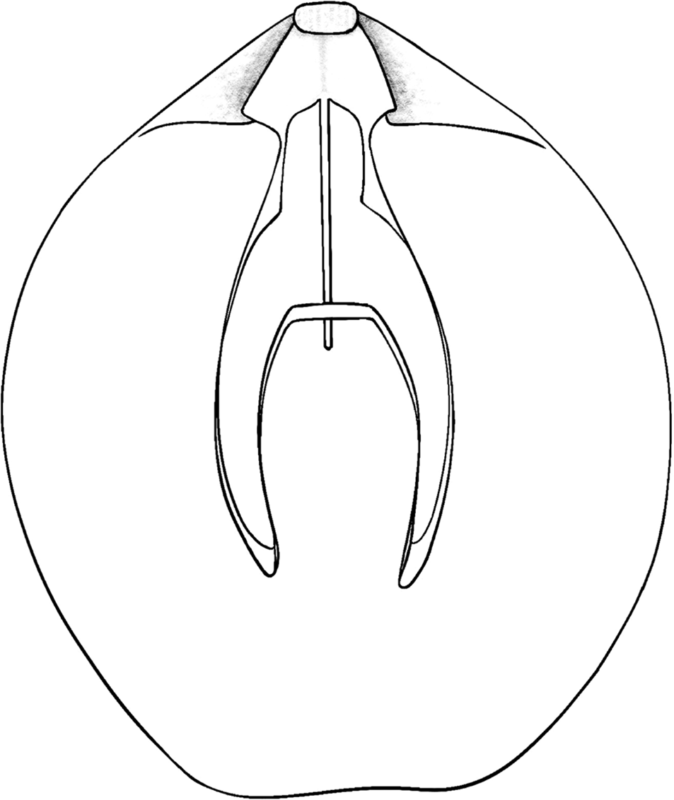
Reconstruction of dorsal interior of Qianothyris qingyanensis (Guo, Chen, and Harper, Reference Guo, Chen and Harper2019), based on BGEG WCP 10099.

Photomicrographs of sectioned specimens showing cardinal process and hinge plates of Qianothyris qingyanensis (Guo, Chen, and Harper, Reference Guo, Chen and Harper2019): (1, 2) BGEG WCP 10096; (3, 4) BGEG WCP 10098. Scale bars = 200 μm.

Figure 11. Long description
A multi-panel photo containing four microscopic cross-sections of brachiopod specimens. Each panel includes a white scale bar in the bottom right corner representing 200 micrometers.
* Panel 1 (Top-Left): Specimen B G E G W C P 10096. A dark, V-shaped cardinal process is centered in a light-colored matrix. The structure shows a distinct heart-shaped central cavity with dark, wing-like extensions reaching laterally.
* Panel 2 (Top-Right): Specimen B G E G W C P 10096 at a different section depth. Two symmetrical, dark-rimmed circular hinge plates are visible, separated by a narrow vertical septum. The interior of the plates is filled with a lighter, granular material.
* Panel 3 (Bottom-Left): Specimen B G E G W C P 10098. Similar to panel 1, it shows a dark, thickened cardinal process with a central triangular opening. The lateral extensions are more robust and darker than in the first specimen.
* Panel 4 (Bottom-Right): Specimen B G E G W C P 10098. This section shows the hinge plates as two large, rounded loops with thick dark borders. The loops are joined at the base, forming a wide U-shape that opens toward the top of the frame.
Reference Wang, Yang and Wang1955 Rhaetina angustaeformis (Boeckh, Reference Böckh1872); Wang, p. 170, pl. 102, figs. 9, 10, 13, 14.
Reference Wang, Jin and Fang1964 Rhaetina angustaeformis; Wang et al., p. 680, pl. 136, figs. 1, 2, 6, 7.
Reference Yang and Xu1966 Rhaetina angustaeformis; Yang and Xu, p. 77, pl. 12, figs. 3–8, text-figs. 75–78.
Reference Liao and Sun1974 Rhaetina angustaeformis; Liao and Sun, p. 351, pl. 184, figs. 18–21.
Reference Feng and Jiang1978 Rhaetina angustaeformis; Feng and Jiang, p. 303, pl. 108, fig. 2.
Reference Chen, Chen and Tong2010 Rhaetina angustaeformis; Chen et al., figs. 5.1–5.4, 6.1, 6.2.
Reference Chen, Tong, Song, Luo, Huang and Xiang2015 Rhaetina angustaeformis; Chen et al., fig. 13.5.
Reference Guo, Chen and Harper2019 Angustothyris qingyanensis Guo, Chen, and Harper, p. 684, figs. 20S–V, 22A–O, 23.
Holotype
BGEG WCP10017 from the Leidapo Member, Qingyan Formation, Wachangpo, Qingyan, Guizhou, southwestern China (Guo et al., Reference Guo, Chen and Harper2019, fig. 22F–I).
Materials
Hundreds of specimens from the Leidapo Member, Qingyan Formation of Wachangpo and Leidapo in Qingyan, Guizhou, southwestern China. Newly registered BGEG LDP 10137–10140, WCP 10089–10101.
Occurrence
Anisian of Guizhou, southwestern China.
Description
Shell small to medium-sized, subpentagonal in outline, longer than wide, greatest width slightly anterior to midlength of shell, anterior margin usually truncated, rarely rounded; ventribiconvex in lateral profile, thickest at or posterior to midlength; anterior commissure rectimarginate to slightly unisulcate.
Ventral valve moderately convex, greatest convexity at midline of valve, forming blunt ridge, lateral flanks flat or slightly convex; beak short, normally slightly curved, rarely moderately curved, without prominent beak ridges; foramen mesothyrid, elongate oval, labiate; deltidial plates conjunct. Dorsal valve convexity lower than that of ventral valve; sulcus narrow and shallow, initiated in umbonal region, widening and deepening anteriorly, as wide as or slightly narrower than anterior margin. Shell ornamented by fine capillae when well preserved.
Pedicle collar prominent; dental plates absent. Cardinal process developed, usually forming shelf above septalium; inner socket ridges low; outer hinge plates normally flat, rarely convex or concave, narrow to wide, slightly inclined medianly, merging with ventral half of inner socket ridges; inner hinge plates of variable width, flat, convex or concave, connected to median septum to form septalium; septalium short, of different depth; median septum low, approximately half of valve length; crural bases located between inner and outer hinge plates, sometimes slightly extending dorsally or ventrally; crural processes moderately high; loop long, teloform in adults, extending to approximately two-thirds of valve length, with slender transverse band and descending and ascending lamellae; loop developing rapidly, with descending lamellae already discrete in a 3.85 mm long specimen.
Remarks
Although the morphology of Qianothyris qingyanensis has been previously illustrated (Yang and Xu, Reference Yang and Xu1966; Guo et al., Reference Guo, Chen and Harper2019), the presence of a well-developed cardinal process (Fig. 11) was not explicitly noted, even though it is visible in previously published serial sections (Yang and Xu, Reference Yang and Xu1966, text-fig. 77; Guo et al., Reference Guo, Chen and Harper2019, fig. 23).
Reconstructing the loop ontogeny of Qianothyris qingyanensis was challenging due to a lack of juvenile specimens, a result of the fossil assemblages having been sorted (Chen et al., Reference Chen, Chen and Tong2010). Available data from a limited number of small specimens indicate rapid early loop development; the descending lamellae are already discrete in a specimen only 3.85 mm long (Fig. 9.1). This contrasts with Angustothyris dagysi, in which a vertical plate persists on the descending lamellae in specimens 4–5 mm long, with traces of the hood still visible in those 6–8 mm in length (Dagys, Reference Dagys1972).
Many previously reported specimens of ‘Angustothyris angustaeformis’ that possess a septalium and outer hinge plates are comparable to Qianothyris qingyanensis. Angustothyris concava Jin, Sun, and Ye in Jin et al., Reference Jin, Ye, Xu and Sun1979 from the Anisian Junzihe Formation of the southern Qilian Mountains, northwestern China was reportedly distinguished from Angustothyris angustaeformis by its subcircular outline and a shallow, wide dorsal sulcus originating anterior to the dorsal umbo (Jin et al., Reference Jin, Ye, Xu and Sun1979). Its serial sections (Jin et al., Reference Jin, Ye, Xu and Sun1979, text-fig. 153) show a lack of dental plates and the presence of a septalium and flat hinge plates, similar to those of Qianothyris n. gen. However, it possesses prominent and subangular beak ridges, a feature absent in Qianothyris n. gen. Recent work by some of the present authors (Guo et al., Reference Guo, Chen, Huang, Chen, Qiu and Guo2022) found no terebratulides lacking dental plates in the Anisian brachiopods from the southern Qilian Mountains, consistent with earlier observations of Xu and Liu (Reference Xu, Liu, Yang, Yin, Xu, Wu, He, Liu and Yin1983). The species Qilianoconcha circularis Guo and Chen in Guo et al., Reference Guo, Chen, Huang, Chen, Qiu and Guo2022 (= ‘Sulcatinella sulcata Dagys, Reference Dagys1974’ of Xu and Liu, Reference Xu, Liu, Yang, Yin, Xu, Wu, He, Liu and Yin1983) has nearly identical external features to Angustothyris concava but possesses dental plates. We therefore speculate that Angustothyris concava and Qilianoconcha circularis are conspecific, with the reported absence of dental plates in the former (Jin et al., Reference Jin, Ye, Xu and Sun1979) likely a preservational artifact. If confirmed, the Qilian species should be renamed Qilianoconcha concava (Jin, Sun, and Ye in Jin et al., Reference Jin, Ye, Xu and Sun1979), making Qilianoconcha circularis a junior synonym. Verification requires re-examination of the type specimens of Angustothyris concava.
Torti and Angiolini (Reference Torti and Angiolini1997) described three species assigned to Angustothyris from the Esino Limestone of Val Parina, northwestern Italy, from an interval attributed to the earliest Ladinian Eoprotrachyceras curionii Biozone (Fantini Sestini, Reference Fantini Sestini1994). The serial sections here re-examined show that these three species possess a septalium, a median septum, and high, flat outer hinge plates similar to those of Qianothyris n. gen. (Fig. 12). Furthermore, the specimens assigned to Angustothyris angustaeformis by Torti and Angiolini (Reference Torti and Angiolini1997) possess a gently sulcate or rectimarginate anterior commissure like those of Qianothyris qingyanensis, but their drop-shaped outline indicates that they could represent a new species of Qianothyris n. gen. Angustothyris sp. aff. A. raxana features a subquadrate outline and rectimarginate anterior commissure, and Angustothyris sp. aff. A. ladina is characterized by a circular outline and a weakly uniplicate anterior commissure. Both lack a dorsal sulcus and differ from all taxa discussed here. New genera might be required to accommodate them once their loop morphologies are known.
Sections of ‘Angustothyris’ species from the early Ladinian of Italy described by Torti and Angiolini (Reference Torti and Angiolini1997), showing distinct hinge plates and septalia: (1, 2) ‘Angustothyris angustaeformis (Böckh, Reference Böckh1872),’ sections 944-F/8-4, 944-F/4-8; (3) ‘Angustothyris sp. aff. A. raxana (Bittner, Reference Bittner1892),’ section 904-M/20-11; (4) ‘Angustothyris sp. aff. A. ladina (Bittner, Reference Bittner1890),’ section 904-M/6-10. Scale bars = 200 μm (1); 500 μm (2–4).

Figure 12. Long description
A four-panel grid of grayscale photomicrographs showing transverse thin sections of brachiopod shells. Each panel includes a black scale bar in the bottom-right corner.
* Panel 1 (Top-Left): Section 944-F/8-4 of Angustothyris angustaeformis. The base shows a thick, curved shell wall. From the center of the base, a vertical septum rises and bifurcates into two symmetrical, upward-curving hinge plates that form a V-shaped septalium. The internal matrix is crystalline and dark. Scale bar is 200 micrometers.
* Panel 2 (Top-Right): Section 944-F/4-8 of Angustothyris angustaeformis. Similar to panel 1 but with a clearer view of the fibrous texture within the hinge plates. The septalium is broader, and the surrounding matrix contains dark, circular mineral inclusions. Scale bar is 500 micrometers.
* Panel 3 (Bottom-Left): Section 904-M/20-11 of Angustothyris species affinity A. raxana. The hinge plates are more elongated and thinner than in the first two panels, extending laterally before curving upward. The central septum is distinct against a fine-grained, grey matrix. Scale bar is 500 micrometers.
* Panel 4 (Bottom-Right): Section 904-M/6-10 of Angustothyris species affinity A. ladina. The V-shaped structure is more compressed horizontally. The hinge plates appear denser, and the crystalline structure of the lower shell wall is highly visible. Scale bar is 500 micrometers.
Popiel-Barczyk and Senkowiczowa (Reference Popiel-Barczyk and Senkowiczowa1983) studied the loop development of juvenile specimens from eastern Poland, which they assigned to Angustothyris angustaeformis. Because these specimens have prominent septalia and median septa, they are not true Angustothyris angustaeformis. Although some juvenile specimens show outer hinge plates like those of Qianothyris n. gen., the lack of mature specimens prevents a confident assignment. Usnarska-Talerzak (Reference Usnarska-Talerzak1988, Reference Usnarska-Talerzak1990) illustrated internal structures of specimens assigned to Angustothyris angustaeformis from western Poland. Hinge plates are prominent in relatively large specimens, whereas in juveniles, the septalium, median septum, and hinge plates are poorly developed. Mature specimens appear to have a short deltiform loop (Usnarska-Talerzak, Reference Usnarska-Talerzak1990, fig. 3), distinguishing them from both Qianothyris qingyanensis and Angustothyris dagysi.
Discussion
Middle Triassic unisulcate terebratulides are globally distributed. Although forms bearing dental plates have been assigned to numerous genera across several superfamilies—e.g., Sulcatinella and Dareithyris Siblík, Reference Siblík1991 (Dielasmatoidea Schuchert, Reference Schuchert and von Zittel1913), Qilianoconcha and Silesiathyris (Zeillerioidea Allan, Reference Allan1940), and Aulacothyropsis Dagys, Reference Dagys1959 (Kingenoidea Elliott, Reference Elliott1948)—those lacking dental plates have historically been grouped under a single genus, Angustothyris. Our re-examination of specimens previously assigned to Angustothyris from Hungary and southwestern China reveals significant differences in their internal structures, indicating previously underestimated diversity. We have established two new genera (Balatonithyris n. gen. and Qianothyris n. gen.) and one new species (Angustothyris aszofoensis n. sp.). Moreover, specimens from other regions, e.g., Italy and Poland, display distinct combinations of external and internal characters, suggesting that they might represent additional new taxa. A comprehensive re-examination of these specimens will likely uncover further hidden genera in this terebratulide group.
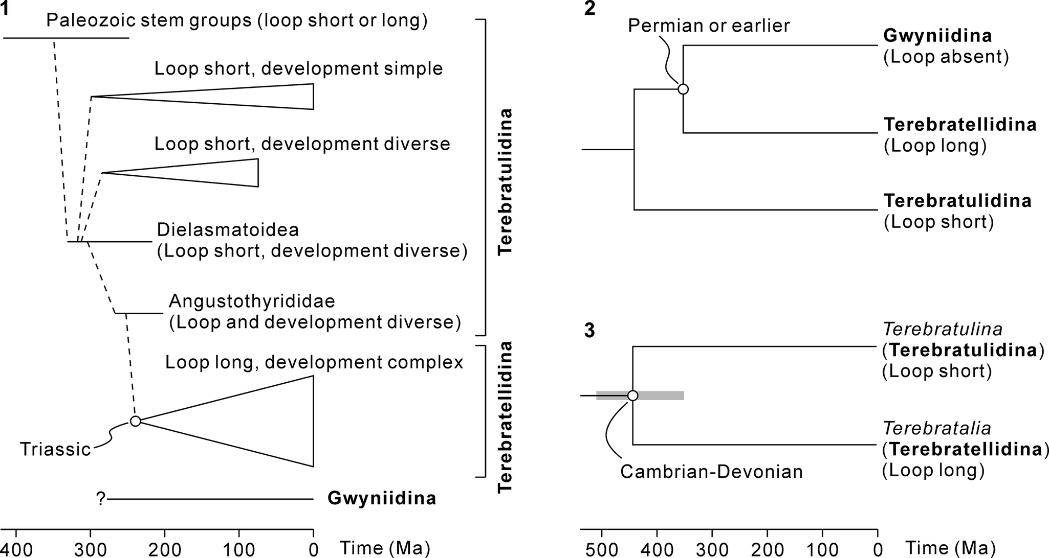
The Angustothyrididae has long been considered a key group in terebratulide evolution (Dagys, Reference Dagys1972, Reference Dagys1974; Smirnova, Reference Smirnova1984, Reference Smirnova2008; Lee et al., Reference Lee, MacKinnon, Smirnova, Baker, Jin, Sun and Kaesler2006), largely based on studies of Angustothyris dagysi from the Caucasus. Although Angustothyris is a terebratulidine with a long-flanged deltiform loop, its cardinalia and complex loop development are strikingly similar to those of long-looped terebratellidines. This led to the hypothesis that Angustothyrididae represents a phylogenetic intermediate between the two suborders (Lee et al., Reference Lee, MacKinnon, Smirnova, Baker, Jin, Sun and Kaesler2006; Fig. 13.1). Our study strengthens this morphological link. We show that some angustothyridids, notably Qianothyris n. gen. and possibly Balatonithyris n. gen., possessed long teloform loops, bridging the morphological gap between short deltiform and long teloform loops, providing a stronger anatomical connection between the Angustothyrididae and Terebratellidina.
Schematic diagrams showing possible phylogenetic relationships within the Terebratulida. (1) Phylogenetic tree based on shell morphology, modified from Lee et al. (Reference Lee, MacKinnon, Smirnova, Baker, Jin, Sun and Kaesler2006). The Terebratellidina is hypothesized to have originated from the Angustothyrididae during the Permian–Triassic transition. Phylogenetic position of the Gwyniidina remains uncertain. (2) Simplified phylogenetic tree based on small subunit rDNA, modified from Cohen (Reference Cohen and Selden2007). Gwynia King, Reference King1859 was assigned to the suborder Gwyniidina, the earliest members of which were reported from the Permian. (3) Dated tree based on molecular-clock analysis of nuclear housekeeping genes and ribosomal genes, modified from Sperling et al. (Reference Sperling, Pisani and Peterson2011). Gray bar indicates 95% posterior distribution. Divergence between Terebratulina d’Orbigny, 1847 in Reference d’Orbigny1847–1851 and Terebratalia Beecher, Reference Beecher1893 was estimated to have occurred in the early–middle Paleozoic.

Figure 13. Long description
A multi-panel diagram containing three phylogenetic trees.
Panel 1 is a large tree on the left. The x-axis at the bottom represents Time in M a from 400 to 0. The tree begins with Paleozoic stem groups with short or long loops. A dashed line descends through several nodes. The first branch leads to Loop short, development simple. The second branch leads to Loop short, development diverse. The third branch leads to Dielasmatoidea with short loops and diverse development. The fourth branch leads to Angustothyrididae with diverse loops and development. A node marked Triassic leads to a large clade labeled Loop long, development complex. A bracket on the right groups the upper branches as Terebratulidina and the lower large clade as Terebratellidina. At the very bottom, a separate line with a question mark leads to Gwyniidina.
Panel 2 is at the top right. It shows a three-branched tree. The top branch is Gwyniidina with loop absent, originating from a node labeled Permian or earlier. The middle branch is Terebratellidina with long loops. The bottom branch is Terebratulidina with short loops.
Panel 3 is at the bottom right. The x-axis represents Time in M a from 500 to 0. A horizontal gray bar indicates a 95 percent posterior distribution around a node labeled Cambrian-Devonian. This node splits into two lineages. The top lineage is Terebratulina within Terebratulidina, characterized by short loops. The bottom lineage is Terebratalia within Terebratellidina, characterized by long loops.
Molecular evidence, however, provides a contrasting phylogenetic narrative. Analysis of small subunit rDNA of living terebratulides recovers Terebratulidina and Terebratellidina as monophyletic clades (Cohen, Reference Cohen and Selden2007). Notably, the morphologically aberrant genus Gwynia King, Reference King1859 (assigned to Terebratellidina by Lee et al., Reference Lee, MacKinnon, Smirnova, Baker, Jin, Sun and Kaesler2006), which lacks a loop and possesses a lophophore supported by submarginal ridges, was suggested as a sister group to all other terebratellidines. Its unique traits prompted its placement in a new suborder, Gwyniidina Halamski and Bitner in Halamski et al., Reference Halamski, Bitner, Kaim, Kolar-Jurkovšek and Jurkovšek2015, the origin of which extends back to the Permian (Halamski et al., Reference Halamski, Bitner, Kaim, Kolar-Jurkovšek and Jurkovšek2015). This implies that the divergence between the Terebratulidina and Terebratellidina likely occurred in the Permian or earlier (Fig. 13.2). This deep divergence is further supported by molecular-clock analyses. Based on analyses of nuclear housekeeping genes and ribosomal genes, Sperling et al. (Reference Sperling, Pisani and Peterson2011) inferred that the split between Terebratulina d’Orbigny, 1847 in Reference d’Orbigny1847–1851 (Terebratulidina) and Terebratalia Beecher, Reference Beecher1893 (Terebratellidina) dates back to the early–middle Paleozoic (Fig. 13.3). This timeline suggests that Mesozoic–Cenozoic terebratellidines could have descended from Paleozoic long-looped ancestors, i.e., the Cryptonelloidea Thomson, Reference Thomson1926.
The apparent conflict between morphology- and molecular-based phylogenies can be interpreted in several ways. First, the similarities in cardinalia and loop ontogeny between the Angustothyrididae and Terebratellidina could be convergent. The involvement of secondary elements in loop development is not exclusive to these groups; it also occurs in other Paleozoic families like Cryptonellidae Thomson, Reference Thomson1926, Compositelasmatidae Smirnova, Reference Smirnova2006, and Pseudodielasmatidae Cooper and Grant, Reference Cooper and Grant1976 (Cooper, Reference Cooper1957; Cooper and Grant, Reference Cooper and Grant1976; Smirnova, Reference Smirnova2001, Reference Smirnova2006, Reference Smirnova2008). This suggests that such an ontogenetic pathway might have evolved multiple times, weakening its value for inferring deep phylogenetic relationships. In this scenario, the Terebratellidina might be a monophyletic group originating in the Paleozoic, with both Paleozoic and post-Paleozoic long-looped forms belonging to the same evolutionary lineage.
An alternative explanation is that post-Paleozoic Terebratellidina (sensu Lee et al., Reference Lee, MacKinnon, Smirnova, Baker, Jin, Sun and Kaesler2006) is polyphyletic. The shared cardinalia and loop development between Angustothyrididae and certain terebratellidines (particularly the Zeillerioidea Allan, Reference Allan1940), reinforced by the teloform loop in Qianothyris n. gen., could indicate a genuine phylogenetic link. It is possible that the Angustothyrididae gave rise only to specific, now-extinct subgroups of post-Paleozoic terebratellidines (sensu Lee et al., Reference Lee, MacKinnon, Smirnova, Baker, Jin, Sun and Kaesler2006), whereas extant terebratellidines belong to separate, older lineages that originated in the early–middle Paleozoic.
Currently, it is difficult to determine which scenario is more plausible, and further studies are needed to determine which morphological features (e.g., cardinalia, loop ontogeny, or adult loop morphology) are more informative for phylogenetic analyses. One conclusion, however, is clear: molecular evidence strongly suggests that the current classification of terebratulides requires revision. Unfortunately, the data necessary to undertake such revisions are insufficient. Additional studies focusing on loop morphology and ontogeny, particularly in late Paleozoic to early Mesozoic taxa, will be essential for improving our understanding of the evolutionary history of terebratulide brachiopods.
Acknowledgments
We are grateful to O. Szives for photographing the holotype of Waldheimia angustaeformis and to L. Angiolini for allowing restudy of the serial sections of Torti and Angiolini (Reference Torti and Angiolini1997). We thank S. Nikolaeva, M. Young, and J. Rong for discussing the issue regarding the type species of Angustothyris. We also thank B.V. Radulović for sharing important literature. L. Su and Z. Zheng are acknowledged for their assistance in processing the specimens. Constructive comments and suggestions from A. Vörös and anonymous reviewers improved the manuscript. This study was funded by a research grant from the National Natural Science Foundation of China (42530209 to ZQC). This is HUN-REN–MTM–ELTE Paleo contribution no. 441.
Competing interests
The authors declare none.