


Introduction
The importance of the gut microbiome in maintaining host health and physiology has been well-established in the literature (Cryan and Dinan, Reference Cryan and Dinan2012; McFall-Ngai et al., Reference McFall-Ngai, Hadfield, Bosch, Carey, Domazet-Lošo, Douglas, Dubilier, Eberl, Fukami, Gilbert, Hentschel, King, Kjelleberg, Knoll, Kremer, Mazmanian, Metcalf, Nealson, Pierce, Rawls, Reid, Ruby, Rumpho, Sanders, Tautz and Wernegreen2013; Luczynski et al., Reference Luczynski, McVey Neufeld, Oriach, Clarke, Dinan and Cryan2016; Wu et al., Reference Wu, Compher, Chen, Smith, Shah, Bittinger, Chehoud, Albenberg, Nessel, Gilroy, Star, Weljie, Flint, Metz, Bennett, Li, Bushman and Lewis2016; Van de Wouw et al., Reference Van de Wouw, Boehme, Clarke, Cryan, Dinan, Lyte, O’Sullivan, Stanton, Strain and Wiley2018; Biesiekierski et al., Reference Biesiekierski, Jalanka and Staudacher2019; Cryan et al., Reference Cryan, O’Riordan, Cowan, Sandhu, Bastiaanssen, Boehme, Codagnone, Cussotto, Fulling, Golubeva, Guzzetta, Jaggar, Long-Smith, Lyte, Martin, Molinero-Perez, Moloney, Morelli, Morillas, O’Connor, Cruz-Pereira, Peterson, Rea, Ritz, Sherwin, Spichak, Teichman, van de Wouw, Ventura-Silva, Wallace-Fitzsimons, Hyland, Clarke and Dinan2019; Leeming et al., Reference Leeming, Johnson, Spector and le Roy2019; Lavelle and Sokol, Reference Lavelle and Sokol2020; Hamamah and Covasa, Reference Hamamah and Covasa2022; Pate et al., Reference Pate, Anthony and Radford-Smith2023; Delgado-Ocaña and Cuesta, Reference Delgado-Ocaña and Cuesta2024; O’Riordan et al., Reference O’Riordan, Moloney, Keane, Clarke and Cryan2025). It is comprised of microorganisms that reside within the host gut and encompasses their complete genetic and metabolic functions (Barko et al., Reference Barko, McMichael, Swanson and Williams2018; Silva et al., Reference Silva, Bernardi and Frozza2020; Sabit et al., Reference Sabit, Tombuloglu, Rehman, Almandil, Cevik, Abdel-Ghany, Rashwan, Abasiyanik and Yee Waye2021). Its overall composition depends on both intrinsic and extrinsic factors (Cho and Blaser, Reference Cho and Blaser2012; Wen and Duffy, Reference Wen and Duffy2017; Cryan, Reference Cryan2025) with diet emerging as a strong predictor in determining gut microbiome composition and diversity (Wu et al., Reference Wu, Chen, Hoffmann, Bittinger, Chen, Keilbaugh, Bewtra, Knights, Walters, Knight, Sinha, Gilroy, Gupta, Baldassano, Nessel, Li, Bushman and Lewis2011; Moles and Otaegui, Reference Moles and Otaegui2020; Redondo-Useros et al., Reference Redondo-Useros, Nova, González-Zancada, Díaz, Gómez-Martínez and Marcos2020; Berding et al., Reference Berding, Vlckova, Marx, Schellekens, Stanton, Clarke, Jacka, Dinan and Cryan2021). Specifically, several studies have shown that consumption of a high-fat Western diet increased the Bacillota/Bacteroidota ratio (Cani et al., Reference Cani, Neyrinck, Fava, Knauf, Burcelin, Tuohy, Gibson and Delzenne2007; Ohland et al., Reference Ohland, Kish, Bell, Thiesen, Hotte, Pankiv and Madsen2013; Patterson et al., Reference Patterson, Cryan, Fitzgerald, Ross, Dinan and Stanton2014; Jørgensen et al., Reference Jørgensen, Hansen, Kihl, Nielsen, Sørensen, Wegener and Winther2015; Velázquez et al., Reference Velázquez, Enos, Bader, Sougiannis, Carson, Chatzistamou, Carson, Nagarkatti, Nagarkatti and Murphy2019; Malesza et al., Reference Malesza, Malesza, Walkowiak, Mussin, Walkowiak, Aringazina, Bartkowiak-Wieczorek and Mądry2021; Marx et al., Reference Marx, Lane, Hockey, Aslam, Berk, Walder, Borsini, Firth, Pariante, Berding, Cryan, Clarke, Craig, Su, Mischoulon, Gomez-Pinilla, Foster, Cani, Thuret, Staudacher, Sánchez-Villegas, Arshad, Akbaraly, O’Neil, Segasby and Jacka2021) and reduced Bifidobacterium spp. (Cani et al., Reference Cani, Neyrinck, Fava, Knauf, Burcelin, Tuohy, Gibson and Delzenne2007; Marx et al., Reference Marx, Lane, Hockey, Aslam, Berk, Walder, Borsini, Firth, Pariante, Berding, Cryan, Clarke, Craig, Su, Mischoulon, Gomez-Pinilla, Foster, Cani, Thuret, Staudacher, Sánchez-Villegas, Arshad, Akbaraly, O’Neil, Segasby and Jacka2021; Schneider et al., Reference Schneider, O’Riordan, Clarke and Cryan2024), while prebiotic supplementation increased the ratio of beneficial microbes (Van de Wouw et al., Reference Van de Wouw, Boehme, Clarke, Cryan, Dinan, Lyte, O’Sullivan, Stanton, Strain and Wiley2018; Marx et al., Reference Marx, Lane, Hockey, Aslam, Berk, Walder, Borsini, Firth, Pariante, Berding, Cryan, Clarke, Craig, Su, Mischoulon, Gomez-Pinilla, Foster, Cani, Thuret, Staudacher, Sánchez-Villegas, Arshad, Akbaraly, O’Neil, Segasby and Jacka2021; Rosell-Cardona et al., Reference Rosell-Cardona, Leigh, Knox, Tirelli, Lyte, Goodson, Kelley-Loughnane, Aburto, Cryan and Clarke2025).
Epidemiological studies have further demonstrated that certain diets can have beneficial effects, modulate levels of microbial metabolites, and prevent the onset of diseases (Gill et al., Reference Gill, Rossi, Bajka and Whelan2021; Horn et al., Reference Horn, Mayer, Chen and Mayer2022; O’Riordan et al., Reference O’Riordan, Collins, Moloney, Knox, Aburto, Fülling, Morley, Clarke, Schellekens and Cryan2022; Heslin and McNulty, Reference Heslin and McNulty2023; Ross et al., Reference Ross, Mayer, Horn, Cryan, del Rio, Randolph, Gill, Gupta, Ross, Stanton and Mayer2024; Schneider et al., Reference Schneider, O’Riordan, Clarke and Cryan2024; Cryan, Reference Cryan2025). For example, diets rich in fibre and legumes (as in the Mediterranean diet) have been linked to reductions in the onset of chronic disease, anti-inflammatory effects, and better mental health (De Filippis et al., Reference De Filippis, Brigidi, Cocolin, Cagno, Ercolini, Ferrocino, Gobbetti, Jeffery, La Storia, Laghi, Lazzi, Neviani, O’Toole, Pellegrini, Serrazanetti, Turroni and Vannini2016; Lassale et al., Reference Lassale, Batty, Baghdadli, Jacka, Sánchez-Villegas, Kivimäki and Akbaraly2019; Asnicar et al., Reference Asnicar, Berry, Valdes, Nguyen, Piccinno, Drew, Leeming, Gibson, le Roy, Khatib, Francis, Mazidi, Mompeo, Valles-Colomer, Tett, Beghini, Dubois, Bazzani, Thomas, Mirzayi, Khleborodova, Oh, Hine, Bonnett, Capdevila, Danzanvilliers, Giordano, Geistlinger, Waldron, Davies, Hadjigeorgiou, Wolf, Ordovás, Gardner, Franks, Chan, Huttenhower, Spector and Segata2021; Jennings et al., Reference Jennings, Kühn, Bondonno, Waniek, Bang, Franke, Kassubek, Müller, Both, Weber, Lieb and Cassidy2024; Rostgaard-Hansen et al., Reference Rostgaard-Hansen, Esberg, Dicksved, Hansen, Pelve, Brunius, Halkjær, Tjønneland, Johansson and Landberg2024; Schneider et al., Reference Schneider, O’Riordan, Clarke and Cryan2024), whereas low-fibre, high-fat, and high-sugar diets have been associated with an increased risk of cardiovascular disease, obesity, and depression (Li et al., Reference Li, Lv, Wei, Sun, Zhang, Zhang and Li2017; Duan et al., Reference Duan, Zeng, Zheng, Song, Li, Kong and Xu2018; Lassale et al., Reference Lassale, Batty, Baghdadli, Jacka, Sánchez-Villegas, Kivimäki and Akbaraly2019; Wali et al., Reference Wali, Jarzebska, Raubenheimer, Simpson, Rodionov and O’Sullivan2020; Wang et al., Reference Wang, Wang, Zhang, Popkin and du2020; Collins et al., Reference Collins, Dash, Allender, Jacka and Hoare2022; Zeng et al., Reference Zeng, Jin and Wang2023; Schneider et al., Reference Schneider, O’Riordan, Clarke and Cryan2024). Expectedly, perturbations in the gut microbiome are often reported in diseased states (Chen et al., Reference Chen, Zhou and Wang2021; De Vos et al., Reference De Vos, Cani, Tilg and Hul2022; Liu et al., Reference Liu, Tan, Cheng, Zhang, Feng and Peng2022; Gan et al., Reference Gan, Chen, Zhong, Liu, Geng, Wang and Wang2024), which emphasises the involvement of diets in shaping the gut microbiome.
The gut microbiome breaks down indigestible fibres from diets via fermentation (Mayer et al., Reference Mayer, Tillisch and Gupta2015; San Mauro Martin et al., Reference San Mauro Martin, Blanco López, Blumenfeld Olivares, Borregon Rivilla, Echeverry López, Elortegui Pascual, García Bernat, Garicano Vilar, Quevedo Santos and Rincón Barrado2018; Liu et al., Reference Liu, Tan, Cheng, Zhang, Feng and Peng2022; Aleman et al., Reference Aleman, Moncada and Aryana2023) to produce metabolites (O’Riordan et al., Reference O’Riordan, Collins, Moloney, Knox, Aburto, Fülling, Morley, Clarke, Schellekens and Cryan2022; Rosell-Cardona et al., Reference Rosell-Cardona, Leigh, Knox, Tirelli, Lyte, Goodson, Kelley-Loughnane, Aburto, Cryan and Clarke2025) that are predominantly made up of short-chain fatty acids (SCFAs) (Kim, Reference Kim2023) that can enter systemic circulation, cross the blood–brain barrier, and ultimately affect the brain (Cryan and Dinan, Reference Cryan and Dinan2012; Mayer et al., Reference Mayer, Tillisch and Gupta2015; Agustí et al., Reference Agustí, García-Pardo, López-Almela, Campillo, Maes, Romaní-Pérez and Sanz2018; Appleton, Reference Appleton2018; Loh et al., Reference Loh, Mak, Tan, Ng, Chan, Yeow, Foo, Ong, How and Khaw2024). However, the exact mechanisms and contributions of these metabolites remain elusive. In previous studies, SCFA treatments were reported to alleviate stress, ameliorate depressive behaviours, increase the growth rate of neural cells, and improve memory (Cryan and Dinan, Reference Cryan and Dinan2012; Mayer et al., Reference Mayer, Tillisch and Gupta2015; Agustí et al., Reference Agustí, García-Pardo, López-Almela, Campillo, Maes, Romaní-Pérez and Sanz2018; Appleton, Reference Appleton2018; Van de Wouw et al., Reference Van de Wouw, Boehme, Clarke, Cryan, Dinan, Lyte, O’Sullivan, Stanton, Strain and Wiley2018; Cryan et al., Reference Cryan, O’Riordan, Cowan, Sandhu, Bastiaanssen, Boehme, Codagnone, Cussotto, Fulling, Golubeva, Guzzetta, Jaggar, Long-Smith, Lyte, Martin, Molinero-Perez, Moloney, Morelli, Morillas, O’Connor, Cruz-Pereira, Peterson, Rea, Ritz, Sherwin, Spichak, Teichman, van de Wouw, Ventura-Silva, Wallace-Fitzsimons, Hyland, Clarke and Dinan2019; Marx et al., Reference Marx, Lane, Hockey, Aslam, Berk, Walder, Borsini, Firth, Pariante, Berding, Cryan, Clarke, Craig, Su, Mischoulon, Gomez-Pinilla, Foster, Cani, Thuret, Staudacher, Sánchez-Villegas, Arshad, Akbaraly, O’Neil, Segasby and Jacka2021; Horn et al., Reference Horn, Mayer, Chen and Mayer2022; Loh et al., Reference Loh, Mak, Tan, Ng, Chan, Yeow, Foo, Ong, How and Khaw2024; Ross et al., Reference Ross, Mayer, Horn, Cryan, del Rio, Randolph, Gill, Gupta, Ross, Stanton and Mayer2024; Schneider et al., Reference Schneider, O’Riordan, Clarke and Cryan2024; Cryan, Reference Cryan2025; O’Riordan et al., Reference O’Riordan, Moloney, Keane, Clarke and Cryan2025; Rosell-Cardona et al., Reference Rosell-Cardona, Leigh, Knox, Tirelli, Lyte, Goodson, Kelley-Loughnane, Aburto, Cryan and Clarke2025). Although these studies emphasise the neuroactive effects of metabolites, many of these treatments were limited to individual SCFAs such as sodium butyrate. The gut microbiome, however, produces numerous metabolites (Topping and Clifton, Reference Topping and Clifton2001; Mayer et al., Reference Mayer, Tillisch and Gupta2015; Postler and Ghosh, Reference Postler and Ghosh2017; San Mauro Martin et al., Reference San Mauro Martin, Blanco López, Blumenfeld Olivares, Borregon Rivilla, Echeverry López, Elortegui Pascual, García Bernat, Garicano Vilar, Quevedo Santos and Rincón Barrado2018; Liu et al., Reference Liu, Tan, Cheng, Zhang, Feng and Peng2022) which likely work in concert. Thus, there is an absence of empirical evidence that illustrates how different diets affect the nervous system.
In this study, we employed different diets and fibre sources based on their well-documented effects on the gut microbiome and association with overall health. The low-fibre Western (LFW) diet includes foods that are highly processed and contain high amounts of added sugars, fats, and salt. This diet is associated with low consumption of fibre and has been linked to many high-risk diseases and disorders, including obesity, diabetes, anxiety, and depression (Li et al., Reference Li, Lv, Wei, Sun, Zhang, Zhang and Li2017; Statovci et al., Reference Statovci, Aguilera, MacSharry and Melgar2017; Duan et al., Reference Duan, Zeng, Zheng, Song, Li, Kong and Xu2018; Lassale et al., Reference Lassale, Batty, Baghdadli, Jacka, Sánchez-Villegas, Kivimäki and Akbaraly2019; Wali et al., Reference Wali, Jarzebska, Raubenheimer, Simpson, Rodionov and O’Sullivan2020; Wang et al., Reference Wang, Wang, Zhang, Popkin and du2020; Shi et al., Reference Shi, Ge, Ma, Zheng, Cui, Pan, Zheng, Yang, Zhang, Hu, Hu, Tang, Zheng, Huang and Yu2021; Collins et al., Reference Collins, Dash, Allender, Jacka and Hoare2022; Zeng et al., Reference Zeng, Jin and Wang2023; Schneider et al., Reference Schneider, O’Riordan, Clarke and Cryan2024). In contrast, the high-fibre Western (HFW) diet follows a largely plant-based diet that often excludes food products of animal origins (Orlich et al., Reference Orlich, Singh, Sabaté, Jaceldo-Siegl, Fan, Knutsen, Beeson and Fraser2013; Xiao et al., Reference Xiao, Zhang, Yu, Tian, Chen and Zhai2022), while the Mediterranean (MED) diet is similar but involves the consumption of fish and poultry products (Davis et al., Reference Davis, Bryan, Hodgson and Murphy2015; De Filippis et al., Reference De Filippis, Brigidi, Cocolin, Cagno, Ercolini, Ferrocino, Gobbetti, Jeffery, La Storia, Laghi, Lazzi, Neviani, O’Toole, Pellegrini, Serrazanetti, Turroni and Vannini2016; Lassale et al., Reference Lassale, Batty, Baghdadli, Jacka, Sánchez-Villegas, Kivimäki and Akbaraly2019; Ventriglio et al., Reference Ventriglio, Sancassiani, Contu, Latorre, di Slavatore, Fornaro and Bhugra2020; Asnicar et al., Reference Asnicar, Berry, Valdes, Nguyen, Piccinno, Drew, Leeming, Gibson, le Roy, Khatib, Francis, Mazidi, Mompeo, Valles-Colomer, Tett, Beghini, Dubois, Bazzani, Thomas, Mirzayi, Khleborodova, Oh, Hine, Bonnett, Capdevila, Danzanvilliers, Giordano, Geistlinger, Waldron, Davies, Hadjigeorgiou, Wolf, Ordovás, Gardner, Franks, Chan, Huttenhower, Spector and Segata2021; Jennings et al., Reference Jennings, Kühn, Bondonno, Waniek, Bang, Franke, Kassubek, Müller, Both, Weber, Lieb and Cassidy2024; Rostgaard-Hansen et al., Reference Rostgaard-Hansen, Esberg, Dicksved, Hansen, Pelve, Brunius, Halkjær, Tjønneland, Johansson and Landberg2024; Schneider et al., Reference Schneider, O’Riordan, Clarke and Cryan2024). Both diets have high fibre contents which is beneficial to the gut microbiome and are linked to better overall health (Orlich et al., Reference Orlich, Singh, Sabaté, Jaceldo-Siegl, Fan, Knutsen, Beeson and Fraser2013; Davis et al., Reference Davis, Bryan, Hodgson and Murphy2015; De Filippis et al., Reference De Filippis, Brigidi, Cocolin, Cagno, Ercolini, Ferrocino, Gobbetti, Jeffery, La Storia, Laghi, Lazzi, Neviani, O’Toole, Pellegrini, Serrazanetti, Turroni and Vannini2016; Lassale et al., Reference Lassale, Batty, Baghdadli, Jacka, Sánchez-Villegas, Kivimäki and Akbaraly2019; Ventriglio et al., Reference Ventriglio, Sancassiani, Contu, Latorre, di Slavatore, Fornaro and Bhugra2020; Asnicar et al., Reference Asnicar, Berry, Valdes, Nguyen, Piccinno, Drew, Leeming, Gibson, le Roy, Khatib, Francis, Mazidi, Mompeo, Valles-Colomer, Tett, Beghini, Dubois, Bazzani, Thomas, Mirzayi, Khleborodova, Oh, Hine, Bonnett, Capdevila, Danzanvilliers, Giordano, Geistlinger, Waldron, Davies, Hadjigeorgiou, Wolf, Ordovás, Gardner, Franks, Chan, Huttenhower, Spector and Segata2021; Xiao et al., Reference Xiao, Zhang, Yu, Tian, Chen and Zhai2022; Jennings et al., Reference Jennings, Kühn, Bondonno, Waniek, Bang, Franke, Kassubek, Müller, Both, Weber, Lieb and Cassidy2024; Rostgaard-Hansen et al., Reference Rostgaard-Hansen, Esberg, Dicksved, Hansen, Pelve, Brunius, Halkjær, Tjønneland, Johansson and Landberg2024; Schneider et al., Reference Schneider, O’Riordan, Clarke and Cryan2024). Finally, the Yanomami (YAN) diet is modelled on the substrate consumption of the Yanomami people of the Amazon, many of whom still subside on fishing, gathering, hunting, and simple horticulture (Mueller et al., Reference Mueller, Noya-Alarcon, Contreras, Appel and Dominguez-Bello2018; Conteville et al., Reference Conteville, Oliveira-Ferreira and Vicente2019). Their diets are remarkably low in salt and fat content and are extremely high in fibre (Mueller et al., Reference Mueller, Noya-Alarcon, Contreras, Appel and Dominguez-Bello2018; Conteville et al., Reference Conteville, Oliveira-Ferreira and Vicente2019), and those Yanomami who still follow traditional subsistence patterns tend to show very high levels of cardiometabolic health and low incidence of chronic diseases (Deehan et al., Reference Deehan, Duar, Armet, Perez-Muñoz, Jin and Walter2017; Mueller et al., Reference Mueller, Noya-Alarcon, Contreras, Appel and Dominguez-Bello2018; Conteville et al., Reference Conteville, Oliveira-Ferreira and Vicente2019), which warrants further investigation. In addition, we also explored the role of fibres by supplementing the LFW diet with soluble and insoluble fibres due to their known benefits and role in modulating the gut microbiome (Deehan et al., Reference Deehan, Duar, Armet, Perez-Muñoz, Jin and Walter2017; Cronin et al., Reference Cronin, Joyce, O’Toole and O’Connor2021). Soluble fibres can be readily metabolised by gut microbes and, therefore, influence the abundance and diversity of the gut microbiota. On the other hand, insoluble fibres are poorly metabolised by gut microbes and instead play a role in bulking waste material in the gut, increasing the transit rate throughout the gut, and potentially decreasing the time available for gut microbes to ferment non-digested food particles (Koh et al., Reference Koh, de Vadder, Kovatcheva-Datchary and Bäckhed2016; Staller et al., Reference Staller, Song, Grodstein, Whitehead, Matthews, Kuo and Chan2018; Gill et al., Reference Gill, Rossi, Bajka and Whelan2021; Guan et al., Reference Guan, Yu and Feng2021).
Given the relationship between diet, fibre, and health, we endeavoured to test the effects of metabolites sourced from different diets and fibres on gene expression in an animal model. We hypothesised that metabolites derived from high-fibre diets would have a positive effect on neurodevelopment and inflammatory gene expression. To test this, we first generated metabolites using a bioreactor that mimics the distal colon to model the different diets and fibre sources and then used these metabolites to treat our germ-free zebrafish model, which we previously demonstrated requires microbial metabolites for a proper nervous system development (Rea et al., Reference Rea, Bell, Ball and van Raay2022; Gianetto-Hill et al., Reference Gianetto-Hill, Vancuren, Daisley, Renwick, Wilde, Schroeter, Daigneault and Allen-Vercoe2023).
Methods
Collection of faecal donor samples
Faecal samples from 10 donors (5 male, 5 female), aged 2 to 6 years old, with a low history of antibiotic use and clinical illnesses and who followed a typical Western diet were evaluated. The samples were collected from participants of the Guelph Family Health Study in accordance with the University of Guelph’s research ethics board approval (REB# 17-07-003) and stored at −80 °C until use. Written informed consent was obtained from each participant or their appropriate guardian.
Shotgun metagenomic sequencing
Faecal DNA was isolated using the QIAamp® PowerFecal® Pro DNA Kit (QIAGEN) and subjected to whole metagenomic shotgun sequencing using Illumina NextSeq 2000 at the University of Pittsburgh’s Microbial Genome Sequencing Center, generating 2x151bp paired-end reads with an average of 7,804,105 forward and reverse reads from all samples. Sequencing quality was evaluated using FastQC v.11.9 (Babraham Bioinformatics, 2010). Sequences that were less than 50 bp in length were removed using Trimmomatic v.0.39 (Bolger et al., Reference Bolger, Lohse and Usadel2014). Forward and reverse reads were joined using Fastq-join v.1.3.1 (Babraham Bioinformatics, 2010). Relative abundance of the microbial taxa was obtained using MetaPhlAn 3 with the ChocoPhlAn 3 database (Beghini et al., Reference Beghini, McIver, Blanco-Míguez, Dubois, Asnicar, Maharjan, Mailyan, Manghi, Scholz, Thomas, Valles-Colomer, Weingart, Zhang, Zolfo, Huttenhower, Franzosa and Segata2021). Bowtie2 was used to align the sequences to clade-specific marker genes (Langmead and Salzberg, Reference Langmead and Salzberg2012). Analyses and visualisation on the resulting microbial composition data were carried out in R v.4.1.2.
Expansion of faecal samples
To provide enough biomass for all diet and fibre experiments, the microbial communities from the selected faecal samples were subsequently expanded using bioreactors (Multifors, Switzerland) under batch conditions for 3 days (McDonald et al., Reference McDonald, Schroeter, Fuentes, Heikamp-deJong, Khursigara, de Vos and Allen-Vercoe2013). Approximately 3.00 g of the selected faecal samples from donors 1 and 2 were used to seed (0.5% w/v) independent bioreactor systems supplied with the HFW diet growth media (McDonald et al., Reference McDonald, Schroeter, Fuentes, Heikamp-deJong, Khursigara, de Vos and Allen-Vercoe2013). Each faecal sample was expanded twice, yielding approximately 1 L of biomass, which was divided into 20 mL aliquots and stored in −80 °C to act as starting inocula for further bioreactor experiments.
Bioreactor set-up, sampling, and maintenance
The bioreactor experiment followed the protocol outlined by Gianetto-Hill et al. (Reference Gianetto-Hill, Vancuren, Daisley, Renwick, Wilde, Schroeter, Daigneault and Allen-Vercoe2023). Growing conditions mimicked the environment of the human distal colon (37 °C, pH 7.0, gentle agitation, and under anaerobic conditions from constant sparging with N2 gas) and were inoculated with 10% v/v of the expansion culture and allowed to grow in the diet growth media for 24 hours (Day 0). On Day 1, media flow was started with the diet growth media at a rate to match a 24-hour retention time. Overall, both faecal donor samples were exposed to 4 different diets and 3 different fibre sources, with a total of 2 replicates per faecal donors. Formulations of the various diet growth media used are detailed in Table 1 (Macfarlane et al., Reference Macfarlane, Macfarlane and Gibson1998; McDonald et al., Reference McDonald, Schroeter, Fuentes, Heikamp-deJong, Khursigara, de Vos and Allen-Vercoe2013). The diet experiments lasted for 21 days and were supplied with either a LFW, HFW, MED, or YAN diet (Figure 1). The LFW diet contains approximately 19 g of fibre, while the HFW diet contains 29 g. The MED diet has approximately 19 g of fibre but also contains legume powder, polyphenols, vegetable peptone, and fish hydrolysate. Finally, the YAN diet contains approximately 33 g of fibre and receives a daily supplementation of 1 g of fibre from plantain flour and cellulose. For the fibre experiments, the bioreactors were supplied with the LFW diet until day 17 and were subsequently provided with supplements of either fruit and vegetable fibre (FVF; 10 g of fibre from pectin), resistant starch (RSF; 9.0 g of fibre from corn starch), cereal fibre (CRF; 9.0 g of fibre from oat fibre and wheat bran) or left with the LFW diet until Day 28 (Figure 1). The bioreactors were sampled in triplicates on Days 0, 1, and 2, then on alternating days and were stored in −80 °C. At the conclusion of each experiment, the vessel contents were harvested, aliquoted, and stored in −80 °C.
Formulations for each diet growth media
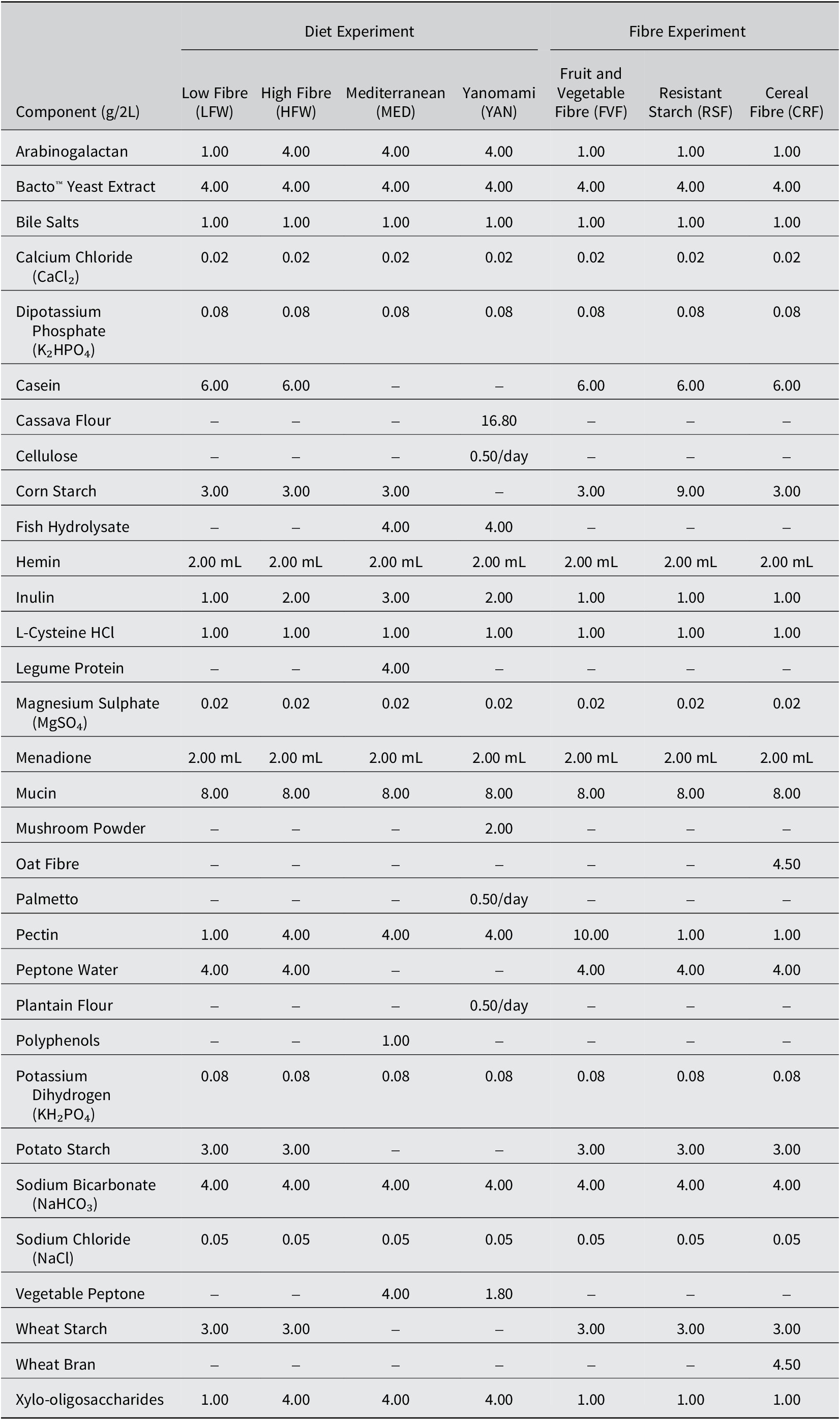
Table 1. Long description
The table lists components in grams per 2 Liters across two main categories: Diet Experiment and Fibre Experiment.
Diet Experiment columns include: Low Fibre (L F W), High Fibre (H F W), Mediterranean (M E D), and Yanomami (Y A N).
Fibre Experiment columns include: Fruit and Vegetable Fibre (F V F), Resistant Starch (R S F), and Cereal Fibre (C R F).
Key component variations include:
* Arabinogalactan: 1.00 for L F W and all Fibre experiments; 4.00 for H F W, M E D, and Y A N.
* Casein: 6.00 for most; absent in M E D and Y A N.
* Cassava Flour: 16.80 only in Y A N.
* Corn Starch: 3.00 for most; 9.00 in R S F; absent in Y A N.
* Fish Hydrolysate: 4.00 only in M E D and Y A N.
* Inulin: Ranges from 1.00 (L F W, Fibre experiments) to 3.00 (M E D).
* Legume Protein: 4.00 only in M E D.
* Oat Fibre: 4.50 only in C R F.
* Pectin: 1.00 for most; 4.00 for H F W, M E D, Y A N; 10.00 for F V F.
* Wheat Bran: 4.50 only in C R F.
* Xylo-oligosaccharides: 1.00 for L F W and Fibre experiments; 4.00 for H F W, M E D, and Y A N.
Standardized components across all diets include Bacto Yeast Extract (4.00), Bile Salts (1.00), C a C l sub 2 (0.02), K sub 2 H P O sub 4 (0.08), Hemin (2.00 m L), L-Cysteine H C l (1.00), M g S O sub 4 (0.02), Menadione (2.00 m L), Mucin (8.00), K H sub 2 P O sub 4 (0.08), N a H C O sub 3 (4.00), and N a C l (0.05).
Flowchart of the bioreactor experiment. All bioreactors were inoculated with the expanded faecal samples on Day 0. Beginning on Day 1, the bioreactors received the diet media at a constant rate. The diet experiments received the same type of diet growth media until Day 21, while the fibre experiments all received the LFW diet until Day 28 or was switched to a different fibre source on Day 17 until Day 28. CRF, cereal fibre; FVF, fruit and vegetable fibre; HFW, high fibre Western; LFW, low fibre Western; MED, Mediterranean; RSF, resistant starch fibre; YAN, Yanomami.

Figure 1. Long description
The flowchart is divided into two horizontal tracks: Diets at the top and Fibres at the bottom.
* Diets Track:
- Starts at Day 0 with Inoculation, represented by a large blue circle.
- Progresses to Day 1 for Feed On, represented by a smaller blue circle.
- A dashed arrow above this track lists the diet types: Low Fibre Western, High Fibre Western, Mediterranean, and Yanomami.
- Ends at Day 21 with Harvesting, represented by a grey circle.
* Fibres Track:
- Starts at Day 0 with Inoculation (large blue circle) and Day 1 with Feed On (small blue circle).
- A dashed arrow indicates the initial diet is Low Fibre Western.
- At Day 17, a red circle marks a Media Change. A dashed arrow following this point lists the new fibre sources: Cereal Fibre, Fruit and Vegetable Fibre, Resistant Starch, and Low Fibre Western.
- Ends at Day 28 with Harvesting, represented by a grey circle.
Vertical arrows connect the shared milestones of Inoculation Day 0 and Feed On Day 1 between the two tracks.
Extraction of bioreactor metabolites
Cell-free metabolites were isolated from the vessel contents of the bioreactor harvested on the last day of each bioreactor experiment (adapted from Ganobis et al., Reference Ganobis, al-Abdul-Wahid, Renwick, Yen, Carriero, Aucoin and Allen-Vercoe2020). In brief, frozen effluents were thawed at sequentially lower temperatures, first from −80 °C to −20 °C for at least 12 hours and then −20 °C to 4 °C for at least 12 hours. Once thawed, the samples were centrifuged at 5,000 × g for 15 minutes at 4 °C to remove crude particulate matter and then again at 14,000 × g for 15 minutes at 4 °C to remove cells and other remaining particulates. The resulting supernatant was filter-sterilised using a 0.22 μM polyether sulfone filter and stored at −20 °C until further processing. Every metabolite sample was plated on a non-selective brain-heart infusion (BHI) plate and incubated at 28.5 °C for 24 hours to confirm sterility (i.e., zero visible colonies).
16S rRNA gene sequencing
Genomic DNA was extracted using the QIAmp® PowerFecal® Pro DNA Kit (QIAGEN) following the manufacturer’s instructions. Partial 16S ribosomal RNA (rRNA) gene sequences of the V3–V4 regions were amplified via PCR and sequenced on the Illumina MiSeq platform (2 × 300 bp, at a depth of 100,000 reads per sample) at the University of Guelph’s Advanced Analysis Centre. The R package DADA2 v1.30.0 was used to process paired end reads and create amplicon sequence variant (ASV) tables. Taxonomy was assigned using the assignTaxonomy function from the DADA2 package with a set of reference sequences via the Silva 138.1 prokaryotic SSU database. Resulting classified ASV data were imported to Phyloseq v1.38.0 for visualisation.
Nuclear magnetic resonance (NMR) spectroscopy and analysis
To determine the metabolic profiles of the bioreactor samples, prepared metabolites were mixed with 10% Chenomx IS-2 standard containing 4.64 mM 3-(Trimethylsilyl)-1-propanesulphonic acid sodium salt in deuterated water and subjected to 1-D 1H NMR using the Bruker Avance 600 MHz NMR spectrometer at the University of Guelph NMR Facility. The concentrations of major SCFAs in the NMR spectra were determined via the Chenomx NMR Suite program, version 8.6.
Zebrafish maintenance and embryo spawning
Adult wild-type Tübingen (TU) zebrafish (Danio rerio) lines (1 to 1.5 years of age) were kept and maintained in 8 L aquaria in mixed sex groups in a dedicated zebrafish rack system (Tecniplast) with automated temperature, pH, and conductivity control in the Hagen Aqualab at the University of Guelph. The system was maintained at 28.5 °C, pH 7.0, 450–550 μS/cm on a 14:10 light/dark simulated photoperiod and fed commercial flake food or brine shrimp 2–3 times daily. Water chemistry was tested weekly and ammonia, nitrate, and nitrite conditions were routinely at 0.0 ppm.
Prior to the end of the photoperiod and preceding the day of embryo collection, adult male and female zebrafish were sexed and sorted into spawning baskets with a barricade to separate the sexes. At the onset of the next photoperiod, the barricades were removed to allow natural spawning. Embryos were collected within 2 hours post-fertilisation (2hpf) and kept in zebrafish embryo medium (EM; 0.00006% w/v Instant Ocean® Sea Salt solution and 0.001% methylene blue in purified distilled water) at 28.5 °C.
Generation and treatment of germ-free zebrafish
To explore the effects of metabolites, zebrafish embryos from pooled clutches were sterilised to remove exogenous bacteria. In brief, at 30% to 50% epiboly (approximately 5hpf), embryos were divided into conventionally raised (CV) and sterilised groups. The CV embryos were kept at room temperature (RT) alongside the embryos being sterilised to ensure similar development progression. Using sterile technique, the embryos were immersed in filter-sterilised gentamicin (100 μg/mL) for 1 hour, followed by three 5-minute washes in 0.003% filter-sterilised hypochlorite solution and sterile EM performed in a sterile biosafety cabinet (adapted from Pham et al., Reference Pham, Kanther, Semova and Rawls2008). Post-sterilisation, sterilised embryos were further divided into treatment groups receiving 1% v/v metabolite treatment or no metabolite treatment (germ-free; GF) (adapted from Rea et al., Reference Rea, Bell, Ball and van Raay2022), with each group having 30–35 embryos each. This concentration was determined to be the highest concentration that did not induce mortality or developmental abnormalities.
All embryos were then incubated at 28.5 C and kept at the same 14:10 light/dark photoperiod. At 1-day post-fertilisation (dpf), one embryo from each treatment group was homogenised in 50 μL of sterile EM. A 20 μL sample was plated on BHI agar plates and then incubated at 28.5 °C for 24 hours to test for sterility. Upon confirmation of sterility (i.e., zero visible colonies), embryos (including the CV embryos) were kept at 28.5 °C in the same media and harvested at 3dpf. Embryos were routinely monitored, with abnormal or dead embryos/larva removed aseptically.
Heat, osmotic, and swirling stressor challenges
Sterilised embryos were assigned to treatment groups as described above. At 3dpf, 10 zebrafish larvae from each treatment group (diets only) were exposed to one stressor challenge lasting 1 hour alongside a non-stressed counterpart where the larvae remained at RT. Therefore, for a single stressor, there were 6 treatment groups (CV, GF, LFW, HFW, MED, and YAN), each with 10 larvae receiving a stressor alongside a non-stressed counterpart.
For the heat stress, larvae were placed in a 50 mL falcon tube containing 20 mL pre-warmed EM and submerged in a 37 °C water bath (adapted from Lim and Bernier, Reference Lim and Bernier2022). For the osmotic stress, larvae were transferred into a 10 cm petri dish containing 20 mL of a 150 mM salt solution and incubated at RT (adapted from Cheng et al., Reference Cheng, Tan, Chua, Tan and Wee2022). Lastly, for the swirling stress, larvae were transferred into a 10 cm petri dish containing 20 mL EM, placed on a high-speed rotator (Stovall Life Sciences, Inc.) and swirled at approximately 5 rotations per second (adapted from Alsop and Vijayan, Reference Alsop and Vijayan2008). Shortly after the stressor, 5 larvae were harvested and macerated in 700 μL Trizol, flash frozen at −80 °C, and processed for qRT-PCR.
Quantitative RT-PCR (qRT-PCR)
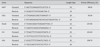
Total RNA from a pool of five 3dpf larvae was extracted with Trizol following the recommendations in the GENEzol™ TriRNA Pure Kit (Geneaid Biotech Ltd.). qRT-PCR was performed on the CFX96 Touch Real-Time Detection System (Bio-Rad) using the Luna Universal One-Step qRT-PCR Kit (New England Biolabs). Primer efficiencies were measured using SYBR Green Master Mix for qPCR (ThermoFisher) and confirmed to be within 95% to 110% (Table 2) using standard curves. Each biological replicate for qRT-PCR included 3 technical replicates and was normalised to the housekeeping gene, ef1a. Gene expressions were calculated using the Livak method and expressed in fold changes.
Details of primer sequences used in qRT-PCR.
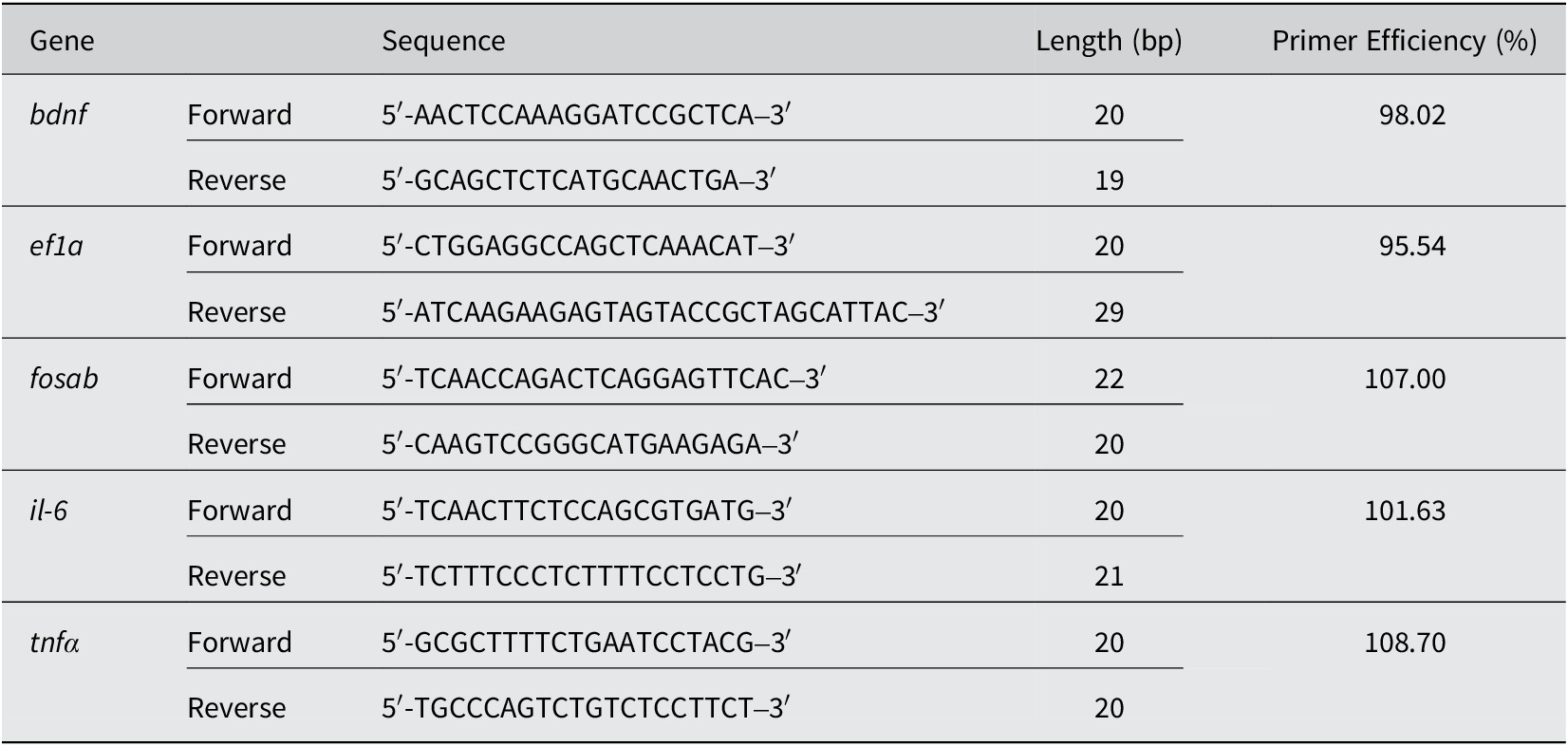
Table 2. Long description
The table consists of five columns: Gene, Direction (Forward or Reverse), Sequence (5 prime to 3 prime), Length in base pairs (b p), and Primer Efficiency percentage.
* b d n f: Forward sequence 5 prime A A C T C C A A A G G A T C C G C T C A 3 prime (20 b p); Reverse sequence 5 prime G C A G C T C T C A T G C A A C T G A 3 prime (19 b p). Efficiency is 98.02 percent.
* e f 1 a: Forward sequence 5 prime C T G G A G G C C A G C T C A A A C A T 3 prime (20 b p); Reverse sequence 5 prime A T C A A G A A G A G T A G T A C C G C T A G C A T T A C 3 prime (29 b p). Efficiency is 95.54 percent.
* f o s a b: Forward sequence 5 prime T C A A C C A G A C T C A G G A G T T C A C 3 prime (22 b p); Reverse sequence 5 prime C A A G T C C G G G C A T G A A G A G A 3 prime (20 b p). Efficiency is 107.00 percent.
* i l - 6: Forward sequence 5 prime T C A A C T T C T C C A G C G T G A T G 3 prime (20 b p); Reverse sequence 5 prime T C T T T C C C T C T T T T C C T C C T G 3 prime (21 b p). Efficiency is 101.63 percent.
* t n f alpha: Forward sequence 5 prime G C G C T T T T C T G A A T C C T A C G 3 prime (20 b p); Reverse sequence 5 prime T G C C C A G T C T G T C T C C T T C T 3 prime (20 b p). Efficiency is 108.70 percent.
Statistical analyses
All data obtained were tested for normality and equal variances. Differences between the treatment groups were analysed by an analysis of variance (ANOVA) or by a t-test (as specified in each figure) with significance set at p < 0.05. Data sets that do not meet normality and variance assumptions were evaluated using non-parametric tests. All analyses were performed using GraphPad Prism version 10.0.0 for Windows. For the diets and fibre source comparisons, LFW was used as the control. For the zebrafish comparisons, the CV group was used as the control. For the stressor challenge, a non-stressed baseline was used as the control. In the figure legend, uppercase “N” refers to the number of biological replicates.
Results
Donor sample selection
Metagenomic shotgun sequencing of the 10 donor samples was used to determine the taxonomic profiles of each faecal sample (Figure 2A). A Principal Component Analysis (PCA) was used to illustrate the variations between donors so that representative samples could be selected. The PCA plot revealed 3 clusters, wherein PC1 and PC2 describe 25.83% and 17.88% of the variation, respectively (Figure 2B). A third PC describes an additional 13.60% variation, but no new clusters were formed. In addition, a k-medoid model plot revealed similar findings in that 3 clusters were formed. In both plots, donors 6 and 8 formed their own clusters, while the rest of the samples clustered together. From these analyses, donors 1 and 2 were chosen as representatives of the pool as these samples were at the centre of its cluster. Donor 1 was a female, 4.3 years old with a history of one antibiotic use (specifically, amoxicillin) and donor 2 was a male, 3 years old with two recorded prior antibiotic use (specifically, amoxicillin and ampicillin). The timing of the antibiotic use is unknown.
Microbial analysis of 10 donor samples. (A) Stacked bar plot showing the top four phyla detected in all 10 donors. The category “Other” describes the phyla with a maximum relative abundance of less than 5% from all samples and was only found in donors 1, 5, 7, 8, and 9. (B) Two-dimensional PCA plot showing the variance between all 10 donors. Enhanced cluster detection using k-medoid identification demonstrated that donors 6 and 8 are separate from the group. From this analysis, donors 1 and 2 were chosen to be the most representative of the group.
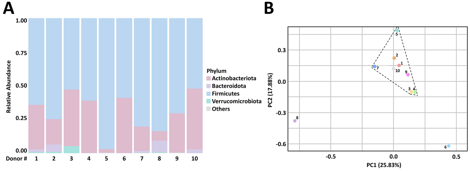
Figure 2. Long description
Panel A is a stacked bar plot. The y-axis is labeled Relative Abundance from 0.00 to 1.00. The x-axis is labeled Donor number with 10 individual bars numbered 1 through 10. Each bar is composed of colored segments representing different phyla. A legend to the right identifies the colors. Firmicutes in light blue is the dominant phylum across all donors. Actinobacteriota in pink is the second most prevalent. Bacteroidota in light purple and Verrucomicrobiota in teal appear in smaller proportions. A light gray segment for Others is visible in donors 1, 5, 7, 8, and 9.
Panel B is a two-dimensional P C A plot. The x-axis is P C 1 (25.83 percent) and the y-axis is P C 2 (17.88 percent). A dashed line polygon encloses a central cluster of eight donors labeled 1, 2, 3, 4, 5, 7, 9, and 10. Donor 8 is located as an outlier in the bottom-left quadrant. Donor 6 is located as an outlier in the bottom-right quadrant.
Diets and fibre source did not impact the microbiota cultured in bioreactors
To determine if different diets affected overall microbial profiles, we submitted samples from each diet/fibre source and donor for 16S rRNA gene sequencing analyses at the end of the bioreactor experiments. Despite being supplied different diets, it was revealed that both donors had similar microbial profiles with no clear shifts in microbial compositions or diversity after exposure to each diet (Figure 3A). The distribution of the microbial compositions was found to be consistent between technical replicates, with the most dominant phyla being Bacillota, Bacteroidota, and Verrucomicrobiota. A small portion of the microbes belongs to the Pseudomonadota and Actinomycetota phyla. We then sought to determine if different fibre sources influenced overall microbial profiles but saw no significant changes despite being supplied with different fibre sources (Figure 3B, Supplementary Figure S1). Collectively, this suggests that the different diets and the different fibre sources did not dramatically alter the microbial composition or diversity of the phyla over the course of the experiment.
Changes in diets or fibre sources do not significantly alter the microbial composition of the gut microbiome. (A) Bioreactors were supplied with either the LFW, HFW, MED, or YAN diet media for 21 days. (B) Bioreactors were supplied with the FVF, RSF, or CRF diet media at Day 17 and grown for another 10 days; LFW had no added fibre. Effluents were collected at the end of each experiment, and an aliquot was subjected to 16S sequencing. The most dominant phyla found were Bacillota, Bacteroidota, and Verrucomicrobiota. Two biological replicates are shown for each donor, except for CRF for donor 1.
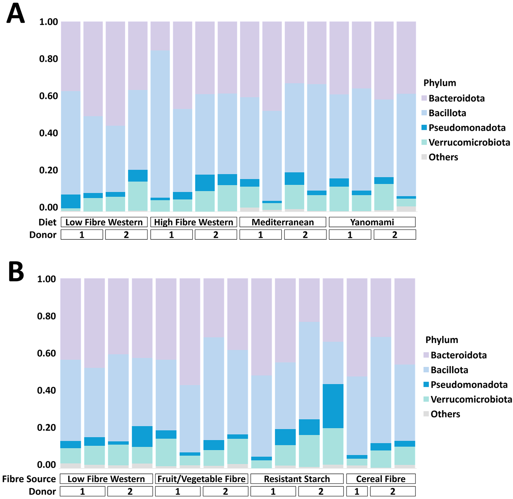
Figure 3. Long description
Panel A is a stacked bar chart with a Y axis ranging from 0.00 to 1.00 representing relative abundance. The X axis is categorized by Diet: Low Fibre Western, High Fibre Western, Mediterranean, and Yanomami. Each diet is further divided into Donor 1 and Donor 2, with two replicates per donor. A legend to the right identifies the phyla from bottom to top as: Others (grey), Verrucomicrobiota (light green), Pseudomonadota (dark blue), Bacillota (light blue), and Bacteroidota (purple). Across all diets, Bacteroidota and Bacillota are the most dominant, typically making up over 80 percent of the composition. Verrucomicrobiota and Pseudomonadota show minor fluctuations but remain low in abundance.
Panel B follows the same Y axis and legend structure but categorizes the X axis by Fibre Source: Low Fibre Western, Fruit forward slash Vegetable Fibre, Resistant Starch, and Cereal Fibre. Data is again split by Donor 1 and Donor 2. The microbial composition remains largely consistent with Panel A, dominated by Bacteroidota and Bacillota. A notable variation occurs in Donor 2 under the Resistant Starch category, where Pseudomonadota and Verrucomicrobiota show a visible increase in relative abundance compared to other samples.
A comparison of the diversity between the raw faecal samples (Figure 2) and the samples collected at the end of both bioreactor experiments (Figure 3) demonstrated that Bacillota remained one of the dominant phyla. We also observed a marked decrease in Actinomycetota and an increase in Bacteroidota after both experiments, suggesting that the media used in both experiments may have favoured the growth of the latter phyla over the former. Nonetheless, the microbial phyla seen at the end of both experiments are consistent with the microbes found in the human gut (Ley et al., Reference Ley, Peterson and Gordon2006; Biagi et al., Reference Biagi, Nylund, Candela, Ostan, Bucci, Pini, Nikkïla, Monti, Satokari, Franceschi, Brigidi and de Vos2010; Arumugam et al., Reference Arumugam, Raes, Pelletier, le Paslier, Yamada, Mende, Fernandes, Tap, Bruls, Batto, Bertalan, Borruel, Casellas, Fernandez, Gautier, Hansen, Hattori, Hayashi, Kleerebezem, Kurokawa, Leclerc, Levenez, Manichanh, Nielsen, Nielsen, Pons, Poulain, Qin, Sicheritz-Ponten, Tims, Torrents, Ugarte, Zoetendal, Wang, Guarner, Pedersen, de Vos, Brunak, Doré, Antolín, Artiguenave, Blottiere, Almeida, Brechot, Cara, Chervaux, Cultrone, Delorme, Denariaz, Dervyn, Foerstner, Friss, van de Guchte, Guedon, Haimet, Huber, van Hylckama-Vlieg, Jamet, Juste, Kaci, Knol, Lakhdari, Layec, le Roux, Maguin, Mérieux, Melo Minardi, M’rini, Muller, Oozeer, Parkhill, Renault, Rescigno, Sanchez, Sunagawa, Torrejon, Turner, Vandemeulebrouck, Varela, Winogradsky, Zeller, Weissenbach, Ehrlich and Bork2011; Tremaroli and Bäckhed, Reference Tremaroli and Bäckhed2012; Vieira-Silva et al., Reference Vieira-Silva, Falony, Darzi, Lima-Mendez, Garcia Yunta, Okuda, Vandeputte, Valles-Colomer, Hildebrand, Chaffron and Raes2016; Rinninella et al., Reference Rinninella, Raoul, Cintoni, Franceschi, Miggiano, Gasbarrini and Mele2019; Salazar et al., Reference Salazar, Angarita, Morillo, Navarro, Martínez, Chacín, Torres, Rajotia, Rojas, Cano, Añez, Rojas and Bermudez2020; Malesza et al., Reference Malesza, Malesza, Walkowiak, Mussin, Walkowiak, Aringazina, Bartkowiak-Wieczorek and Mądry2021), indicating that the bioreactor is indeed a good model to mimic the environment of the gut.
Diets and fibre source have unique effects on microbial metabolite output
Despite the lack of appreciable microbial changes between diets or fibre sources, it is still possible that the metabolic profiles between the diets and/or fibre sources are different. To test this, we performed 1-D 1H NMR to evaluate changes in the concentrations of the 3 major SCFAs: acetate, butyrate, and propionate. For the diet experiment, the SCFA concentrations remained relatively stable for the duration of the bioreactor runs (Figure 4A). Here, acetate concentrations were the highest across all diets, followed by propionate then butyrate concentrations; however, in the YAN diet, acetate concentrations were followed by butyrate instead of propionate concentrations. For the fibre experiment, steady increases in SCFA concentrations were observed following the addition of fibres at Day 17 (Figure 4B), with FVF having a meaningful effect on acetate levels. Overall, there were no significant differences in SCFA concentrations between donors (Supplementary Figures S2 and S3) and the changes observed over time were consistent between donors (data not shown).
SCFA concentrations reach stability over time. Donors and biological replicates are pooled. (A-B) Metabolites were harvested at the indicated time points and submitted for 1-D 1H NMR analysis. In the diet experiment, stability appears to be reached at Day 14, while in the fibre experiment where fibre was added at Day 17, stability appears to be reached at approximately Day 26. In both experiments, acetate concentrations were consistently the highest, followed by propionate then butyrate concentrations (except for YAN). Acetate concentrations were significantly higher on Day 28 when compared to Day 16 in the FVF fibre source (t (6) = 4.16, p = 0.025). (C-D) There was a significant effect of diet on acetate (F (3, 11) = 11.61, p = 0.001) and butyrate (F (3, 11) = 10.35, p = 0.003) in the diet experiment and a post-hoc test showed that acetate concentrations in HFW (p = 0.03) and YAN (p = 0.0006) and butyrate concentrations in YAN (p = 0.0042) were significantly different from LFW. A significant effect of fibre was found on acetate (F (3, 10) = 11.31, p = 0.0001), with a post-hoc revealing a significant difference between FVF and LFW acetate concentrations (p = 0.004). Error bars represent standard deviation (SD). * p < 0.05, ** p < 0.01, *** p < 0.001 using a one-way ANOVA or Kruskal-Wallis test.
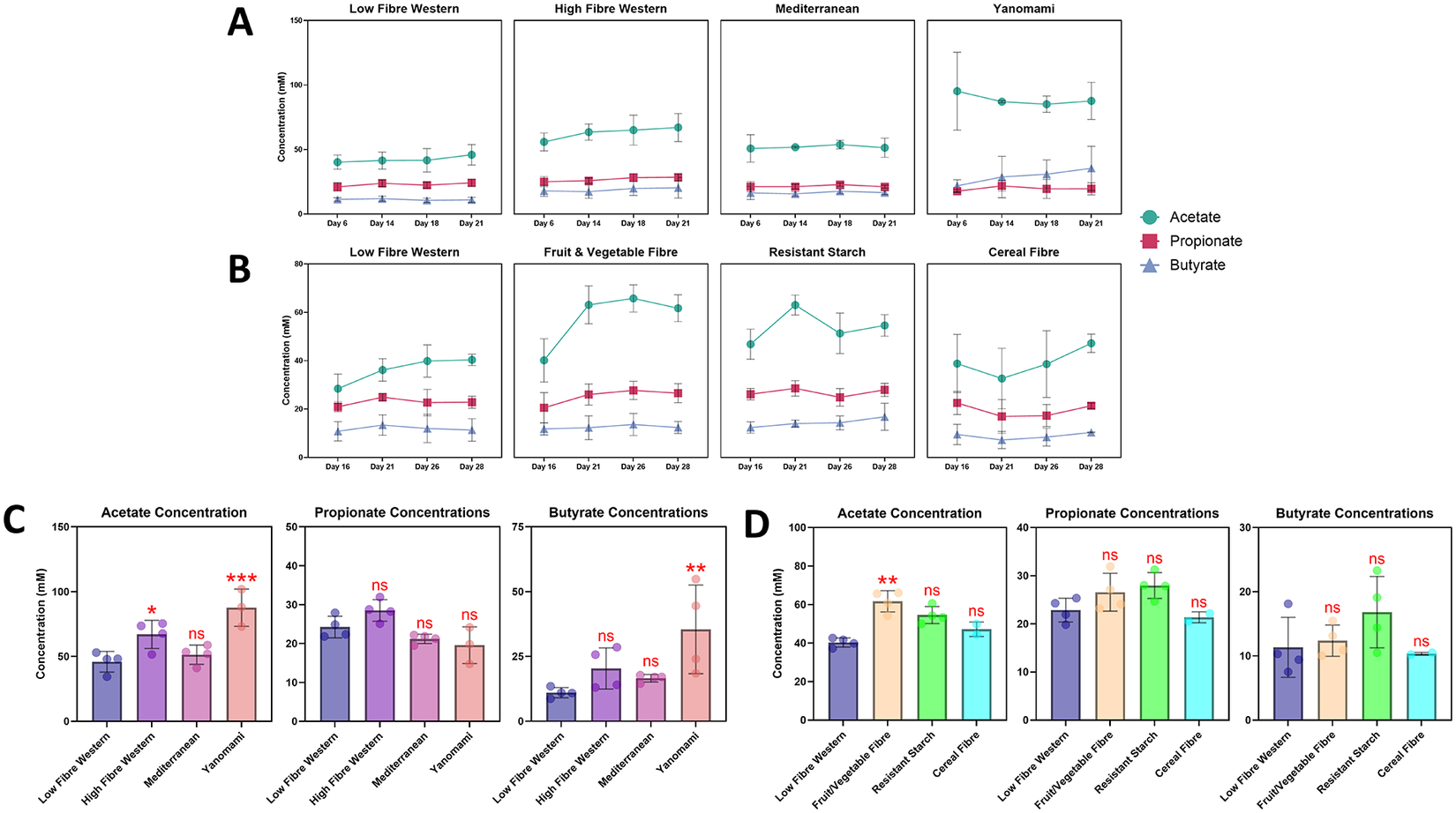
Figure 4. Long description
The figure consists of four main sections labeled A through D.
Row A: Four line graphs showing concentration in m M from Day 6 to Day 21 for Low Fibre Western, High Fibre Western, Mediterranean, and Yanomami diets. Acetate (teal circles) is highest, ranging from 40 to 100 m M. Propionate (pink squares) and Butyrate (blue triangles) remain lower and stable between 10 and 30 m M.
Row B: Four line graphs showing concentration from Day 16 to Day 28 for Low Fibre Western, Fruit and Vegetable Fibre, Resistant Starch, and Cereal Fibre. Acetate shows a sharp increase in the Fruit and Vegetable Fibre and Resistant Starch groups between Day 16 and Day 21, peaking near 60 m M.
Row C: Three bar charts comparing Acetate, Propionate, and Butyrate concentrations across the four diets from Row A. Acetate shows a significant increase in High Fibre Western (one asterisk) and Yanomami (three asterisks) compared to Low Fibre Western. Butyrate shows a significant increase in Yanomami (two asterisks).
Row D: Three bar charts comparing Acetate, Propionate, and Butyrate concentrations across the four fibre types from Row B. Acetate is significantly higher in Fruit and Vegetable Fibre (two asterisks) compared to Low Fibre Western. Other comparisons are marked n s for non-significant.
Legend: Teal circles represent Acetate. Pink squares represent Propionate. Blue triangles represent Butyrate. Error bars indicate S D.
We then compared the SCFA concentrations at the end of each experiment (Day 21 for the diets, Day 28 for the fibres) as these metabolites would be used for the subsequent experiments. Although we could not perform statistical tests for each donor due to the limited number of biological replicates, we did observe some trends (Supplementary Figures S2 and S3). For the diet experiment, acetate and butyrate concentrations in both donors tended to be higher in HFW, MED, and YAN when compared to LFW. Propionate concentrations in both donors were highest in HFW but were similar in MED and YAN when compared to LFW (Supplementary Figure S2). For the fibre experiment, most SCFAs saw modest increases in concentrations following the addition of the fibres (Supplementary Figure S3). Overall, both donors had similar metabolite profiles and follow the general 60:20:20 ratio for the 3 major SCFAs. When both donors and replicates are pooled, it was revealed that acetate concentrations from the HFW (p < 0.05) and YAN (p < 0.001) diets, as well as the butyrate concentrations from the YAN diet (p < 0.01), were significantly different from the LFW diet (Figure 4C). Likewise, the acetate concentrations in the FVF fibre source were found to be much higher on Day 28 compared to Day 16 (Figure 4B) but were also significantly different from LFW on Day 28 (Figure 4D; p < 0.01). Collectively, these results suggest that diets and fibre impact microbial metabolite output while the microbial composition remains relatively stable.
The source of metabolites influences gene expression in the developing zebrafish
To better understand the contributions of diet on the developing nervous system, we took advantage of the zebrafish model where we previously demonstrated that bacterially derived metabolites are both necessary and sufficient for proper nervous system development (Rea et al., Reference Rea, Bell, Ball and van Raay2022). In the current paradigm, we added cell-free metabolites generated from the bioreactors to GF zebrafish embryos at 30 to 50% epiboly and then pooled 5 zebrafish larvae at 3dpf for qRT-PCR to assess changes in the expression of key neural and inflammatory markers (Figure 5).
The effects of metabolites on the expression of neurodevelopmental and pro-inflammatory genes were limited, but distinct between donors. GF embryos were treated with metabolites at approximately 5hpf, harvested at 3dpf, and subjected to qRT-PCR. The expression of bdnf, fosab, il-6, and tnfα were measured, normalized to ef1α, and made relative to the CV group that had received no treatment. GF embryos that did not receive metabolite treatment were also evaluated. (A) Metabolites derived from diets did not seem to affect the expression of bdnf and fosab. Although no significant effect of metabolites treatment was observed, a post-hoc pairwise comparison revealed that the expression of il-6 in YAN for donor 1 was significantly different compared to the CV group (p = 0.032). (B) Metabolites derived from different fibre source had minimal effects on the expression of the targeted genes, with the exception of il-6 (F(4, 9) = 9.70, p = 0.013), noting a significant difference between CV and RSF groups (p = 0.034). N = 3. Error bars represent standard error of means (SEM). *p < 0.05 using a one-way ANOVA or Kruskal-Wallis test.
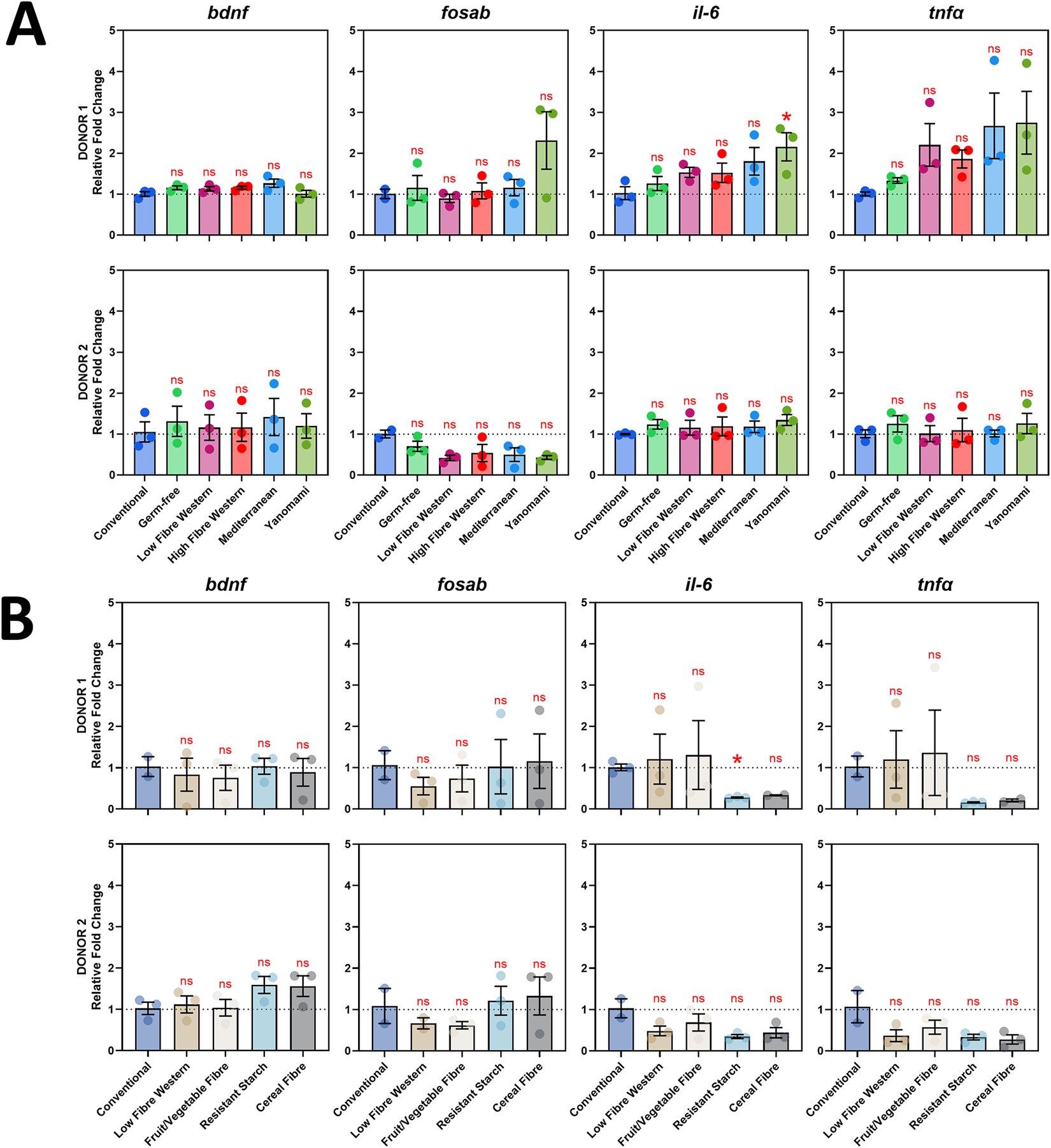
Figure 5. Long description
A multi-panel set of bar graphs divided into sections A and B. All graphs share a Y-axis labeled Relative Fold Change ranging from 0 to 5, with a horizontal dashed line at 1 representing the baseline. The X-axis lists treatment groups.
Section A: Diet-derived metabolites. The top row shows Donor 1 and the bottom row shows Donor 2. Columns from left to right represent genes b d n f, f o s a b, i l-6, and t n f alpha.
* For Donor 1, i l-6 shows a significant increase in the Yanomami group marked with a red asterisk. Other treatments including Conventional, Germ-free, Low Fibre Western, High Fibre Western, and Mediterranean show non-significant (n s) changes.
* For Donor 2, all genes across all treatments remain near baseline with n s labels.
Section B: Fibre source metabolites. The layout mirrors Section A but with different X-axis categories: Conventional, Low Fibre Western, Fruit forward slash Vegetable Fibre, Resistant Starch, and Cereal Fibre.
* For Donor 1, i l-6 shows a significant decrease in the Resistant Starch group marked with a red asterisk.
* For Donor 2, all gene expressions across all fibre treatments are marked n s, indicating no significant difference from the baseline.
Error bars on all graphs represent S E M. Individual data points are overlaid on the bars as semi-transparent dots.
Despite the changes observed in the metabolic profiles, this did not translate into significant changes in the expression of the four genes we evaluated; however, we did observe unique expression patterns between donors. When assessing metabolites derived from different diets, the expression of both il-6 and tnfα tended to be higher in zebrafish embryos treated with donor 1 metabolites when compared to CV zebrafish embryos, while fosab expression was lower in zebrafish embryos treated with donor 2 metabolites (Figure 5A). For metabolites derived from different fibre sources, we observed that the expression of the four genes evaluated remained relatively consistent between donors, but with a trend of reduced il-6 and tnfα expression in the RSF and CRF fibre sources for both donors. Unlike the diet metabolites, the pattern of expression observed between donors in the fibre experiment was comparable (Figure 5B). Taken together, these results indicate that the metabolites used in these experiments have limited impact on gene expression in the developing zebrafish nervous and immune systems, at least for the four genes we evaluated.
Metabolites can ameliorate some of the effects of stress
Given the neuroactive properties of diets high in fibre, we wanted to determine if the different diets can ameliorate the effects of stressors. We hypothesised that diets rich in fibre (HFW, MED, and YAN) would be able to assuage the effects of stress when compared to the diets LFW. To test this, we chose 3 stressors commonly used in zebrafish studies: swirling, osmotic, and heat (Alsop and Vijayan, Reference Alsop and Vijayan2008; Cheng et al., Reference Cheng, Tan, Chua, Tan and Wee2022; Lim and Bernier, Reference Lim and Bernier2022). First, we evaluated how the stressors affected gene expression in CV and GF zebrafish larvae at 3dpf. For the CV group, the swirling stress caused a significant increase in the expression of bdnf (Figure 6A, Controls; p < 0.01) and tnfα (Supplementary Figure S4A; p < 0.05). The osmotic and heat stress significantly increased the expression of tnfα only (Supplementary Figure S4B and S4C; p < 0.05 and p < 0.01, respectively). We then predicted that the GF group, devoid of microbes and metabolites, would have exacerbated gene expression; however, we only observed significant increases in bdnf (Figure 6A, Controls; p < 0.05) and fosab expression (Supplementary Figure S5A; p < 0.01) for the swirling stress, while we saw significant increases in bdnf (Figure 6B, Controls; p < 0.05) and fosab expression (Supplementary Figure S5C; p < 0.05) for the osmotic and heat stress, respectively. There were no significant changes in il-6 expression for CV or GF under any of the stressors (Supplementary Figure S6). These results indicate that the expression of bdnf seems to be the most sensitive to the three stressors, with the swirling stressor having a greater effect than the other two stressors.
The metabolites can ameliorate the effects of stressors on bdnf expression. (A) A significant increase in bdnf expression was observed following an hour-long swirling stressor in the CV and GF groups. Similar findings were seen in the LFW, MED, and YAN groups for donor 1, but these were reduced to non-significant levels for donor 2. (B) Only the GF group saw a significant increase in bdnf expression following the osmotic stressor. The CV and all metabolite-treated groups were non-significant. (C) The heat stressor did not affect the expression of bdnf in all groups. N = 6 for controls, N = 3 for metabolite treatment groups. Error bars represent standard deviation * p < 0.05, ** p < 0.01, *** p < 0.001 using an unpaired t-test or Mann–Whitney test.
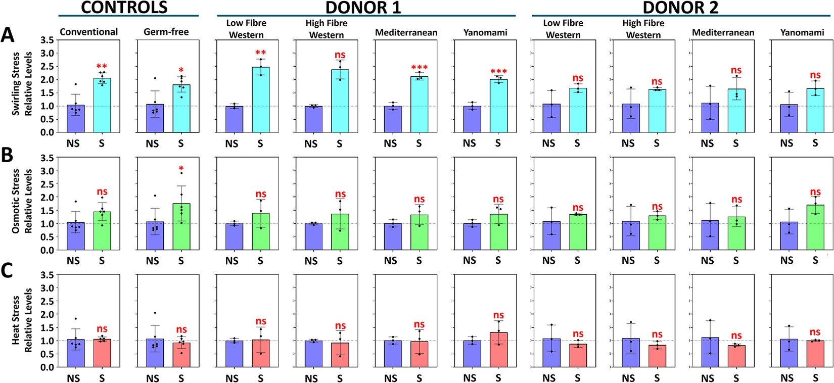
Figure 6. Long description
A grid of 30 bar graphs organized into three rows (A, B, C) and three main columns (Controls, Donor 1, Donor 2). The Y-axis for all graphs represents Relative Levels from 0.0 to 3.5. The X-axis for each graph compares N S (No Stress, blue bars) to S (Stress, colored bars).
Row A: Swirling Stress (light blue stress bars). In Controls, both Conventional and Germ-free show significant increases (double asterisk and single asterisk). In Donor 1, all four groups (Low Fibre Western, High Fibre Western, Mediterranean, Yanomami) show increases, with Mediterranean and Yanomami being highly significant (triple asterisk). In Donor 2, all groups show non-significant (n s) increases.
Row B: Osmotic Stress (green stress bars). Only the Germ-free control shows a significant increase (single asterisk). All other groups across Controls, Donor 1, and Donor 2 show non-significant (n s) changes.
Row C: Heat Stress (red stress bars). All groups across Controls, Donor 1, and Donor 2 show non-significant (n s) changes, with bars remaining near the baseline level of 1.0.
Statistical significance is marked above the S bars: n s for non-significant, asterisk for p less than 0.05, double asterisk for p less than 0.01, and triple asterisk for p less than 0.001. Error bars indicate standard deviation.
Next, we added the different metabolites to GF embryos and evaluated gene expression under the same stressors. While we observed a significant effect of diets, contrary to our hypothesis, we did not observe any diet-specific effects and the LFW diet-affected gene expression similar to the other diets (Figure 6). More specifically, we observed that the bdnf expression was rescued (i.e., reduced to non-significant levels) by metabolites derived from donor 2, but not donor 1 in the swirling stress (Figure 6A). In contrast, metabolites from both donors were able to rescue bdnf expression from the effects of osmotic stress (Figure 6B). Curiously, the heat stress did not seem to impact bdnf expression (Figure 6C). After evaluating the expression of fosab, il-6, and tnfα, it was revealed that the fosab had variable and inconsistent results (Supplementary Figure S5), while both il-6 (Supplementary Figure S6) and tnfα (Supplementary Figure S4) seemed to be relatively stable following all three stressors. Overall, no discernible differences between diets could be identified. Nonetheless, we saw that bdnf expression appears to be amenable to both stressor and metabolites and, likewise, follows a donor-specific pattern of expression consistent with our previous results.
Discussion
Gut-derived microbial metabolites have gained increasing attention for their ability to influence the brain and behaviour (Cryan and Dinan, Reference Cryan and Dinan2012; Mayer et al., Reference Mayer, Tillisch and Gupta2015; Agustí et al., Reference Agustí, García-Pardo, López-Almela, Campillo, Maes, Romaní-Pérez and Sanz2018; Appleton, Reference Appleton2018; Van de Wouw et al., Reference Van de Wouw, Boehme, Clarke, Cryan, Dinan, Lyte, O’Sullivan, Stanton, Strain and Wiley2018; Cryan et al., Reference Cryan, O’Riordan, Cowan, Sandhu, Bastiaanssen, Boehme, Codagnone, Cussotto, Fulling, Golubeva, Guzzetta, Jaggar, Long-Smith, Lyte, Martin, Molinero-Perez, Moloney, Morelli, Morillas, O’Connor, Cruz-Pereira, Peterson, Rea, Ritz, Sherwin, Spichak, Teichman, van de Wouw, Ventura-Silva, Wallace-Fitzsimons, Hyland, Clarke and Dinan2019; Marx et al., Reference Marx, Lane, Hockey, Aslam, Berk, Walder, Borsini, Firth, Pariante, Berding, Cryan, Clarke, Craig, Su, Mischoulon, Gomez-Pinilla, Foster, Cani, Thuret, Staudacher, Sánchez-Villegas, Arshad, Akbaraly, O’Neil, Segasby and Jacka2021; Horn et al., Reference Horn, Mayer, Chen and Mayer2022; Loh et al., Reference Loh, Mak, Tan, Ng, Chan, Yeow, Foo, Ong, How and Khaw2024; Ross et al., Reference Ross, Mayer, Horn, Cryan, del Rio, Randolph, Gill, Gupta, Ross, Stanton and Mayer2024; Schneider et al., Reference Schneider, O’Riordan, Clarke and Cryan2024; Cryan, Reference Cryan2025; O’Riordan et al., Reference O’Riordan, Moloney, Keane, Clarke and Cryan2025; Rosell-Cardona et al., Reference Rosell-Cardona, Leigh, Knox, Tirelli, Lyte, Goodson, Kelley-Loughnane, Aburto, Cryan and Clarke2025) but empirical evidence is lacking. Previous work in our lab demonstrated that metabolites are both necessary and sufficient for timely neurodevelopment. We found that, in the absence of microbes and their metabolites (i.e, germ-free), broad neural gene expression was delayed but was partially rescued by the addition of gut-derived zebrafish metabolites (Rea et al., Reference Rea, Bell, Ball and van Raay2022). Predicated on this work, here we used the GF zebrafish model to evaluate diet-derived metabolites on nervous system development.
Following the expansion of our selected donor faecal samples, we observed a decrease in Actinomycetota and an increase in Bacteroidota phyla. Although these phyla are common in typical gut microbiomes (Dethlefsen et al., Reference Dethlefsen, Huse, Sogin and Relman2008; Turnbaugh et al., Reference Turnbaugh, Vanessa, Jeremiah, Federico, Rob and Jeffrey2009; Ubeda et al., Reference Ubeda, Taur, Jenq, Equinda, Son, Samstein, Viale, Socci, van den Brink, Kamboj and Pamer2010; Walker et al., Reference Walker, Ince, Duncan, Webster, Holtrop, Ze, Brown, Stares, Scott, Bergerat, Louis, McIntosh, Johnstone, Lobley, Parkhill and Flint2011; Wu et al., Reference Wu, Chen, Hoffmann, Bittinger, Chen, Keilbaugh, Bewtra, Knights, Walters, Knight, Sinha, Gilroy, Gupta, Baldassano, Nessel, Li, Bushman and Lewis2011; David et al., Reference David, Materna, Friedman, Campos-Baptista, Blackburn, Perrotta, Erdman and Alm2014; David et al., Reference David, Maurice, Carmody, Gootenberg, Button, Wolfe, Ling, Devlin, Varma, Fischbach, Biddinger, Dutton and Turnbaugh2014; Sonnenburg and Bäckhed, Reference Sonnenburg and Bäckhed2016; Wipperman et al., Reference Wipperman, Fitzgerald, Juste, Taur, Namasivayam, Sher, Bean, Bucci and Glickman2017; De Gunzburg et al., Reference De Gunzburg, Alavoine, Andremont, Armand-Lefevre, Augustin, Burdet, Chachaty, Ducher, Duval, Ghozlane, Kennedy, Chatelier, Levenez, Mentré, Pons, Ruppé, Sablier-Gallis and Varastet2018; Palleja et al., Reference Palleja, Mikkelsen, Forslund, Kashani, Allin, Nielsen, Hansen, Liang, Feng, Zhang, Pyl, Coelho, Yang, Wang, Typas, Nielsen, Nielsen, Bork, Wang, Vilsbøll, Hansen, Knop, Arumugam and Pedersen2018; Valdes et al., Reference Valdes, Walter, Segal and Spector2018; Cabral et al., Reference Cabral, Wurster, Korry, Penumutchu and Belenky2020; Anthony et al., Reference Anthony, Wang, Sukhum, D’Souza, Hink, Cass, Seiler, Reske, Coon, Dubberke, Burnham, Dantas and Kwon2022), this shift may have made the microbial community less responsive to changes in diet or fibre supplementation (De Filippo et al., Reference De Filippo, Cavalieri, Collini, Paola, Lionetti, Massart, Pieraccini, Poullet and Ramazzotti2010; Ou et al., Reference Ou, Carbonero, Zoetendal, DeLany, Wang, Newton, Gaskins and O’Keefe2013; Wu et al., Reference Wu, Compher, Chen, Smith, Shah, Bittinger, Chehoud, Albenberg, Nessel, Gilroy, Star, Weljie, Flint, Metz, Bennett, Li, Bushman and Lewis2016; Biesiekierski et al., Reference Biesiekierski, Jalanka and Staudacher2019; Leeming et al., Reference Leeming, Johnson, Spector and le Roy2019; Ard et al., Reference Ard, Lewis, Cohen, Rothberg, Coburn, Loper, Matarese, Pories and Periman2020; Ostojic, Reference Ostojic2020). Alternatively, the gut microbiome of healthy individuals is typically characterised by its resilience to perturbations (Dethlefsen et al., Reference Dethlefsen, Huse, Sogin and Relman2008; Bäckhed et al., Reference Bäckhed, Fraser, Ringel, Sanders, Sartor, Sherman, Versalovic, Young and Finlay2012; Relman, Reference Relman2012; Manrique et al., Reference Manrique, Dills and Young2017), suggesting that the use of healthy donor samples may explain the lack of significant changes we observed. Because our study was limited to only two healthy donors, future iterations of this experiment should expand the number of donor faecal samples to increase both the sample size and statistical power, as well as include samples from diseased or unhealthy donors. In support of this, preliminary data from our lab has shown that GF zebrafish embryos treated with metabolites from an autistic donor have markedly different effects on gene expression when compared to non-autistic controls.
Despite the lack of microbial distinctions, we found several differences in the metabolic profiles between the diets and fibre sources in a pooled analysis. Specifically, differences in SCFA levels between LFW and the other diets and fibre sources were found, indicating that high-fibre diets or the supplementation of fibre can alter the metabolic profiles of the gut microbiome. Combined with the sequencing results, the differences in SCFA concentrations are strikingly similar to the observations reported by Wu et al. (Reference Wu, Compher, Chen, Smith, Shah, Bittinger, Chehoud, Albenberg, Nessel, Gilroy, Star, Weljie, Flint, Metz, Bennett, Li, Bushman and Lewis2016). In their study, they report that healthy vegans and omnivores had similar gut microbial profiles. While they did not see significant differences in faecal SCFA concentrations after 10 days of dietary intervention, they attributed this to a high intersubject variability of baseline SCFA concentrations, suggesting that SCFA concentrations depend on the subject, consistent with the donor effects observed in our study. Further, work by Fragiadakis et al. (Reference Fragiadakis, Wastyk, Robinson, Sonnenburg, Sonnenburg and Gardner2020) revealed that dietary interventions caused substantial changes in participant gut microbiomes, but their microbiomes eventually returned near its baseline status despite maintaining the dietary interventions. Together, these studies corroborate our results, underscoring the resilience of the gut microbiome.
The gross similarities we observed in the microbial profiles between donors 1 and 2, along with the differences we saw in their metabolic profiles, suggest that subtleties in the microbiome allow individuals to respond differently to the diets and fibres. Given the neuroactive properties of metabolites (Cryan and Dinan, Reference Cryan and Dinan2012; Mayer et al., Reference Mayer, Tillisch and Gupta2015; Agustí et al., Reference Agustí, García-Pardo, López-Almela, Campillo, Maes, Romaní-Pérez and Sanz2018; Appleton, Reference Appleton2018; Van de Wouw et al., Reference Van de Wouw, Boehme, Clarke, Cryan, Dinan, Lyte, O’Sullivan, Stanton, Strain and Wiley2018; Cryan et al., Reference Cryan, O’Riordan, Cowan, Sandhu, Bastiaanssen, Boehme, Codagnone, Cussotto, Fulling, Golubeva, Guzzetta, Jaggar, Long-Smith, Lyte, Martin, Molinero-Perez, Moloney, Morelli, Morillas, O’Connor, Cruz-Pereira, Peterson, Rea, Ritz, Sherwin, Spichak, Teichman, van de Wouw, Ventura-Silva, Wallace-Fitzsimons, Hyland, Clarke and Dinan2019; Marx et al., Reference Marx, Lane, Hockey, Aslam, Berk, Walder, Borsini, Firth, Pariante, Berding, Cryan, Clarke, Craig, Su, Mischoulon, Gomez-Pinilla, Foster, Cani, Thuret, Staudacher, Sánchez-Villegas, Arshad, Akbaraly, O’Neil, Segasby and Jacka2021; Horn et al., Reference Horn, Mayer, Chen and Mayer2022; Loh et al., Reference Loh, Mak, Tan, Ng, Chan, Yeow, Foo, Ong, How and Khaw2024; Ross et al., Reference Ross, Mayer, Horn, Cryan, del Rio, Randolph, Gill, Gupta, Ross, Stanton and Mayer2024; Schneider et al., Reference Schneider, O’Riordan, Clarke and Cryan2024; Cryan, Reference Cryan2025; O’Riordan et al., Reference O’Riordan, Moloney, Keane, Clarke and Cryan2025; Rosell-Cardona et al., Reference Rosell-Cardona, Leigh, Knox, Tirelli, Lyte, Goodson, Kelley-Loughnane, Aburto, Cryan and Clarke2025), we hypothesised that diets high in fibre would positively affect the nervous system compared to diets low in fibre. To test this, we evaluated the expression of brain-derived neurotrophic factor (bdnf) and fosab (a zebrafish homolog of mammalian fos), which are excellent markers for neurodevelopment (Scharfman et al., Reference Scharfman, Goodman, Macleod, Phani, Antonelli and Croll2005; Rossi et al., Reference Rossi, Angelucci, Costantin, Braschi, Mazzantini, Babbini, Fabbri, Tessarollo, Maffei, Berardi and Caleo2006; Appleyard, Reference Appleyard2009; Bath et al., Reference Bath, Akins and Lee2012; Bathina and Das, Reference Bathina and Das2015; Colucci-D’Amato et al., Reference Colucci-D’Amato, Speranza and Volpicelli2020; Agnihotri and Mohajeri, Reference Agnihotri and Mohajeri2022; Aparicio et al., Reference Aparicio, Fierro, Abreu, Cárdenas, Hernández, Ávila, Durán, Aguilar, Denes, Chi-Castañeda and Estudillo2022; Pisani et al., Reference Pisani, Paciello, del Vecchio, Malesci, de Corso, Cantone and Fetoni2023; Delgado-Ocaña and Cuesta, Reference Delgado-Ocaña and Cuesta2024), as well as interleukin-6 (il-6) and tumour necrosis factor alpha (tnfɑ) which are reliable markers for stress and immune function (Sullivan et al., Reference Sullivan, Gabor, Jurczyszak, Kim, Matty, Millard and Tobin2017; Liao et al., Reference Liao, Lin, Jia, Cai, Yu and Li2021; Miao et al., Reference Miao, Kim, Meara, Qin and Feng2021; Franza et al., Reference Franza, Varricchio, Alloisio, de Simone, di Bella, Ascenzi and di Masi2024). Overall, our results demonstrated that the metabolites have limited effects on the expression of the four genes evaluated; however, we observed donor-specific effects, particularly in the expression of il-6 and tnfa. These results suggest that the effects of the metabolites are less dependent on the diet or fibre source but more influenced by its source microbiome (i.e., the donors).
Next, we subjected a pool of zebrafish larvae to one of three stressors and found a significant increase in bdnf expression in CV and GF larvae following an osmotic or swirling stress. When treated with metabolites, we found that all four diets were successful in rescuing bdnf expression in both donors following an osmotic stressor, but this rescue was only seen in larvae treated with donor 2 metabolites following a swirling stressor. Notably, the same patterns are not seen in the other genes evaluated. A study by Pate et al. (Reference Pate, Anthony and Radford-Smith2023) found reductions of baseline fos in the prefrontal cortex of GF animals, whereas Yu et al. (Reference Yu, Xiao, Jayaraman, Yen, Lee, Pettersson and Je2025) saw an increase in fos expression in the amygdala and hypothalamus of GF mice. Therefore, the variability of fosab expression in our experiments may be due to the use of the whole embryo instead of just the brain or specific brain regions.
While our results did not support our hypothesis that diets high in fibre would ameliorate the effects of stress, it did demonstrate that metabolites, in general, can potentially limit the effects of stress and that its source plays a larger role in determining its effects. More detailed gene-expression analyses may reveal significant subtleties between the diets. Additionally, our results demonstrated that while the microbial component of the gut microbiome is resilient to changes in diet or fibre source, their metabolic output is not, arguing that it is more likely the collective genetic complement of the gut microbiome that matters and not the individual microbial species. Despite the lack of distinctions between the different diets or fibre sources, our study has demonstrated that gut-derived microbial metabolites can modulate gene expression in response to specific stressors and that these effects are more dependent on the source of the metabolites rather than the composition of the diet.
Supplementary material
The supplementary material for this article can be found at http://doi.org/10.1017/gmb.2026.10033.
Data availability statement
The data that support the findings of this study are available from the corresponding author upon reasonable request. The data analysed during this study are publicly available in the NCBI BIOProject accession number PRJNA1391537.
Acknowledgements
We would like to thank Hagan AquaLab staff, Matt Cornish and Mike Davies, for their assistance with our zebrafish colony. Thanks to members of the Van Raay lab and thanks to members of the Allen-Vercoe lab for their assistance with everything microbial. The authors acknowledge the families who participated in the Guelph Family Health Study (GFHS), Angela Annis and Madeline Nixon for their work in coordinating the GFHS, and Adam Sadowski and Amar Laila for their data analytical expertise.
Author contributions
Conceptualisation: T.J.V.R. and E.A.V; Methodology: J.D.R.C., T.B., S.J.V., E.A.V., and T.J.V.R.; Investigation: J.D.R.C., B.K., M.S.H., P.N.G.T., T.B., S.C., S.J.V., and T.J.V.R.; Formal analysis: J.D.R.C., S.C., and T.J.V.R.; Visualisation: J.D.R.C and T.J.V.R.; Supervision: J.D.R.C., S.J.V., and T.J.V.R.; Writing/editing: J.D.R.C. and T.J.V.R.; Funding acquisition: D.W.L.M., E.A.V., and T.J.V.R.
Funding
We are grateful for the generosity of the Weston Family Foundation for their support on this project.
Ethics and animal approval
The samples were collected from participants of the Guelph Family Health Study in accordance with the University of Guelph’s research ethics board approval (REB# 17-07-003). The University of Guelph’s Animal Care Committee has approved the care and use of the animals for this study (Animal User Protocol #4871).




 Open access

