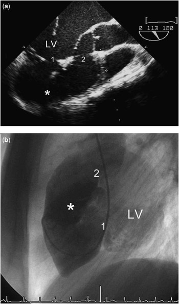
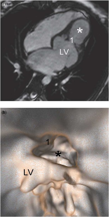
A 17-year-old asymptomatic girl was referred to our institution because of frequent ventricular ectopics with right bundle branch block pattern in her electrocardiogram. Transoesophageal echocardiography revealed a well contractile left ventricle connecting to an accessory large chamber anterior and inferior to the left ventricle (Fig 1a). There was an apical and subaortic connection between the left ventricle and the accessory chamber with laminar bidirectional flow across on colour flow Doppler. Angiography confirmed synchronous contraction of the accessory chamber with the left ventricle (Fig 1b). Left atrial and left ventricular end-diastolic pressures were elevated up to 15 millimetres of mercury without pressure differences between the left ventricle and the accessory chamber. Magnetic resonance imaging displayed the apical connection just below the papillary muscles (Fig 2a). The accessory chamber consisted of a thin muscular wall with similar appearance to the wall of the left ventricle, which had a normal muscular thickness. Hence, the accessory chamber fulfilled the criteria of a diverticulum owing to its synchronous contractility and muscular texture.Reference Marijon, Ou and Fermont 1 Virtual endoscopy was performed based on a magnetic resonance four-dimensional true fast imaging with steady-state precession short-axis data set. Inner views of the left ventricle were reconstructed in order to visualise the inlet part. The apical connection within the ventricular wall leading into the diverticulum could be demonstrated in detail (Fig 2b).
(a) Transoesophageal echocardiography displays the left ventricle (LV) with an apical (1) a subaortic and (2) connection into the diverticulum (star). (b) Angiography of the diverticulum with the catheter coming from the left ventricle passed through the apical connection.

(a) Magnetic resonance imaging shows the apical connection (1) between the left ventricle (LV) and the diverticulum (star). The atrial septum bulges to the right as a consequence of elevated left atrial and left ventricular end-diastolic pressures. (b) Virtual endoscopy based on magnetic resonance imaging delineates the muscular texture of the left ventricle and the diverticulum with the exact margins of the apical connection between the two chambers.

The patient presented at 38 weeks of gestation 1 year later. Caesarean section was performed without complications. Subsequent magnetic resonance imaging proved constant morphology of the diverticulum.
To our knowledge, a double orifice left ventricular diverticulum has not been described to date. The cause of the two connections between the left ventricle and the diverticulum remains unclear; however, it supports the hypothesis that congenital weakness within the ventricular muscle could have caused gradual outpouching from high ventricular pressures. The muscular contractile wall of the large diverticulum is able to resist the significant volume load during pregnancy, and conservative follow-up is likely to be appropriate. Magnetic resonance imaging-based virtual endoscopy enables to compute views for better anatomical understanding.