Introduction
Freshwater fishes of the genus Brycon (Characiformes) are well distributed in Central and South America and inhabit most Brazilian rivers. This genus comprises about 40 species (Zaniboni-Filho et al., Reference Zaniboni-Filho, Reynalte-Tataje and Weingartner2006), six of which, including the pirapitinga (Brycon nattereri Günther 1864), are on the national list of fish species that are threatened with extinction (Rosa & Lima, Reference Rosa, Lima, Machado, Drummond and Paglia2008). Fishes of this genus have a silvery-grey colour and are important to commercial and subsistence fishing (Gomiero & Braga, Reference Gomiero and Braga2007). Brycon nattereri is endemic to the upper Paraná, São Francisco and upper Tocantins River basins and its populations are declining due mainly to deforestation, water pollution and river damming (Rosa & Lima, Reference Rosa, Lima, Machado, Drummond and Paglia2008). Actions such as the maintenance and restoration of natural habitats and research on reproductive biology have been highlighted as the best ways to ensure the conservation of B. nattereri (Lima et al., Reference Lima, Albrecht, Pavanelli and Vono2007).
Spring/summer is the predominant spawning season of species of the genus Brycon, except for B. petrosus, which reproduces in winter (Kramer, Reference Kramer1978), and B. opalinus, which has two reproductive peaks – one in spring/summer and another in autumn (Gomiero & Braga, Reference Gomiero and Braga2007). Dependent on the river location, the spawning season for B. nattereri occurs in autumn or winter (dry season; Lima et al., Reference Lima, Albrecht, Pavanelli and Vono2007). As with most fish in captivity, the collection of B. nattereri gametes for artificial reproduction requires hormone induction (Oliveira et al., Reference Oliveira, Viveiros, Maria, Freitas and Izaú2007; Viveiros et al., Reference Viveiros, Maria, Amaral, Orfão, Isaú and Verissimo-Silveira2012).
Studies have been carried out on male reproductive characteristics for B. nattereri (Oliveira et al., Reference Oliveira, Viveiros, Maria, Freitas and Izaú2007; Viveiros et al., Reference Viveiros, Maria, Amaral, Orfão, Isaú and Verissimo-Silveira2012), but to our knowledge no similar studies on female reproductive characteristics can be found in the literature. Knowledge on the histological and ultrastructural features of oocytes can help optimize reproduction management and increase the efficiency of artificial reproduction techniques (Isaú et al., Reference Isaú, Rizzo, Amaral, Mourad and Viveiros2013). Moreover, carbohydrate histochemistry contributes to the understanding of egg adhesiveness. Thus, the aim of the present study was to characterize biopsied and stripped oocytes from Brycon nattereri using histochemical and morphological analyses.
Materials and methods
Females were obtained from the Fish Culture Station of the Minas Gerais Power Company (CEMIG) in the city of Itutinga, state of Minas Gerais, Brazil. This experiment was carried out during the spawning season for Brycon nattereri (May to August). The region is characterized by a dry winter with very low rainfall (nearly zero in some years) and temperatures between 14 and 26°C.
Histochemical and morphological analysis of biopsied oocytes
All fish were handled following the guidelines for animal experimentation described in Van Zutphen et al. (Reference Van Zutphen, Baumans and Beynen2001). During the experimental period, all females were examined weekly and those with a swollen abdomen and reddish genital pore were selected (n = 19). Ovarian biopsies were performed to determine oocyte diameter and for carbohydrate histochemical evaluation. For these techniques, fish were anesthetized with benzocaine (ethyl aminobenzoate; 60 mg/l of water) and a sample of approximately 60 biopsied oocytes/female was collected with the aid of a plastic urethral catheter inserted into the urogenital papilla. Half of each sample was fixed in Gilson's solution (50 ml of 60% ethanol, 440 ml of distilled water, 7 ml of nitric acid, 10 g of mercuric chloride and 9 ml of glacial acetic). The diameter of each oocyte was measured under a stereomicroscope with a micrometric ocular, following the method described by Isaú et al. (Reference Isaú, Rizzo, Amaral, Mourad and Viveiros2013). The frequency distribution of oocyte diameters was calculated in classes of 100 μm. The remaining oocytes in the samples were fixed in Bouin solution for 12 h and subjected to histological and carbohydrate histochemical analyses. For this purpose, oocytes were embedded in paraffin, cut to a thickness of 3–5 μm and stained with periodic acid-Schiff (PAS) stain in order to detect neutral polysaccharides and Alcian blue (AB) at pH 2.5 for the detection of acidic polysaccharides (Pearse, Reference Pearse1985). Reactivity of the oocyte structures (follicular cells, zona radiata, cortical alveoli and yolk globules) to carbohydrate histochemistry was classified as follows: negative reaction (–), positive reaction (+) and strongly positive reaction (++).
Induction of spawning and morphological analysis of stripped oocytes
All 19 females selected for reproduction were transferred from a pond to an aquarium with a water temperature of 18 ± 1 °C and oxygen at 7–8 mg/l 48 h prior to hormone induction. Each female received two intramuscular injections of carp pituitary extract (cPE; Argent Chemical Laboratory, Redmond, Washington, USA) at 0.4 and 4.0 mg/kg body weight with a 12-h interval. Between 14 and 18 h after the second dose, all females were hand-stripped. For the fish that responded to cPE treatment and released oocytes (n = 10 females), the following variables were determined: body weight, standard length, total length, ova weight, spawning index (ova weight × 100/body weight), number of oocytes/g of ova, number of oocytes/female and number of oocytes/g body weight.
A sample of stripped oocytes was collected and fixed in Gilson's solution for 30 min for the measurement of oocyte diameter, which was determined as described for the biopsied oocytes. Oocytes were assessed for adhesiveness under macroscopic analysis and classified as adhesive (when oocytes stuck firmly to each other and formed a coherent egg mass), weakly adhesive (when oocytes adhered to each other, but became free under slight agitation) and non-adhesive (when oocytes were completely free) (Rizzo et al., Reference Rizzo, Sato, Barreto and Godinho2002). A third sample of stripped oocytes from each female was fixed in modified Karnovsky solution (2.5% glutaraldehyde, 2.5% paraformaldehyde in 50 mM sodium cacodylate buffer, pH 7.2, 1 mM CaCl2). The oocytes were post-fixed in 1% osmium tetroxide for 4 h at room temperature, washed in 0.1 M cacodylate buffer (pH 7.4), dehydrated through an increasing gradient of acetone solutions (25, 50, 75, 90 and 100%), dried with CO2 in a PELCO CPD 030 critical-point dryer (Leica Microsystems, Wetzlar, Germany), coated with gold under vacuum conditions with SEM Coating Unit SCD 050 (Leica Microsystems) and examined with a scanning electron microscope (LEO EVO 40 XVP ESC, Carl Zeiss SMT, LEO Electron Microscopy Group, Oberkochen, Germany) equipped with a digital camera, and following the method described by Isaú et al. (Reference Isaú, Rizzo, Amaral, Mourad and Viveiros2013). Digital images of the oocytes were used for the morphometric analysis of the structures. Micropyle diameter (n = 18 oocytes), thickness of the zona radiata (n = 21 oocytes) and number of pore canals/μm2 (n = 30 oocytes) were determined using the LEO-SRV32 software (Microsoft Windows version).
Results
Histochemical and morphological analysis of biopsied oocytes
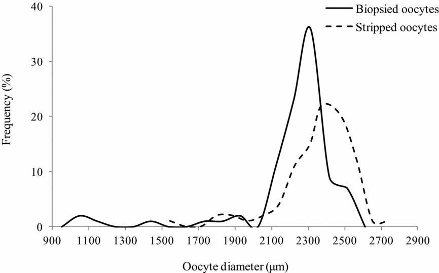
A unimodal frequency distribution was found for the biopsied oocyte diameter among the 19 females used in the present study. Oocyte diameter ranged from 1.000–2.563 mm, with a mean of 2.225 ± 0.262 mm and a mode at 2.312 mm (Fig. 1).
Frequency distribution of biopsied (—) and stripped (—) oocytes diameter of pirapitinga Brycon nattereri after carp pituitary extract treatment.

In the histological analysis, most biopsied oocytes had complete vitellogenesis, yolk globules occupied most of the ooplasm, different sizes of cortical alveoli were aligned in layers, there were central or slightly eccentric nucleus with multiple peripheral nucleoli, zona radiata with a thick inner layer and thin outer layer, and a squamous follicular cell layer supported by a basal membrane. Perinucleolar oocytes with finely basophilic cytoplasm, central nucleus and several peripheral nucleoli were also observed in the ovarian sections.
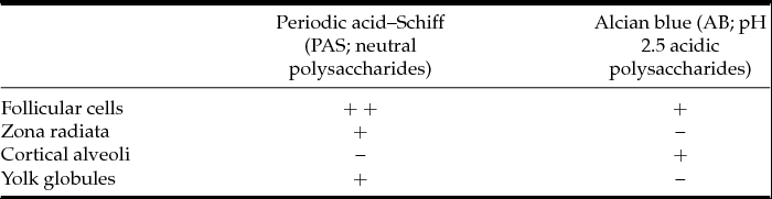
In the carbohydrate histochemical analysis, neutral polysaccharides were detected in the follicular cells, zona radiata and yolk globules (PAS positive) and acidic polysaccharides were detected in the follicular cells and cortical alveoli (AB positive; Table 1).
Reactivity of biopsied oocyte structures in carbohydrate histochemical analysis of Brycon nattereri

–, negative reaction; +, positive reaction; ++, strongly positive reaction.
Induction of spawning and morphological analysis of stripped oocytes
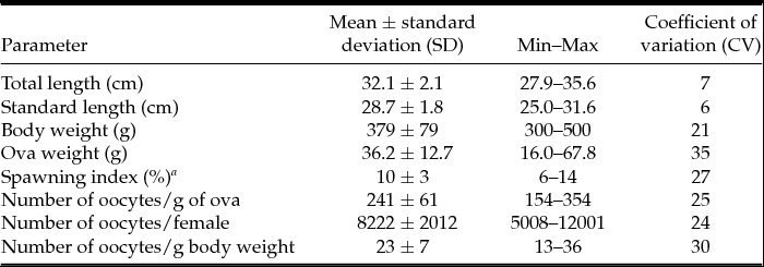
Ten out of the 19 females that had been treated with cPE subsequently released oocytes. These females had a mean of body weight of 379 g, standard length of 28.7 cm and total length of 32.1 cm. Stripping occurred 292 ± 39 degree-hours after the second dose of cPE and led to a mean spawning weight of 36.2 g, 10% spawning index, 241 oocytes/g of ova, 8222 oocytes/female and 23 oocytes/g of body weight (Table 2). The diameter of the stripped oocytes ranged from 1.344–2.781 mm, with mean of 2.330 ± 0.222 mm and a mode at 2.375 mm (Fig. 1). The oocytes adhered to each other, but became free under mild agitation and were thus classified as weakly adhesive. Oocyte coloration ranged from wine to brown.
Body length and weight of Brycon nattereri and spawning quality after treatment with carp pituitary extract (n = 10)

a Spawning index = ova weight × 100/body weight.
Under scanning electron microscopy, stripped oocytes were devoid of a gelatinous cover, had a single micropyle and exhibited pore canals that were distributed along the surface of the zona radiata. The micropyle was characterized by a conical vestibule and a micropylar canal that crossed the zona radiata (Fig. 2 A). An inner layer of folds formed grooves in the micropylar vestibule. The outer opening of the micropylar vestibule had an oblong shape with the largest diameter at 20.4 μm and the smallest at 18.7 μm (Table 3). The zona radiata was smooth, (Fig. 2 B) with pore canals (Fig. 2 C) and were devoid of specialized structures such as adhesive filaments. At the vegetative pole, the surface of the zona radiata exhibited a uniform distribution of eight pore canals/μm2 with a greater diameter compared with the animal pole (Fig. 2 D). The plasma membrane had long, thin filaments (microvilli) that formed a dense coat (Fig. 2 E). The cortical alveoli were aligned in layers that were located in the cortical cytoplasm immediately below the plasma membrane and more internally, with yolk granules that occupied most of the oocyte. The presence of pore canals on the surface of the plasma membrane and impressions left by the cortical alveoli after rupture were also observed (Fig. 2 F).
Morphometry of stripped oocytes from Brycon nattereri

a Pore canals on surface of zona radiata at vegetative pole.
b External micropyle opening.
Scanning electron micrographs of stripped oocytes of Brycon nattereri. (A) Details of micropyle with vestibule (v) and micropylar canal (c). (B) External oocyte surface at animal pole. (C) Fractured zona radiata (Zr) with pore canals at the oocyte surface (o). (D) Pore canals in the surface of the zona radiate at the vegetative pole. (E) Cross-section of the oocyte, showing the plasma membrane (M), cortical alveoli (CA) and yolk globules (YG). insertion: microvilli of the plasma membrane. (F) Outer surface of the oocyte plasma membrane showing the pore canals left by the cortical alveoli (arrowheads). Scale bars = 20 μm (A), 2 μm (B–E); and 10 μm (F).

Discussion
The present study offers a first analysis of female reproductive parameters and oocyte morphology in Brycon nattereri. Relative fecundity was low (23 oocytes/g body weight) and the biopsied oocytes had a large diameter (2.312 mm). Among females of this genus, B. opalinus had a mean of 31 oocytes/g body weight and a mean oocyte diameter of 1.90 mm (Narahara et al., Reference Narahara, Andrade-Talmelli, Kavamoto and Godinho2002; Gomiero & Braga, Reference Gomiero and Braga2007), B. insignis had a mean of 60 oocytes/g body weight and an oocyte diameter of 1.25 mm (Andrade-Talmelli et al., Reference Andrade-Talmelli, Kavamoto, Narahara and Fenerich-Verani2002) and B. orthotaenia had 105 oocytes/g body weight and an oocyte diameter of 1.479 mm (Sato et al., Reference Sato, Fenerich-Verani, Godinho, Godinho and Godinho2003). The distribution pattern of the diameter of biopsied oocytes has been successfully used to identify females that were suitable for reproduction, as this variable indicates the degree of ovarian development (West, Reference West1990).
In the present study, unimodal distribution was found in the oocyte diameter of B. nattereri from mid autumn to mid winter, when the most oocytes were at the same maturation stage, with complete vitellogenesis and a central or slightly eccentric nucleus, a finding that suggested the presence of a group-synchronous type of oocyte development. In the final stage of oocyte maturation, the germinal vesicle (nucleus) migrates from a central position to the periphery of the oocyte before the resumption of meiosis and breakdown of the germinal vesicle (Grier & Neidig, Reference Grier, Neidig, Tiersch and Green2011).
Vitellogenic oocytes from B. nattereri were coated with a smooth zona radiata with pore canals and neutral polysaccharides. These characteristics constitute a less complex apparatus of egg binding to the substrate and do not ensure a strong degree of adhesiveness (Riehl & Patzner, Reference Riehl and Patzner1998). Similarly, other species of Characiformes have non-adhesive (Isaú et al., Reference Isaú, Rizzo, Amaral, Mourad and Viveiros2013) or weakly adhesive oocytes (Rizzo et al., Reference Rizzo, Sato, Barreto and Godinho2002; Rizzo & Godinho, Reference Rizzo, Godinho, Godinho and Godinho2003). In contrast, acidic polysaccharides have been commonly detected on the surface of the zona radiata in adhesive oocytes (Rizzo & Bazzoli, Reference Rizzo and Bazzoli1991; Riehl & Patzner, Reference Riehl and Patzner1998; Gomes et al., Reference Gomes, Scarpelli, Arantes, Sato, Bazzoli and Rizzo2007; Weber et al., Reference Weber, Arantes, Sato, Rizzo and Bazzoli2012). In the wild, B. nattereri females spawn in areas in the water with low light such as under rocks or at the river edge. The eggs then become slightly adhered to substrates, such as roots, are covered by leaves and abandoned by their parents (Viveiros et al., Reference Viveiros, Maria, Amaral, Orfão, Isaú and Verissimo-Silveira2012).
The biopsied oocytes from B. nattereri exhibited a histochemical pattern, with acidic polysaccharides found in the cortical alveoli; this pattern is similar to that of species of the families Characidae (Brycon lundii, Brycon orbignyanus, Salminus maxillosus, Salminus brasiliensis and Salminus hilarii; Bazzoli & Godinho, Reference Bazzoli and Godinho1994) and Erythrinidae (Hoplerythrinus unitaeniatus, Hoplias lacerdae and Hoplias malabaricus; Gomes et al., Reference Gomes, Scarpelli, Arantes, Sato, Bazzoli and Rizzo2007). However, the oocytes of female Characiformes from the families Anostomidae and Curimatidae have been found to contain only neutral polysaccharides in the cortical alveoli (Bazzoli & Godinho, Reference Bazzoli and Godinho1994). In addition to species-specific variations in carbohydrate content in the cortical alveoli, variations have also been found in the composition of these alveoli during oocyte maturation (Ohta et al., Reference Ohta, Iwamatsu, Tanaka and Yashimoto1990). Acidic polysaccharides in the cortical alveoli are released into the perivitelline space during the cortical reaction, when these substances interact with the zona radiata and may contribute to blocking polyspermy (Tyler & Sumpter, Reference Tyler and Sumpter1996).
The stripped oocytes from B. nattereri were spherical and had a diameter with a mode at 2.375 mm. The oocyte surface exhibited a single, funnel-like shaped micropyle and pore canals, similar to that found for other species of Characiformes (Rizzo et al., Reference Rizzo, Sato, Barreto and Godinho2002; Ganeco & Nakaghi, Reference Ganeco and Nakaghi2003; Ganeco et al., Reference Ganeco, Franceschini-Vicentini and Nakaghi2009; Alexandre et al., Reference Alexandre, Ninhaus-Silveira, Veríssimo-Silveira, Buzollo, Senhorini and Chaguri2010; Isaú et al., Reference Isaú, Rizzo, Amaral, Mourad and Viveiros2013). The micropyle is a concave region that is located on the oocyte surface and is composed of a continuous vestibule with an internal canal that narrows progressively toward the plasma membrane of the egg (Ganeco & Nakaghi, Reference Ganeco and Nakaghi2003) to allow the entrance of a single spermatozoon during fertilization and thus blocking polyspermy.
In conclusion, Brycon nattereri females display group-synchronous-type oocyte development, and exhibit characteristics that differ from other species of the genus such as fecundity, egg size and egg adhesiveness. The findings of the current study provide essential information for a better understanding of the reproductive biology of B. nattereri and the establishment of conservation measures for this threatened species.
Acknowledgements
The authors wish to thank G.A. Azarias and J.M. Silva (CEMIG Itutinga Unit, MG) for assistance during oocyte collection; the Brazilian fostering agency FAPEMIG (project CVZ-1609/06) for financial support; and Dr E. Alves for allowing the use of the Electron Microscopy and Ultrastructural Analysis Laboratory of UFLA.