Tetrapods include all living four-limbed backboned animals, and some that have secondarily lost limbs, as well as stem members, which comprise a group of extinct Palaeozoic fishes. The group, in total, is known as Tetrapodomorpha. Here we use the term ‘tetrapod' to include all digitate tetrapodomorphans, and ‘stem tetrapods', which include fishes, on the stem leading to tetrapods. Their ancestry is deeply rooted within the sarcopterygians, a clade of lobe-finned osteichthyans containing lungfishes and related taxa (Dipnomorpha), coelacanths (Actinistia), plus extinct groups such as the Onychodontiformes and psarolepids, which first appeared in the Late Silurian of China (Zhu et al. Reference Zhu, Zhao, Jia, Lu, Qiao and Qu2009; Long Reference Long2011).
Tetrapodomorph fishes first appeared in the Early Devonian. The oldest taxon, Tungsenia, comes from the Pragian of Yunnan, China, dated at around 409Ma (Lu et al. Reference Lu, Zhu, Long, Zhao, Senden, Jia and Qiao2012), but is only represented by scant remains (ethmosphenoid, lower jaw, cheek and cleithrum). The only other taxon known from this clade of Early Devonian age is Kenichthys, also from China. Kenichthys, from the Emsian of Yunnan, is known from relatively complete cranial remains (Chang & Zhu Reference Chang and Zhu1993; Zhu & Ahlberg Reference Zhu and Ahlberg2004). By the Middle Devonian, the group was widespread across the globe, with faunas well-documented from Britain and Europe (Jarvik Reference Jarvik1948; Gross Reference Gross1956; Jessen Reference Jessen1966), the Middle East (Janvier et al. Reference Janvier, Clément and Cloutier2007), the USA (Thomson Reference Thomson1969; Rackoff Reference Rackoff and Panchen1980), Australia (Thomson Reference Thomson1973; Long Reference Long1985a, Reference Longb, Reference Longc, Reference Long, Vickers-Rich, Monaghan, Baird and Rich1991; Ahlberg & Johanson Reference Ahlberg1998; Johanson & Ahlberg Reference Johanson and Ahlberg1998) and Antarctica (Young et al. Reference Young, Long and Ritchie1992).
Middle Devonian tetrapodomorph faunas are dominated by Osteolepis-like taxa (family ‘Osteolepididae', probably paraphyletic, but here also including megalichthyines), with tristichopterids and rhizodontids appearing by the latter half of the Middle Devonian. In the Late Devonian, these faunas are dominated by tristichopterids like Eusthenopteron and Mandageria (Jarvik Reference Jarvik1980; Johanson & Ahlberg Reference Johanson and Ahlberg1997), osteolepidids such as Gogonasus (Long et al. Reference Long, Barwick and Campbell1997) and the elpistostegalians, which include highly derived tetrapodomorph fishes like Tiktaalik (Daeschler et al. Reference Daeschler, Shubin and Jenkins2006; Shubin et al. Reference Shubin, Daeschler and Jenkins2006; Downs et al. Reference Downs, Daeschler, Jenkins and Shubin2008; Shubin et al. Reference Shubin, Daeschler and Jenkins2014), plus the first digitate tetrapods, such as Ichthyostega (Save-Soderbergh Reference Save-Soderbergh1932; Jarvik Reference Jarvik1996; Clack Reference Clack2012), which are more prevalent in the Fammenian. The Carboniferous tetrapodomorph fishes include mainly megalichthyines (e.g., Megalichthys, Claradosymblema, Cope Reference Cope1882), rhizodopsids (Rhizodopsis, Williamson Reference Williamson1837) and rhizodontids, the latter including the largest sarcopterygians known, such as Rhizodus, which had an estimated total length close to 6m (Miall Reference Miall1875). The end Permian marks the extinction of the clade, with the last tetrapodomorph fishes represented by a few taxa of megalichthyines (e.g., Ectosteorhachis, Romer Reference Romer1937).
The oldest representatives of tetrapods are from the upper Frasnian (ca.372–380 mya); these include isolated skeletal elements of taxa such as Elginerpeton from Scotland (Ahlberg Reference Ahlberg1995; Ahlberg Reference Ahlberg1998), as well as a jaw from Australia known as Metaxygnathus (Campbell & Bell Reference Campbell and Bell1997). Well-preserved trackways demonstrating that digitate tetrapods had appeared are known from the early Middle Devonian of Zalchemie, Poland (Niedźwiedzki et al. Reference Niedźwiedzki, Szrek, Narkiewicz, Narkiewicz and Ahlberg2010), the upper Middle Devonian of Ireland (Stössel Reference Stössel1995; Stössel et al. Reference Stössel, Williams and Higgs2016; plus Long, pers. obs. 2016) and the Late Devonian of South Eastern Australia (Warren & Wakefield Reference Warren and Wakefield1972; Clack Reference Clack1997). It is clear from these few remains, and other more complete records of Late Devonian (Famennian) tetrapods from East Greenland (Clack Reference Clack1988; Clack Reference Clack2012), Russia (Lebedev & Coates Reference Lebedev1995; Lebedev Reference Lebedev2004), Latvia (Ahlberg et al. Reference Ahlberg, Clack, Luksevics, Blom and Zupins2008), South Africa (Gess & Ahlberg Reference Gess and Ahlberg2018) and scant remains in North America (Daeschler et al. Reference Daeschler, Clack and Shubin2009) and China (Zhu et al. Reference Zhu, Ahlberg, Zhao and Jia2002), that tetrapods were widespread across the globe by the end of the Devonian.
In the Carboniferous, tetrapods occupied a number of specialist niches (Clack Reference Clack2012; Clack et al. Reference Clack, Bennett, Carpenter, Davies, Fraser, Kearsey, Marshall, Millward, Otoo, Reeves and Ross2017; Pardo et al. Reference Pardo, Szostakiwskyj, Ahlberg and Anderson2017), with the first amniotes appearing during this time (Hylonemus, ca.330 mya), signalling the beginning of the conquest of land freed from dependence on water for reproduction.
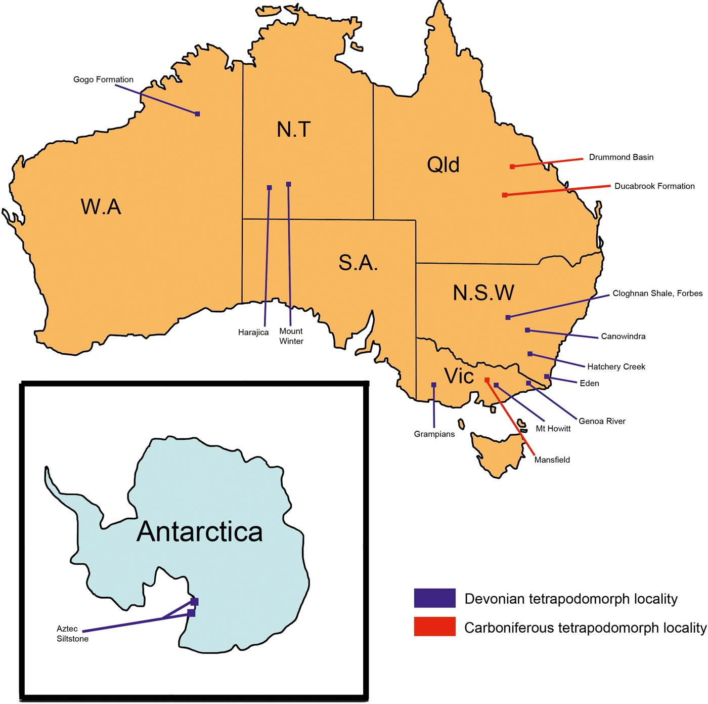
The early radiation of tetrapods, especially the tetrapodomorph fishes in the Devonian, is of particular interest as only Gondwanan taxa tend to show a high degree of endemism during their early diversification. In this paper we present a detailed historical review of the Devonian tetrapod record of East Gondwana (Australasia, Antarctica; fossil sites referred to herein are shown in Fig. 1). We herein highlight new anatomical and geological data relating to the radiation of tetrapodomorphans and the revised dating associated with certain key taxa, especially the endemic canowindrids (Long Reference Long1987; Young et al. Reference Young, Long and Ritchie1992). We also present some preliminary findings based on synchrotron scan data of one of the best preserved member of this group, Koharalepis jarviki, from the Middle Devonian Aztec Siltstone of Antarctica (Young et al. Reference Young, Long and Ritchie1992).
Locations of notable fossil discoveries of Eastern Gondwanan tetrapodomorphs from the Devonian to early Carboniferous (modified after Young et al. Reference Young, Burrow, Long, Turner and Choo2010).

1. Historical review of East Gondwana mid-Palaeozoic stem tetrapods
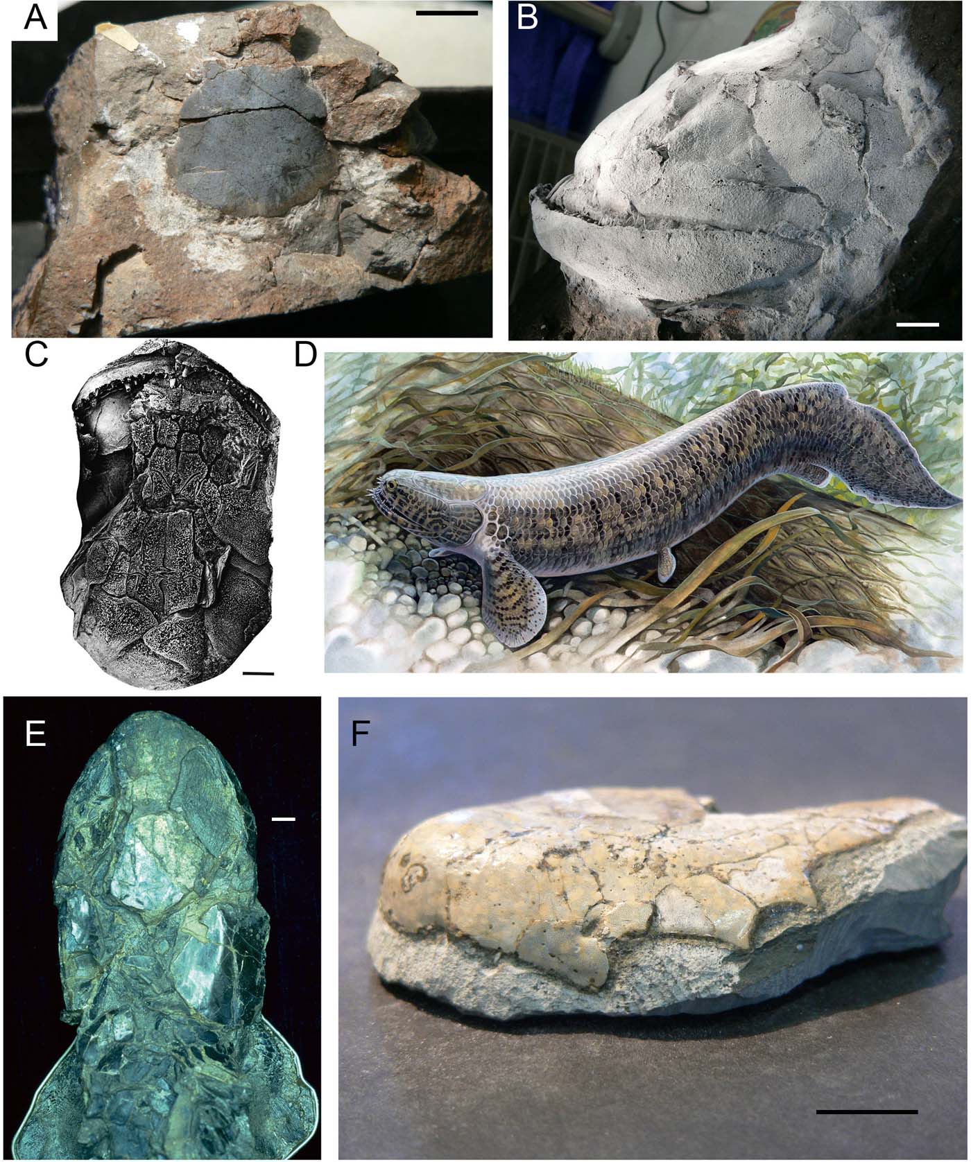
The first record of a tetrapodomorph fish from Eastern Gondwana was the publication by Woodward (Reference Woodward1906) of the Mansfield fauna of central Victoria, where the rhizodontid ‘Strepsodus' decipiens (renamed Barameda, Long Reference Long1989; Fig. 2c, d) was described based on an large isolated jaw and shoulder girdle material, plus scales. The fauna comprised typical elements of the Northern Hemisphere Carboniferous continental fishes, like gyracanths, Acanthodes, elonichthyiform palaeoniscoids and ctenodid-like lungfish, so it was not thought indicative, at the time, of any endemism in Australian faunas. Later in the 20th Century it would be demonstrated that all the fossil osteichthyan taxa that were assigned to Northern Hemisphere generic identifications by Woodward turned out to be new genera unique to East Gondwana (Long & Campbell Reference Long and Campbell1985; Long Reference Long1988, Reference Long1989).
Various basal tetrapodomorph fishes from Eastern Gondwana. (A) Postparietal shield from an undescribed taxon from the Early Devonian (Emsian) Hatchery Creek Group, Wee Jasper, New South Wales. (B) Whitened skull cast of Goologongia loomesi, a basal rhizodont from the Late Devonian Mandagery Sandstone, Canowindra, New South Wales. (C) Skull in dorsal view of Barameda mitchelli, a derived rhizodont from the Early Carboniferous Snowy Plains Formation, Mansfield, Victoria. (D) Life reconstruction of Barameda mitchelli. (E) Cladarosymblema narrienense, a megalichthyid from the Early Carboniferous Raymond Formation, Drummond Basin, Queensland. Skull and anterior body in dorsal view. (F) Mahalalepis resima, a megalichthyid from the Middle–Late Devonian Aztec Siltstone, Antarctica. Fronto-ethmoidal shield in dorso-lateral view. (Photos by John Long, illustration by Peter Schouten, with permission.) Scale bars=1cm.

The next tetrapodomorph record from East Gondwana was also the first report of Devonian fishes from Antarctica. An indeterminate osteolepidid-like fish, based on a scale and cheek plate, was described by Woodward (Reference Woodward1921) from collections retrieved from the ill-fated 1910–1912 Terra Nova Expedition led by Robert Falcon Scott. This was a highly significant paper as it showed the first appearance of a fish fauna of typically northern hemisphere composition (containing taxa like Bothriolepis, ‘Phyllolepis' osteolepids and gyracanths) to be found in the Southern Hemisphere, well before the days of accepted plate tectonic theory. At the time of this publication, the dating of this succession was placed as Upper Devonian based on these fishes known to occur in the Late Devonian of Europe, Russia and the USA. Since then, revised dating based on extensive description of the fish fauna (>70 species; Young & Long Reference Young and Long2014) and biostratigraphical comparisons with well-dated faunas from Australia (Young Reference Young and Long1993; Young et al. Reference Young, Burrow, Long, Turner and Choo2010), means the base of this assemblage could be potentially as old as Eifelian (Young Reference Young2006; Young et al. Reference Young, Burrow, Long, Turner and Choo2010). However, as the sequence is over 200m thick, it most likely represents a Givetian and upper Eifelian age range.
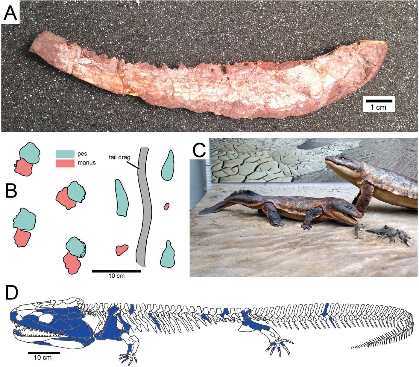
In the early 1970s, Jim Warren and Norman Wakefield from Monash University, Victoria, announced they had found unambiguous sets of tetrapod trackways from a Devonian site in eastern Victoria (Warren & Wakefield Reference Warren and Wakefield1972). The discovery made the cover of Nature, as it was not only the first record of tetrapods from the Palaeozoic of Australia, but also because it occurred in strata of possible Frasnian age, based on an associated plant assemblage, which made it the oldest record of tetrapods known at that time. Clack (Reference Clack1997) redescribed these trackways, confirming their tetrapod affinity and suggested that two different track makers might have been present (Fig. 6b).
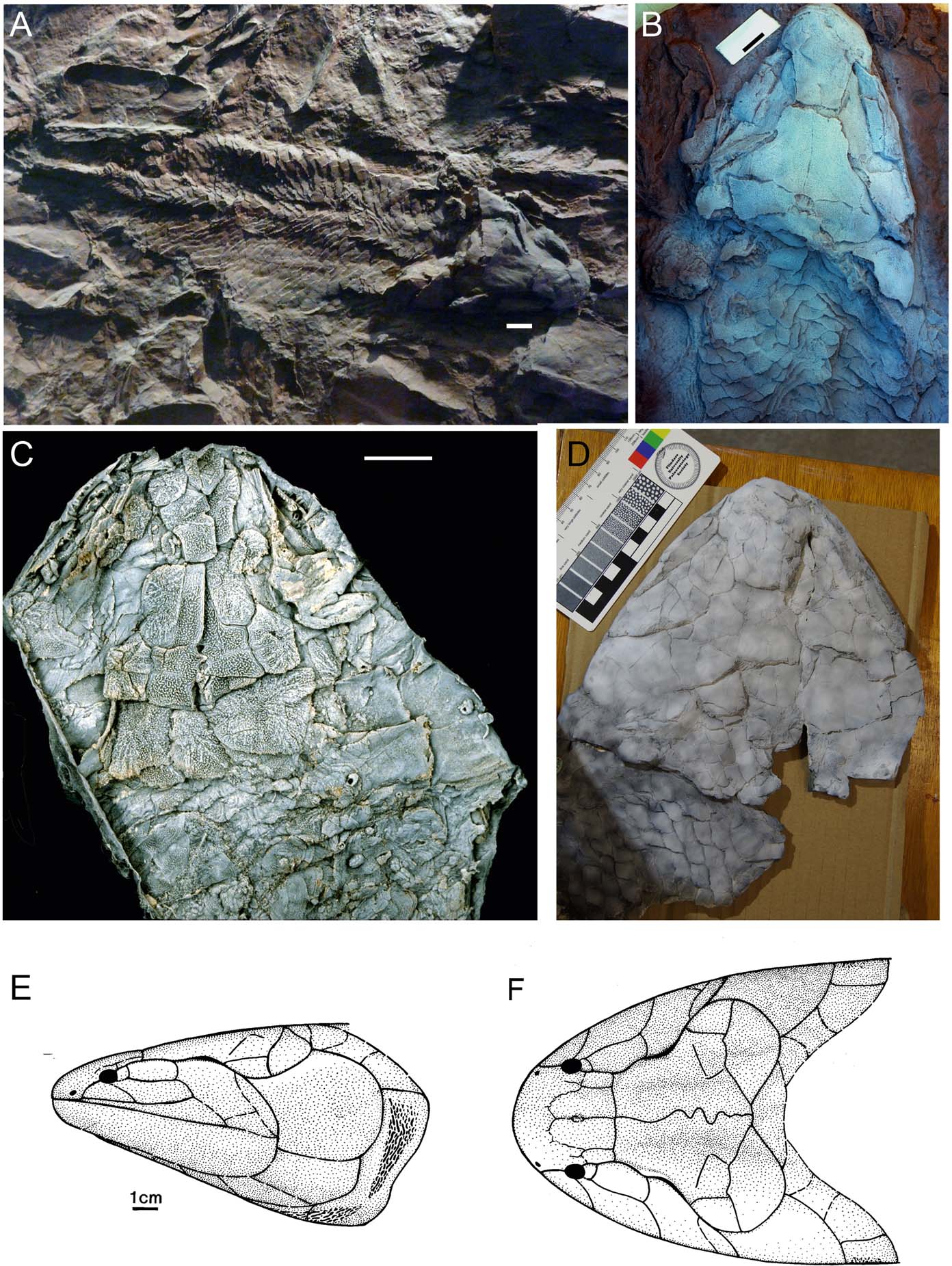
In the mid-1950s, the widening of a road near the New South Wales town of Canowindra saw the discovery of a slab of rock covered in placoderm fishes with one large sarcopterygian in the centre (reconstructed in Fig. 5d). Keith Thomson (Reference Thomson1973) described this enigmatic form as Canowindra grossi based on a cast of the entire fish preserved as a mould in the sandstone (Fig. 4a, b). At the time he did not classify it as an ‘osteolepiform' but said it was of uncertain affinity. Its broad postparietal shield and very small orbits suggested it was like a porolepiform. Long (Reference Long1985a) redescribed Canowindra grossi based on new cleaned casts of the specimen and identified features which were thought to represent osteolepiform synapomorphies, like an upright bar-like preoperculum, large dermal anocleithrum and the specific cheek plate arrangement.
In the mid-1970s a complete lower jaw of a tetrapod was found in Late Devonian Cloughnan Shale near Jemalong, New South Wales (Fig. 3a), associated with an assemblage of placoderm fishes such as Bothriolepis, Remigolepis and a phyllolepid. It was described as a new taxon of tetrapod, Metaxygnathus denticulatus (Campbell & Bell Reference Campbell and Bell1997; Fig. 6a). Later controversy grew around this specimen when Schultze & Arsenault (Reference Schultze and Arsenault1985) suggested it belonged to an advanced osteolepiform fish. This was resolved by further detailed study of stem tetrapod jaws (Ahlberg & Clack Reference Ahlberg and Clack1998), confirming it as a definite tetrapod jaw.
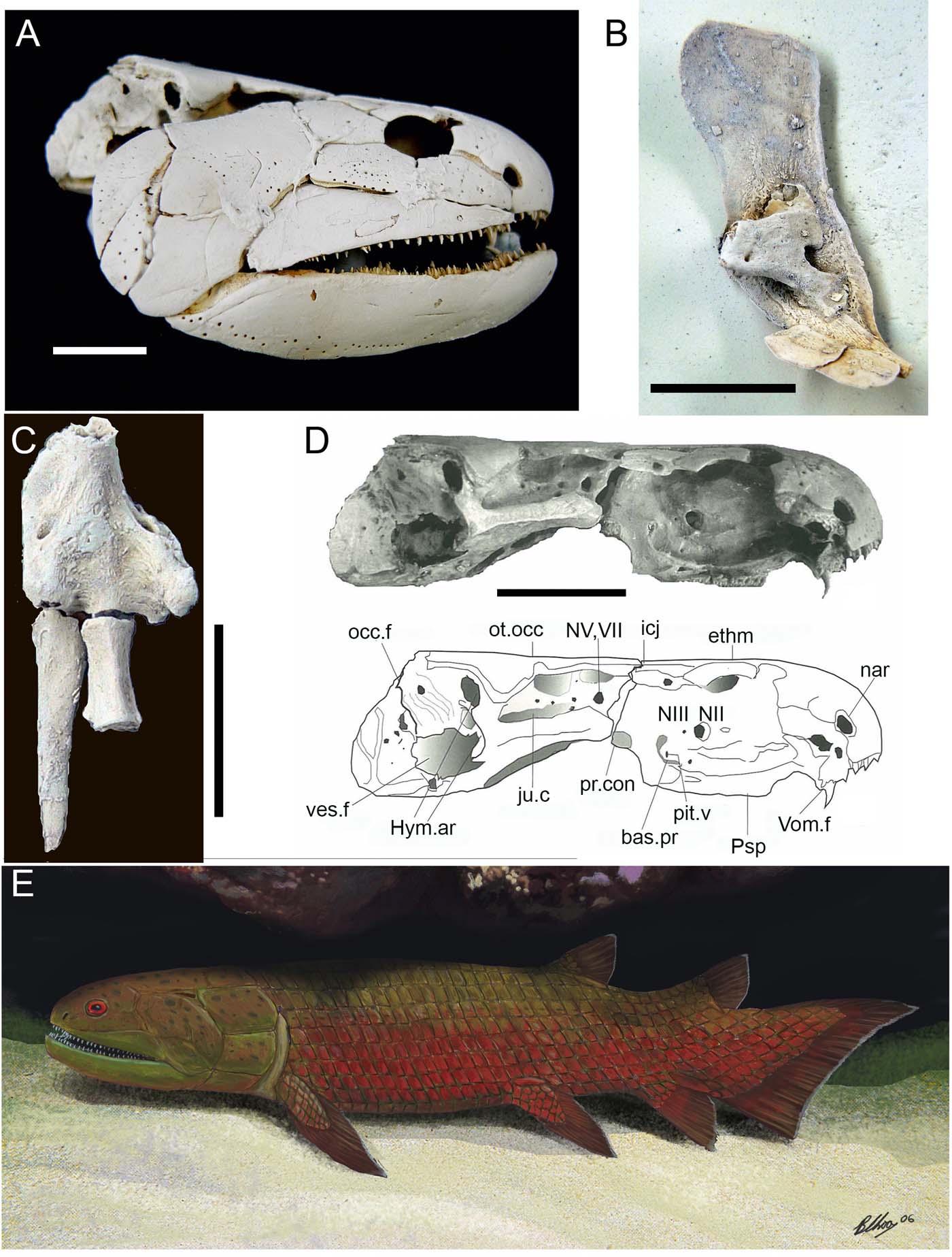
Gogonasus andrewsae, an unusual marine tetrapodomorph from the Late Devonian Gogo Formation, Western Australia. (A) Skull in left lateral view. (B) Left cleithrum in viscera view. (C) Proximal three bones of the pectoral fin, showing humerus, radius and ulna. (D) Photograph and line drawing of braincase with anatomical features indicated. (E) Life reconstruction of Gogonasus. (Photos by John Long, illustration by Brian Choo, with permission). Scale bars = 1cm. Abbreviations: bas.pr = basipterygoid process; ethm = ethmosphenoid; Hym.ar = hyomandibular articulations; icj = intracranial joint; ju.c = jugular canal; nar = narial opening; NV,VII = foramen for facial and trigeminal nerves; occ.f = occipital fissure; ot.occ = oticco-occipital; pit.v = foramen for pituitary vein; pr.con = processus connectdens; Psp = Parasphenoid; ves.f = vestibular fontanelles; Vom.f = vomerine fangs.

In 1985 the total number of Devonian ‘osteolepiform' fishes formally named and described from Australia greatly increased when two new taxa were added to the literature. Gogonasus andrewsae was described from a single three-dimensional (3D)-preserved ethmosphenoid (Long Reference Long1985b) found on the 1967 British Museum–Hunterian Museum–WA Museum expedition (Fig. 4). In this paper, the ‘Osteolepiform problem', concerning the taxonomic definition of the group, was discussed with a case for osteolepiform monophyly presented, and synapomophies defined to support at least one osteleopiform clade, the Megalichthyinae, as monophyletic. A new form of basal ‘tristichopterid', Marsdenichthys longioccipitus (Fig. 2c), was also described that year from the Givetian Mount Howitt site in Victoria (Long Reference Long1985c). This paper included the first published cladistic analysis of tetrapodomorphan fishes based on manual character analysis. Further description of Marsdenichthys longiocciptus was presented based on a new specimen from Mount Howitt by Holland et al. (Reference Holland, Long and Snitting2010).
Representatives of clade Canowindridae. (A) Cast of the holotype of Canowindra grossi, from the Late Devonian Mandagery Sandstone, New South Wales, surrounded by specimens of the antiarchs Remigolepis and Bothriolepis. (B) Cast of the skull of Canowindra in dorsal view (photo by John Long, with permission). (C) Whitened latex peel of the holotype of Marsdenichthys longioccipitus, a problematic form from Middle–Late Devonian of Mount Howitt, Victoria, that may represent an unusual canowindrid. (D) Whitened latex peel of the holotype of Koharalepis jarvicki from the Middle–Late Devonian Aztec Siltstone, Antarctica. (E) Line drawings of the skull and pectoral girdle of Beelargonia patrichae from the Middle–Late Devonian of Mount Howitt. (Photos by John Long and Brian Choo, with permission). Scale bars = 1cm.

In 1987 another taxon from the Mount Howitt assemblage was described, Beelarongia patrichae (Long Reference Long1987), based on latex peels of the impression of a large skull and the shoulder girdle of a fish with large cosmoid rhombic scales (Fig. 2e). It's resemblance to Canowindra grossi was noted in its broad postparietal shield, small orbits and cheek having an extra postorbital bone, and it was suggested in that paper that these taxa represented an endemic East Gondwana clade of tetrapodomorph fishes.
Woodward's Carboniferous rhizodontid from Mansfield was soon after redescribed as Barameda decipiens (Long Reference Long1989) based on new material uncovered in the collections of Museum Victoria (Fig. 4e, f). Although Andrews (Reference Andrews1985) had described a juvenile articulated rhizodontid from Scotland, much of the cranial anatomy in this specimen was unclear. The new skull from Mansfield enabled the first detailed description of the skull and palate of a rhizodontid, and a manual cladistic analysis suggested that rhizodontids might be the sister group to the Choanata (‘Osteolepiformes, Panderichthyida + Tetrapoda'). The paper originally identified paired nostrils in Barameda, implying a choana was absent; this interpretation was later shown to be incorrect and a new interpretation of the snout presented (Long & Ahlberg Reference Long and Ahlberg1999). In most subsequent analyses of tetrapodomorph relationships, rhizodontids are still a clade outside all of the other stem tetrapods, except for the basal forms from China such as Kenichthys and Tungsenia (Young et al. Reference Young, Long and Ritchie1992; Lu et al. Reference Lu, Zhu, Long, Zhao, Senden, Jia and Qiao2012; Swartz Reference Swartz2012). An exception is that of the unusual fish, Hongyu, from Ningxia, China, which raises alternative possible phylogenetic positions for rhizodontids (Zhu et al. Reference Zhu, Ahlberg, Zhao and Jia2017).
In 1986, a controversial trackway assigned to a tetrapod was described from the Late Silurian–Early Devonian of the Grampians ranges, Victoria, by Anne Warren et al. (Reference Warren, Jupp and Bolton1986). Today, much debate continues over the identity of this track-maker, with several workers dismissing its tetrapod affinity (Clack Reference Clack1997; Clack Reference Clack2012). A new investigation is currently underway to determine its affinity using mathematical parameters to objectively distinguish arthropod and tetrapod trackways and hopefully settle this controversy.
An assemblage of tetrapodomorph taxa from the Aztec Siltstone was described by Young et al. (Reference Young, Long and Ritchie1992), which included three cosmine-covered taxa, Koharalepis (Figs 4d, 7–10), Mahalalepis (Fig. 2f) and Platyethmoidea, one tristichopterid, Notorhizodon (first thought to be a rhizodontid by Young et al. Reference Young, Long and Ritchie1992) and a rhizodontid, Aztecia (Johanson & Ahlberg Reference Johanson and Ahlberg2001), plus the remains of several indeterminate taxa (Young et al. Reference Young, Long and Ritchie1992). More significantly, this paper formally recognised the endemic clade erected as the family Canowindridae to include Canowindra, Beelarongia and Koharalepis (Fig. 2). In this manual cladistic analysis, the clade Canowindridae was placed between Rhizodonts as the most basal tetrapodomorphans, and the rest of the group comprising Osteolepis-like forms, Megalichthyinae, tristichopterids (‘Eusthenopteridae' in the paper), Panderichthyidae and Tetrapoda (the latter two groups, including certain basal members of the digitate tetrapods, are now contained within the Elpistostegalia).
Between 1995 and 1997, two major monographic works appeared detailing the anatomy of 3D acid-prepared tetrapodomorph fishes from Australia. Fox et al. (Reference Fox, Campbell, Barwick and Long1995) described a lower Carboniferous (? Upper Visean) megalichthyinid, Cladarosymblema narrienensis, based on an articulated head and trunk area of the Holotype plus several isolated skulls and dermal bones (Fig. 2e). Long et al. (Reference Long, Barwick and Campbell1997) described the complete cranial and pectoral anatomy and histology of teeth and scales, based on two new specimens of Gogonasus ansdrewsae (Fig. 4), showing relatively complete cranial remains collected on the 1986 and 1990 expeditions to the Gogo Formation.
New excavations at the Canowindra fish kill site by Alex Ritchie through the 1990s revealed many new taxa present (Fig. 6), including two unusual tristichopterids, Mandageria fairfaxi and Cabonnichthys burnsi (Ahlberg & Johanson Reference Ahlberg and Johanson1997; Johanson & Ahlberg Reference Johanson and Ahlberg1997). The site also yielded relatively complete remains of a basal rhizodontid, Goologongia loomesi (Johanson & Ahlberg Reference Johanson and Ahlberg1998; Fig. 2b). Study of this specimen has been integral to refining the phylogenetic relationships of the Tetrapodomorpha and suggesting an East Gondwanan centre of origin for the group. Further isolated remains of rhizodontid pectoral fin and shoulder girdles were described from central Queensland, and assigned to Strepsodus by Parker et al. (Reference Parker, Warren and Johanson2005) and Johanson et al. (Reference Johanson and Ritchie2000). Further attention on Victorian Carboniferous rhizodontid material saw two more papers describing new material of Barameda (Garvey et al. Reference Garvey, Johanson and Warren2005) and a new species erected, B. mitchelli, for the smaller complete skull described by Long (Reference Long1989) in a revision of the genus by Holland et al. (Reference Holland, Warren, Johanson, Long, Parker and Garvey2007).
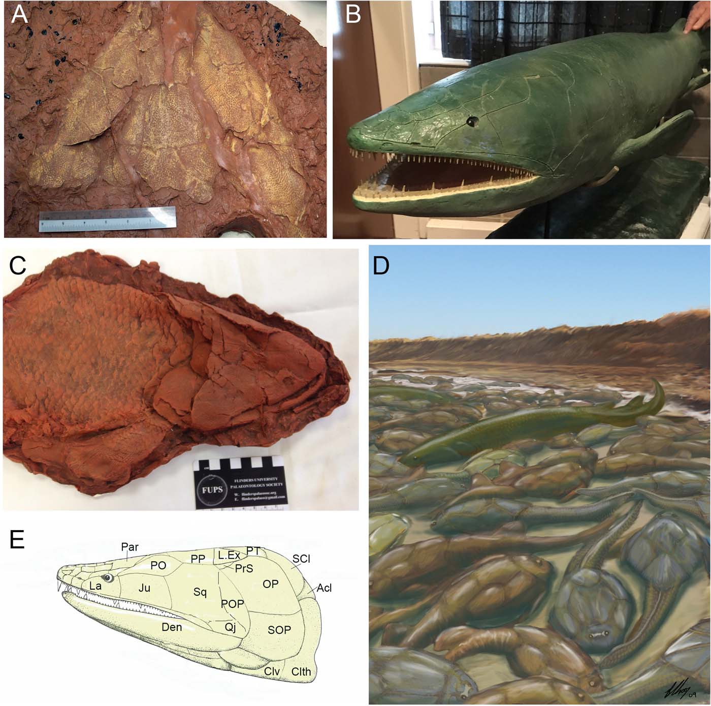
Late Devonian tristichopterid remains described from isolated bones found in the Late Famennian Hunter Siltstone of New South Wales as Eusthenodon gavini also include mention of a possible tetrapodomorphan Yambira thomsoni, known largely from scales and fragmentary shoulder girdle remains (Johanson & Ritchie Reference Johanson and Ritchie2000). Young (Reference Young2008) reviewed the anatomical features of the two Canowindra tristichopterids and concluded that they were separated from the Northern Hemisphere taxon as they share unique scale features and palatal bones called accessory vomers. He united Mandageria (Fig. 5a, b, e) and Cabonnichthys (Fig. 5c) in the family Mandageriidae (Fig. 5). Edenopteron keithcrooki (Fig. 5b) is another very large mandageriid, possibly 2 m in length, described from the Late Famennian coastal Devonian red beds (Worange Point Formation) exposed south of Eden, New South Wales (Young et al. Reference Young, Dunstone, Senden and Young2013).
Representative of clade Mandageriinae, a subgroup of tristicopterids endemic to the Devonian of Eastern Gondwana. (A) Cast of the skull of Edenopteron keithcrooki, an enormous Late Devonian form discovered at Eden, New South Wales (photo by Brian Choo, with permission). (B) Life-sized sculpture of Mandageria fairfaxi from the Late Devonian Mandagery Sandstone, New South Wales (photo by John Long, with permission). (C) Cast of Cabonnichthys burnsi from the Mandagery Sandstone (cast. Miyess Mitri). (D) Reconstruction of the Canowindra site during the Devonian, showing Cabonnichthys and numerous antiarchs (Bothriolepis yeungae and Remigolepis walkeri) perishing after the evaporation of an ephemeral pool (illustration by Brian Choo, with permission). (E) Lateral reconstruction of the skull of Mandageria with anatomical features indicated. Abbreviations: Acl = anocleithrum; Clth = cleithrum; Clv = clavicle; Den = dentary; Ju = jugal; L.Ex = lateral extrascapular; La = lacrymal; OP = operculum; Par = parietal; PO = postorbital; POP = preoperculum; PP = postparietal; PrS = prespiracular; PT = post-temporal; Qj = quadratojugal; SCl = supraclaithrum; SOP = suboperculum; Sq = squamosal.

An isolated cosmine-covered skull roof and ethmosphenoid named Owensia chooi was described from the Middle Devonian Kevington Creek Formation in the South Blue Range succession of central Victoria, by Holland (Reference Holland and Long2009). While its affinity is unclear, it has overall shape similarity to Koharalepis, and a weak orbital notch suggesting the presence of very small orbits, so it was suggested by Holland that it could be a canowindrid. If so, it provides the first description of part of the neurocranium for this group.
Bones of a digitate tetrapod from the Lower Carboniferous Ducabrook site in Queensland were first discussed by Thulborn et al. (Reference Thulborn, Warren and Turner1996) and described in detail by Warren & Turner (Reference Warren and Turner2004) and Warren (Reference Warren2007). Ossinodus pueri (Fig. 6d) is known from relatively complete yet isolated remains, well preserved in 3D form. It has been placed phylogenetically either as a stem tetrapod immediately above the node of known Devonian tetrapod taxa (Warren & Turner Reference Warren and Turner2004), or higher up the tree nestled within other stem tetrapod clades (Bernardi et al. Reference Bernardi, Petti, Piñuela, García-Ramos, Avanzini, Lockley, Landman and Harries2016).
Fossil evidence of early limbed tetrapods from Australia. (A) The lower jaw of Metaxygnathus denticulatus from the Late Devonian of New South Wales (photo by John Long, with permission). (B) Schematic diagrams of two distinct tetrapod trackways from the Late Devonian Genoa River Beds, Victoria (modified after Warren & Wakefield Reference Warren and Wakefield1972). (C) Life-sized sculptures of limbed stem tetrapods on display at Państwowy Instytut Geologiczny, Warsaw – the Gondwanan forms were likely very similar in appearance (photo by Brian Choo, with permission). (D) Reconstructed skeleton of Ossinodus pueri from the Early Devonian Ducabrook Formation, Queensland, with blue elements represented bones preserved, modified from an illustration by Adam Yates in Warren (Reference Warren2007).

New material of Gogonasus found on the 2005 Gogo Expedition described in Long et al. (Reference Long, Young, Holland, Senden and Fitzgerald2006) shows the presence of a large spiracle on top of the skull, and shows the complete 3D pectoral girdle skeleton of this taxon (Fig. 4a–c). Previously described Gogonasus skulls did not allow complete cranial reconstruction due to minor damage on parts of the oticco-occipital regions, so had not revealed the presence of large spiracles. Later papers emanating from the study of this new specimen, the first to be micro-CT (computerised tomography) scanned, include a review of the phylogenetic position of Gogonasus (Holland & Long Reference Holland and Long2009), and a description of the shoulder girdle and scapulocoracoid (Holland Reference Holland2013) and the endocranium (Holland Reference Holland2014). Work currently in press shows details of the caudal, anal and posterior dorsal fins of Gogonasus based on the Museum Victoria specimen (Long & Trinajstic Reference Long and Trinajstic2018).
On-going research continues to investigate taxa such as Gogonasus, based on a new complete specimen found in 2011 on the Gogo Expedition led by Kate Trinajstic of Curtin University. Neutron beam imaging at Lucas Heights, Australia's Nuclear Science and Technology Organisation (ANSTO), has been applied to this specimen to resolve features still encased in the rock. CT scanning and further manual preparation by G. Young and J. Lu at the Australian National University (ANU) is revealing much new information on the basal tetrapodomorph taxon (Fig. 2a) from the Eifelian Hatchery Creek Conglomerate (earlier assigned to ‘Gyroptychius? australis', Young & Gorter Reference Young and Gorter1981). Other finds under investigation include a new genus of basal canowindrid found at the Stokes Pass site (Givetian Harajica Sandstone) in the Northern Territory. Earlier finds at the site by Gavin Young and Alex Ritchie have been recently augmented by a joint Flinders–ANU expedition in 2016 that uncovered a fairly complete new specimen, plus additional specimens showing postcranial remains.
Field work completed in late 2017 in Victoria will enable a more complete assessment of the palaeoenvironment and context of possible tetrapod trackways from the Grampians site, and a description of the associated fish fauna and age assessment of the Genoa River trackways.
2. New anatomical information on the canowindrid Koharalepis jarviki
The Holotype skull of Koharalepis jarviki (AM F54325), collected in 1971 from the Middle Devonian Aztec Siltstone at Mount Crean in Antarctica (Young et al. Reference Young, Long and Ritchie1992), was scanned at the Australian Synchrotron facility on the Imaging and Medical Beamline (IMBL, ANSTO), using 60KeV and with a resultant voxel size of 32 microns. 3D segmentation and modelling was undertaken using Mimics v.19 and Drishti v.2.3. The specimen is filled with fine-grained siltstone, but yielded reasonable results from the scans to show new information on the anatomy of the lower jaws, braincase and palate. Some preliminary observations made using Drishti are presented herein. A more comprehensive anatomical investigation and description is currently in preparation.
2.1. Description of new cranial features
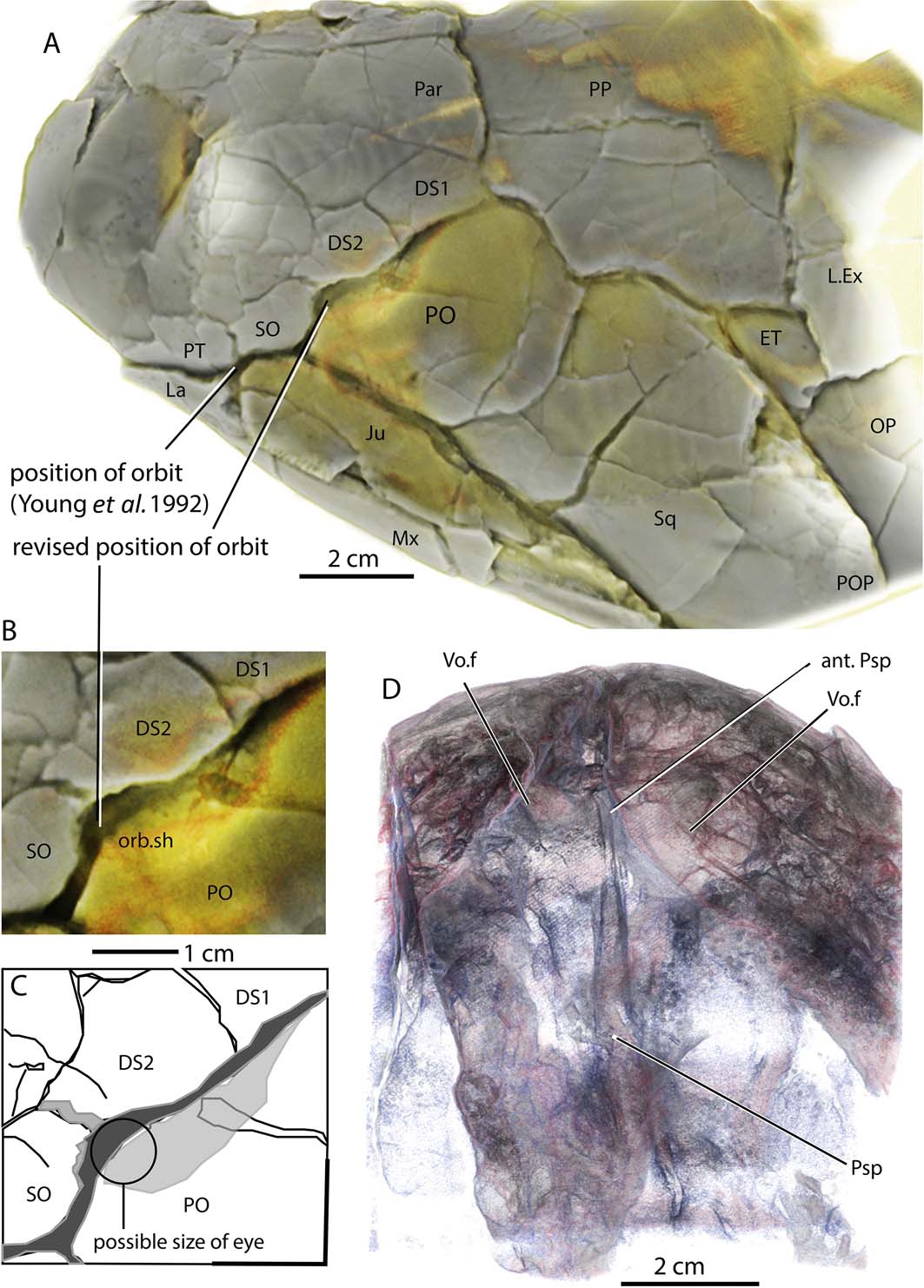
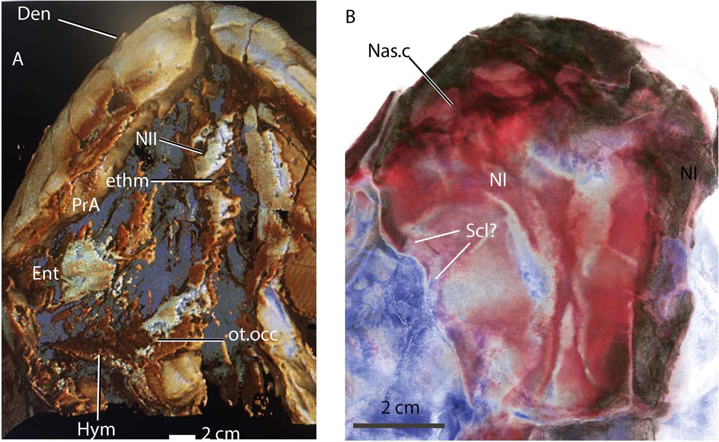
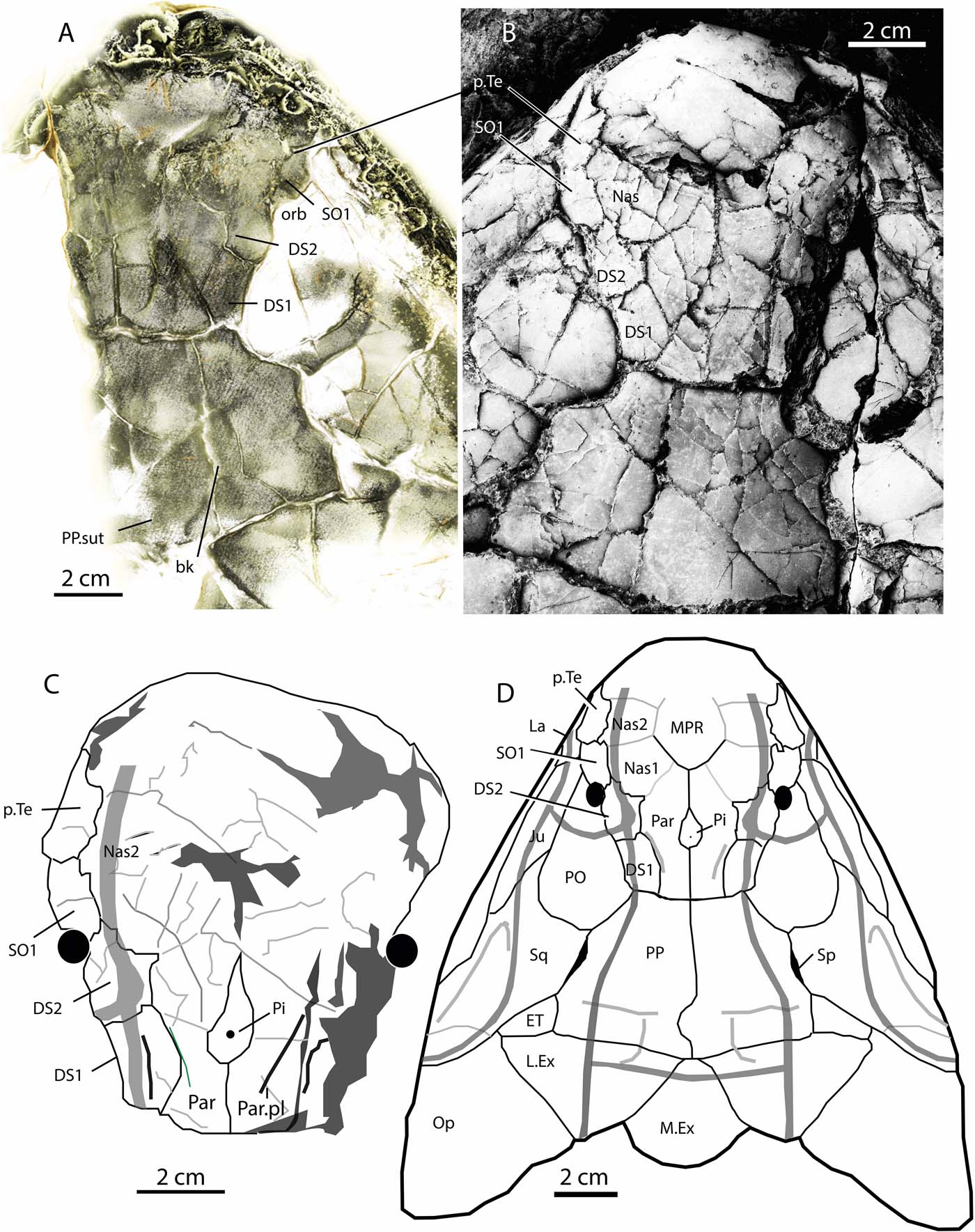
The skull roof, when shown as a high contrast tomogram, reveals new information on the position of the orbit (Fig. 7a–c). Our new data show that Young et al. (Reference Young, Long and Ritchie1992) placed the orbit too far ventrally. We agree that two dermosphenotic bones were present, as the anterior-most element bears the branch of the infraorbital sensory-line canal (Fig. 8b). We can now confirm the orbit lies in the same position as in other tetrapodomorph fishes such as Gogonasus (Long et al. Reference Long, Barwick and Campbell1997) at the junction of the postorbital, dermosphenotic and supraorbital bones (Fig. 7b, c). The tomograms clearly show the embayed dorsal margin of the orbit, and that the posterior of the orbit is bound by a weakly depressed shelf (Fig. 7b) on the large postorbital bone (Fig. 7a–c). Further study of the CT images under varied phase-contrast settings show the presence of a series of small rectangular bones below the orbital margin of the supraorbital bone, which we interpret as a series of possible sclerotic elements (Fig. 8b). It thus follows that the eyeball must have been very small, on a similar scale as that for most porolepiforms.
Koharalepis elucidated from synchrotron data, interpreted using Drishti 2.6. (A) Skull roof and cheek, left side. (B, C) Interpretation of orbital region. (D) Palate. Abbreviations: ant.Psp = anterior parasphenoid; DS1,2 = dermosphenotic elements; ET = extra temporal; Ju = jugal; La = lacrymal; Mx = maxilla; OP = operculum; orb.sh = orbital shelf; Par = parietal; PO = postorbital; POP = preoperculum; PP = postparietal; Psp = parasphenoid; PT = post-temporal; SO = supraorbital; Sq = squamosal; Vo.f = vomerine fangs.

Koharalepis elucidated from synchrotron data showing braincase details. (A) Drishti 2.6 rendition of the braincase and hyomandibular, ventral view. (B) Detail of ethmosphenoid showing position of nasal capsule and possible sclerotic ring elements. Abbreviations: Den = dentary; Ent = entopterygoid; ethm = ethmosphenoid; Hym = hyomandibular; Nas.c = nasal capsule; NI = olfactory nerve; NII = optic nerve; ot.occ = oticco-occipital; PrA = prearticular; Scl? = possible sclerotic elements.

The braincase is difficult to discern from the scan data, although it is ossified and visible from some cross sections seen in the tomographic slices. A Drishti image taken from the day of scanning, photographed on the computer screen (Fig. 8a), shows the outline and lateral walls of the ethmosphenoid (ethm, Fig. 8a) and oticco-occipital (Fig. 8a) divisions of the braincase. A stout rod-like element, the hyomandibular, comes out at a right angle to, and articulates with, the left side of the lateral commissure of the oticco-occipital (Hym, Fig. 8a). The same image shows parts of the entopterygoid (Ent, Fig. 8a) and prearticular (PrA, Fig. 8a). A large, circular foramen clearly visible on the left lateral wall of the ethmosphenoid is interpreted as the opening for the optic nerve (NII, Fig. 8a).
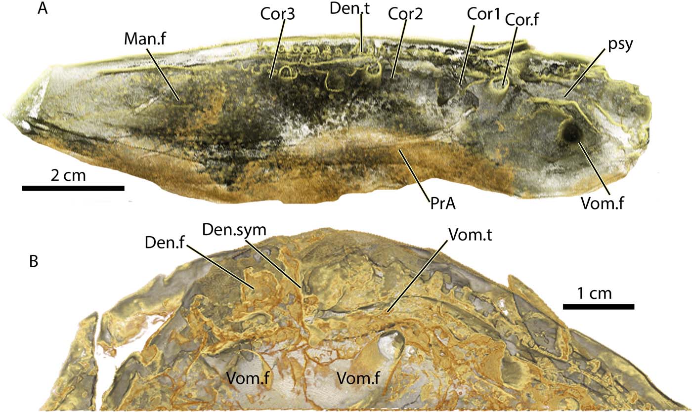
The palate (Figs 7d, 9b) is clearly visible from the tomograms. The large pentagonal-shaped vomers bear very large fangs (Vom.f, Figs 7d, 9b), and a row of smaller laniary teeth along their anterior crista (Vom.t, Fig. 9b). They are separated from contact with each other by the anterior stalk of the parasphenoid (Fig. 7d). In this respect, the palate shows resemblance to basal sarcopterygians like Youngolepis (Chang Reference Chang1982) or Tungsenia, where the clear depressions for attachment of the vomers indicate they are not in contact (Lu et al. Reference Lu, Zhu, Long, Zhao, Senden, Jia and Qiao2012, fig. 2b, f). It is quite unlike the evenly broad, lozenge-shaped parasphenoid seen in typical osteolepidids like Gogonasus (Long et al. Reference Long, Barwick and Campbell1997; Holland Reference Holland2014) or Medoevia (Lebedev Reference Lebedev1995). Instead, it is more reminiscent of the narrow lanceolate shape, which broadens posteriorly, found in megalichthyinids like Ectosteorhachis, Megalichthys (Thomson Reference Thomson1964, Jarvik Reference Jarvik1966) and Megistolepis (Vorobyeva Reference Vorobyeva1977).
Koharalepis elucidated from synchrotron data showing details of mandible (A) and anterior of palate (B). Drishti 2.6 rendition. Abbreviations: Cor.f = coronoid fang; Cor1,2,3 = coronoids; Den.f = dentary fangs; Den.sym = dentary symphysis; Den.t = dentary tooth; Man.f = mandibular fossa; PrA = Prearticular; psy = parasymphysial; Vom.f = vomerine fangs; Vom.t = vomerine teeth.

The external features of the lower jaw were described by Young et al. (Reference Young, Long and Ritchie1992), and the internal features can be seen by Drishti-rendered images (Fig. 9a), although imprinting of the palatal features through the tomograms needs be taken into account when interpreting the key features of the jaw. Figures 9a, b show that large dentary fangs are present (Den.f), almost as large as (but still visibly smaller than) the first coronoid fang (Cor.f, Fig. 9a). The parasymphsial plate is finely denticulated, and forms a broad shelf similar to that seen in Gogonasus or Medoevia. The coronoids (Cor1,2,3) are of almost equal length, with the largest fangs present on the anterior-most element (Cor.f, Fig. 9a). The prearticular (PrA, Figs 8a, 9a) is finely denticulated and there is no sign of any enlarged teeth or denticles along its dorsal edge.
A new restoration of the dermal skeleton of Koharalepis is presented in Figure 10d, based on a reinterpretation of the features mainly on the parietal-ethmoidal shield (Fig. 10).
Koharalepis elucidated from synchrotron data, image using Drishti 2.6, showing skull roof details (A, B). (C) Ethmoidal shield details, taken from images above. (D) New restoration of the skull in dorsal view. Abbreviations: bk = broken bone; DS1,2 = dermosphenotic elements; ET = extra temporal; Ju = jugal; L.Ex = lateral extrascapular; La = lacrymal; M.Ex = median extrascapular; MPR = median postrostral; Nas = nasal bones; Op = operculum; Orb = orbit; p.Te = posterior tectal; Par = parietal; Par.pl = parietal pit-line; Pi = pineal plate; PO = postorbital; PP = postparietal; PP.sut = postparietal suture; SO = supraorbital; Sp = spiracular notch; Sq = squamosal.

3. Phylogenetic and biogeographic implications of East Gondwana stem tetrapods
The timing and centre of origin of tetrapods has been resolved in recent years based on many discoveries of well-preserved elpistostegalian fishes and digitate tetrapods, which share a strong suite of synapomophies (Ahlberg & Johanson Reference Ahlberg and Johanson1998; Daeschler et al. Reference Daeschler, Shubin and Jenkins2006; Ahlberg et al. Reference Ahlberg, Clack, Luksevics, Blom and Zupins2008; Boisvert et al. Reference Boisvert, Mark-Kurik and Ahlberg2008; Zhu et al. Reference Zhu, Ahlberg, Zhao and Jia2017). Ahlberg & Johanson (Reference Ahlberg and Johanson1998) published one of the first comprehensive computer-generated cladistic analyses of stem tetrapods. They suggested that Kenichthys was the most basal member of the group, with rhizodontids forming the next node up clade, meaning rhizodonts were the sister taxa to all other stem tetrapods. Their work highlighted an earlier finding by Young et al. (Reference Young, Long and Ritchie1992) that the canowindrids formed an endemic East Gondwana clade. Whilst Young et al. (Reference Young, Long and Ritchie1992) suggested the canowindrid clade to be relatively basal, sitting one node above rhizodontids, Ahlberg & Johanson (Reference Ahlberg and Johanson1998) placed them more crownward within a paraphyletic cluster of four other osteolepidid-like groups.
Swartz (Reference Swartz2012), Lu et al. (Reference Lu, Zhu, Long, Zhao, Senden, Jia and Qiao2012) and Zhu et al. (Reference Zhu, Yu, Lu, Qiao, Zhao and Jia2012) show the significance of the East Gondwanan taxa in elucidating major radiations of the total group. Swartz (Reference Swartz2012) resolved the most basal member of the tetrapodomorpha to be Kenichthys from China, then rhizodonts, then canowindrids (here including Marsdenichthys as the sister taxon to the clade ‘Beelarongia + Koharalepis + Canowindra'. The rest of the group contained osteolepids in various positions, a monophyletic Megalichthyidae (with Medoevia) and monophyletic Tristichopteridae (including Australian ‘mandageriid' taxa as an end clade).
Recent phylogenies of stem tetrapod fishes have shown a commonality to the pattern in placing Tungsenia and Kenichthys as the most basal members. Both of these taxa come from the early Devonian of China (Chang & Zhu Reference Chang and Zhu1993; Lu et al. Reference Lu, Zhu, Long, Zhao, Senden, Jia and Qiao2012). The next node crownward from these taxa is the clade containing rhizodontids (Ahlberg & Johanson Reference Ahlberg and Johanson1998; Johanson & Ahlberg Reference Johanson and Ahlberg1998; Lu et al. Reference Lu, Zhu, Long, Zhao, Senden, Jia and Qiao2012; Swartz Reference Swartz2012; Zhu et al. Reference Zhu, Ahlberg, Zhao and Jia2017). The enigmatic Chinese taxon Hongyu, known from partial skull material and axial skeleton remains, is unresolved between two positions on the tree; one as sister taxon to all other rhizodontids, the other with rhizodontids in a polytomy with elpistostegalians. The latter position is supported by the unusual cleithrum, which is shortened ventrally to expose the scapulocoracoid laterally, a condition seen in some early tetrapods (e.g., Ventastega, Ahlberg et al. Reference Ahlberg, Clack, Luksevics, Blom and Zupins2008). Additional material of this peculiar taxon is required to resolve its phylogenetic affinity. The rest of the trees generated by the aforementioned analyses tend to conform to the same pattern – a spread of many branches of ‘osteolepid'-like taxa in the middle, a monophyletic clade of megalichthyinids and a crownward cluster defined by tristichopterids forming the sister group to elpistostegalians and tetrapods.
The East Gondwana clades are of special interest here biogeographically. Whilst Young et al. (Reference Young, Long and Ritchie1992) suggested canowindridoids were basal to the radiation of the osteolepidid-like groups, a position supported by Swartz (Reference Swartz2012), others saw the canowindrids crownward of the osteolepid-like clades (including megalichthyinids), placing them closer to the node defining the Tristichopterid-Elpistostegalid clade (Ahlberg & Johanson Reference Ahlberg and Johanson1998; Lu et al. Reference Lu, Zhu, Long, Zhao, Senden, Jia and Qiao2012). Zhu et al. (Reference Zhu, Ahlberg, Zhao and Jia2017) place canowindrids in a central position within the spread of ‘osteolepid'-like taxa.
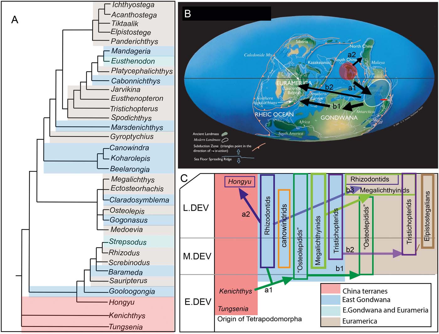
We have colour-coded taxa according to their geographical location (Fig. 11a) using the most recently published phylogenetic analysis of tetrapodomorphan interrelationships (involving 169 characters scored for 33 taxa by Zhu et al. (Reference Zhu, Ahlberg, Zhao and Jia2017), and their 50 % majority-rule strict consensus tree; Zhu et al. Reference Zhu, Ahlberg, Zhao and Jia2017, fig. 5b). This clearly shows that if the centre of origin of the group was the South China terrane in the Early Devonian, migration of the group out of China most likely took place via East Gondwana (Fig. 11b, c).
(A) A recent phylogenetic strict consensus tree (50 % majority rule) of stem tetrapods from Zhu et al. (Reference Zhu, Ahlberg, Zhao and Jia2017) with taxa colour-coded according to geographic region (see key in part C). (B) Palaeogeographic map of the Early Devonian taken from Scotese (Reference Scotese1991), with possible migration routes for stem tetrapod diversification routes as discussed in text. (C) Possible migratory routes for major groups of stem tetrapod fishes, as discussed in text.

Supporting this hypothesis is the fact that the oldest non-Euramerican tetrapodomorph taxa are possibly those from the upper Emsian–early Eifelian of Australia, as represented by remains of possible rhizodontids from the Wuttagoonaspis fauna of Georgina Basin, Cravens Peak Beds (Young & Goujet Reference Young and Goujet2003) and remains found in the Hatchery Creek Conglomerate (Young & Gorter Reference Young and Gorter1981). The oldest osteolepidid-like remains found in Euramerica appear to come from the basal Eifelian Parnu beds in the Baltic Succession, with Scottish forms like Thursius macrolepidotus appearing near the base of the Eifelian in the Lower Caithness Flagstones (Dineley & Loeffler Reference Dineley, Loeffler and Long1993). The majority of Scottish Old Red Sandstone osteolepid taxa begin to appear in the Achanarras horizon, which is upper Eifelian in age.
The coding of East Gondwana taxa in Figure 11a suggests it is likely that an early radiation of stem tetrapod fishes may have taken place somewhere within the China–East Gondwana region, including the appearance of the first rhizodontids. This is where the phylogenetically most basal rhizodontids (Hongyu, Goologongia) and the geologically oldest remains of rhizodontids are known. These include remains found in the Wuttagoonaspis fauna, of Emsian–Eifelian age (Young & Goujet Reference Young and Goujet2003), and also Aztecia from the Eifelian–Givetian Aztec Siltstone sequence in Antarctica (Johanson Reference Johanson, Arratia, Wilson and Cloutier2004). Two possibilities here are that rhizodontids originated in South China and first spread to Northern China (Ningxia is part of the Hexizoulang terrane) as shown in Figure 11b by route a2. Alternatively, if rhizodontids had their origin in East Gondwana, the spread of basal tetrapodomorphans out of China by the end of the Early Devonian would have been by a migration to East Gondwana (Fig. 11b, route a1).
The next radiation of fishes includes the canowindrid clade (East Gondwanan) as sister group to the tristichopterid-elpistostegalian clade, with one Euramerican taxon, Gyroptychius, positioned between them. The other clade of osteolepid-like taxa includes a clade containing megalichthyinids and a spread of other ‘osteolepids' (represented here by Osteolepis, Gogonasus and Medoevia). Megalichthyinids are well supported as a monophyletic group by several synapomophies (Long Reference Long1985b). This clade is represented by the oldest known taxa in East Gondwana (Mahalalepis, Eifelian–Givetian; Young et al. Reference Young, Long and Ritchie1992) and West Gondwana (Sengoerichthys, Frasnian; Janvier et al. Reference Janvier, Clément and Cloutier2007). In Euramerica, the megalichthyinids, as represented by Megalichthys, Ectosteorhachis (Thomson Reference Thomson1964) and Palatinichthys (Witzmann & Schoch Reference Witzmann and Schoch2012), plus Askerichthys (Borgen & Nakrem Reference Borgen and Nakrem2017), are only known from post-Devonian deposits.
The clade containing tristichopterids has Marsdenichthys from East Gondwana as the sister taxon to the clade containing well-known Northern Hemisphere taxa (Spodichthys, Tristichopterus, Eusthenopteron, etc.). Whilst Young argued for an endemic clade of East Gondwanan tristichopterids, the mandageriids, the analysis of Zhu et al. (Reference Zhu, Ahlberg, Zhao and Jia2017) failed to recover this group unless Eusthenodon (also found in Australia; Johanson & Ritchie Reference Johanson, Turner and Warren2000) is reconciled as part of this endemic group. At present, the description of Eusthenodon from East Greenland fails to show the synapomorphies designated by Young (Reference Young2006) to define mandageriids.
The only reliable records of elpistostegalians come from the Euramerican terrane comprising parts of Russia, Europe and North America. One report of a possible elpistostegalian from the Middle Devonian of Mount Howitt, Victoria (Long & Holland Reference Long and Holland2008), is based on a partial body with robust non-cosmine scales, and external features of the pectoral fin cannot be confirmed unless more complete material is found. Alternatively, the fish named Howittichthys could be a Glypotopomus-like osteolepidid. Whilst the evidence for the Euramerican origin of tetrapods is strong and remains largely uncontested based on phylogenetic similarities between the fishes Panderichthys, Tiktaalik and Elpistostege and early digitate tetrapods, an alternative case for an East Gondwanan origin of tetrapods can be put forward based on alternative criteria.
Panchen (Reference Panchen, Hecht, Goody and Hecht1977) first suggested that tetrapods might have first appeared in the East Gondwana region based on the early occurrence of the Genoa River tetrapod trackways (Fig. 3b). Long (Reference Long1990) used the older and phylogenetically more basal position of some key tetrapodomorph fish groups to frame a case supporting Panchen (Reference Panchen, Hecht, Goody and Hecht1977) for an East Gondwana radiation of tetrapodomorph fishes. The inference of this argument is that early tetrapods might have originated in East Gondwana, before migrating in the late Middle Devonian to the Northern Hemisphere. The inconsistency with this scenario is the lack of elpistostegalian fishes from Gondwana, which can be interpreted as a real gap in the known fossil record of that region, or alternatively seen as a real absence, which has biogeographic implications supporting the Euramerican origin of the group.
Recent evidence based on trackway data (Genoa River and the problematic Grampians Glenisla tracks) suggests that if tetrapods evolved earlier in East Gondwana, then there are two possible interpretations of the current phylogenetic signal. Firstly there could be a long undetected ghost lineage, based on absence of the fossil record of elpistostegalians from East Gondwana. Alternatively, but far less plausible in theoretical terms (and less parsimonious), is the possibility that digitate tetrapods could have appeared in east Gondwana as an independent radiation, not necessarily related to Northern elpistostegalians. The loss and reappearance of digits in different lineages of living amniote vertebrates shows that this feature can be quite plastic (e.g., in Bachia, Kohlsdorf & Wagner Reference Kohlsdorf and Wagner2006). This lizard shows strong evidence for the re-evolution of digit numbers in derived species, and that evidence is stronger for the development of hindlimb digits compared to forelimb digits.
4. Comments on the Glenisla trackway
Let us now look briefly at one of the most controversial records, the Glenisla trackway from the Grampians in western Victoria. The age of the controversial Glenisla trackways (Clack Reference Clack2012, p. 125) is constrained by the volcanic rocks dating the top of the Grampians sequence, capping it as no younger than Pragian (Simpson & Woodfall Reference Simpson and Woodfall1994). The fish-bearing horizon in the Silverband Formation, immediately above the trackway-bearing unit in the Major Mitchell Sandstone, contains microremains of thelodont scales and chondrichthyan spines, supporting a Late Ludlow age (Turner Reference Turner1986; Burrow Reference Burrow2003). Thus, the age range for the trackways is constrained between approximately 410 and 425 mya. Two recent independent analyses, one using molecular data from 13 proteins sequenced from 17 amphibian species to calculate divergence times (George & Blieck Reference George and Blieck2011), and the other from two independent statistical methods looking at predicted nodes for tetrapod divergence (Friedman & Brazeau Reference Friedman and Brazeau2010), support an Early Devonian time frame for the predicted origin of tetrapods. Another new trackway with ladder-like impressions made by broad-foot-like appendages and claw-like digit marks, has also been recently described from the Late Silurian Tumblagooda Sandstone of Western Australia by McNamara (Reference McNamara2014). McNamara referred to this trace as a possible tetrapod track. This new data also supports the possibility that the Glenisla trackway from Australia could be a genuine tetrapod track, even though digit impressions are lacking. Niedźwiedzki et al. (Reference Niedźwiedzki, Szrek, Narkiewicz, Narkiewicz and Ahlberg2010, p. 47) conclude at the end of their paper: ‘For the present the timing of the fish-tetrapod transition is best regarded as uncertain, though it clearly pre-dates the early Eifelian; an Early Devonian date seems likely, but even earlier potential tetrapod ichnofossils such as the Silurian Glenisla track should not be dismissed out of hand.'
Clack (Reference Clack2012, p. 125) alternatively suggests that the Glenisla tracks are similar to the ladder-like markings made by the placoderm Bothriolepis, as reported from the Late Devonian of East Greenland. This idea would have merit if the Glenisla tracks were Late Devonian; however, the only known placoderms from the Late Silurian and basal Early Devonian that have bone-covered appendages are the yunnanolepidoids. These have short, robust, non-segmented pectoral appendages. Such morphology could not make a ladder-like track, only sharp impressions in the substrate if we assume they had the ability to move the pointed appendage that far ventrally. Such a track would have to include a large central drag of the ventral body armour and a possible tail drag overprinting that trace. Whilst the degree of movement is unknown in the yunanaolepidoid pectoral fin, it was certainly limited in these fishes as constricted by the pectoral joint on the anterior ventrolateral plate (Janvier Reference Janvier, Clément and Cloutier1995). We agree after examining the original specimen that there are no digit impressions seen in the Glenisla trackways (Warrenn et al. Reference Warren, Jupp and Bolton1986). However, of the types of fishes living in this environment (agnathans, sharks and possibly placoderms, although none of the latter have been found in the sequence), none would be expected to make such trackways. Such fishes have rigid pectoral spines that, if present, usually make the kinds of traces represented by a series of narrow sinusoidal drag marks from the fin spines. Undichna septemsulcata is a typical example of this kind of trace found in the Early Devonian of Spitzbergen (Wisshak et al. Reference Wisshak, Volohonsky and Blomeier2004) and Undichna species found in the Lochkovian of Scotland (Trewin & Davidson Reference Trewin and Davidson1995). Thus, while the Glenisla trackways have not been confirmed to be tetrapod (due to lacking digit impressions), they have not yet been adequately explained as belonging to any other animal.
If the Glenisla tracks were made by a tetrapod, it implies a possible dual origin for tetrapods, with an extinction event of one line in East Gondwana, followed by a migratory event between Euramerica and Gondwana in the end of the Middle Devonian (Fig. 11b). This event, which was first suggested by Young (Reference Young1981) and supported by new evidence from studies of placoderm biogeography (Young Reference Young2006, Reference Young2007; Young et al. Reference Young, Burrow, Long, Turner and Choo2010), would involve the migration of advanced stem tetrapods, including tristichopterids and possibly elpistostegalians, sometime at the start of the Late Devonian.
The trends discussed so far, based on phylogenetic models and biogeography (Fig. 11), are supportive of an East Gondwana early radiation of stem tetrapods at the start of the Middle Devonian, and a migratory spread of the group through western Gondwana to Euramerica by the late Middle Devonian (Fig. 11b, route b1). This model accords well with the appearance of elpistostegalians in the Euramerican region by the latter part of the Middle Devonian. The lack of actual tetrapod body fossils or trackways from western Gondwana in the Late Devonian could support an alternative model – that if tetrapods did first evolve in East Gondwana, they arrived in Euramerica by the end of the Middle Devonian or start of the Late Devonian via a direct coastal route across the top of Gondwana (Fig. 11a, route b2).
The report of a single, incomplete Late Devonian tetrapod jaw from northern China (Sinostega, Zhu et al. Reference Zhu, Ahlberg, Zhao and Jia2002) highlights that the biogeography of Devonian tetrapods is not as simple as these linear migratory models might suggest. Nonetheless, as the fossil record of Devonian tetrapods outside of Euramerica is scant, we can only aspire to find more complete remains of them from Gondwana regions, as has been recently achieved in South Africa (Gess & Ahlberg Reference Gess and Ahlberg2018), before a clearer vision of tetrapod origins might be possible.
5. Acknowledgements
We would like to thank Anton Maksimenko and Vincent Dupret for their assistance during the synchrotron scanning session with AM F10403, and The Australia Museum for access to this specimen. We would also like to thank the editors for the invitation to contribute to this volume. This work was supported by the Australian Research Council grant DP 160102460.