Introduction
Fibrous dysplasia is a slowly progressive bone disorder of unknown origin in which normal bone is replaced with fibrotic tissue and disorganised bone trabeculae. It was first described by McCune and Bruch in 1937.Reference McCune and Bruch1 The disease is classified into three variants: monostotic (involving one bone), polyostotic (involving more than one bone) and McCune–Albright syndrome (associated with endocrinopathies and skin hyperpigmentation).Reference Nager, Kennedy and Kopstein2 Fibrous dysplasia of the temporal bone is very rare. Most involvement of the temporal bone is unilateral and associated with conductive hearing loss attributable to compression of the external auditory canal. Sensorineural hearing loss (SNHL) is usually due to otic capsule involvement by fibrous dysplasia or secondary cholesteatoma. However, the progression of the disease is so slow that it takes years before patients become symptomatic.
This report presents a patient with fibrous dysplasia of the temporal bone incidentally found on a routine computed tomography (CT) scan.
Case report
A 62-year-old man consulted the Department of Neurosurgery after a transient episode of syncope and a slight headache. He was referred to the ENT department with an abnormal shadow on the right temporal bone.
There was no external deformity of the contour of the patient's head and no skin lesions. Otoscopic evaluation did not demonstrate any stenosis of the right external auditory canal and the tympanic membrane was normal. Cranial nerve function and cerebellar function were intact. Hearing in both ears was within normal limits by pure tone audiometry examination. Caloric responses did not demonstrate canal paresis on either side. There was no family history of bone disorders, early puberty, or other endocrine or genetic abnormalities.
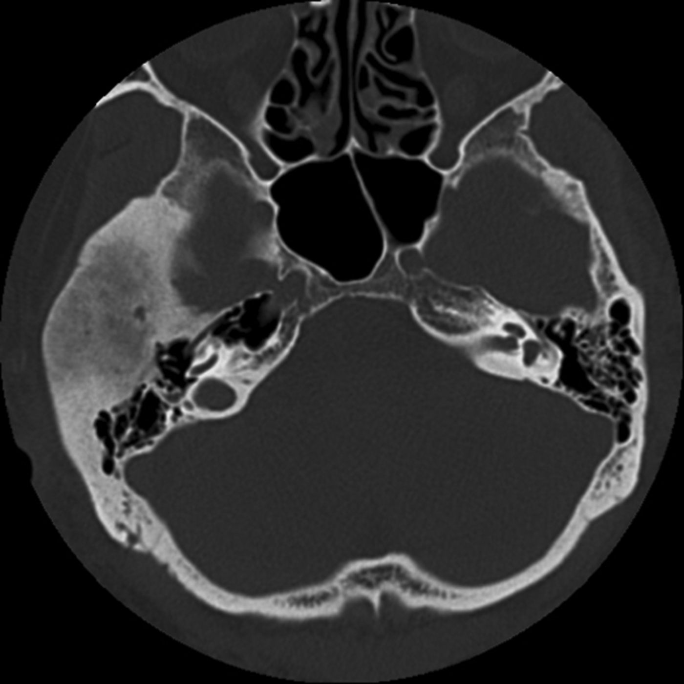
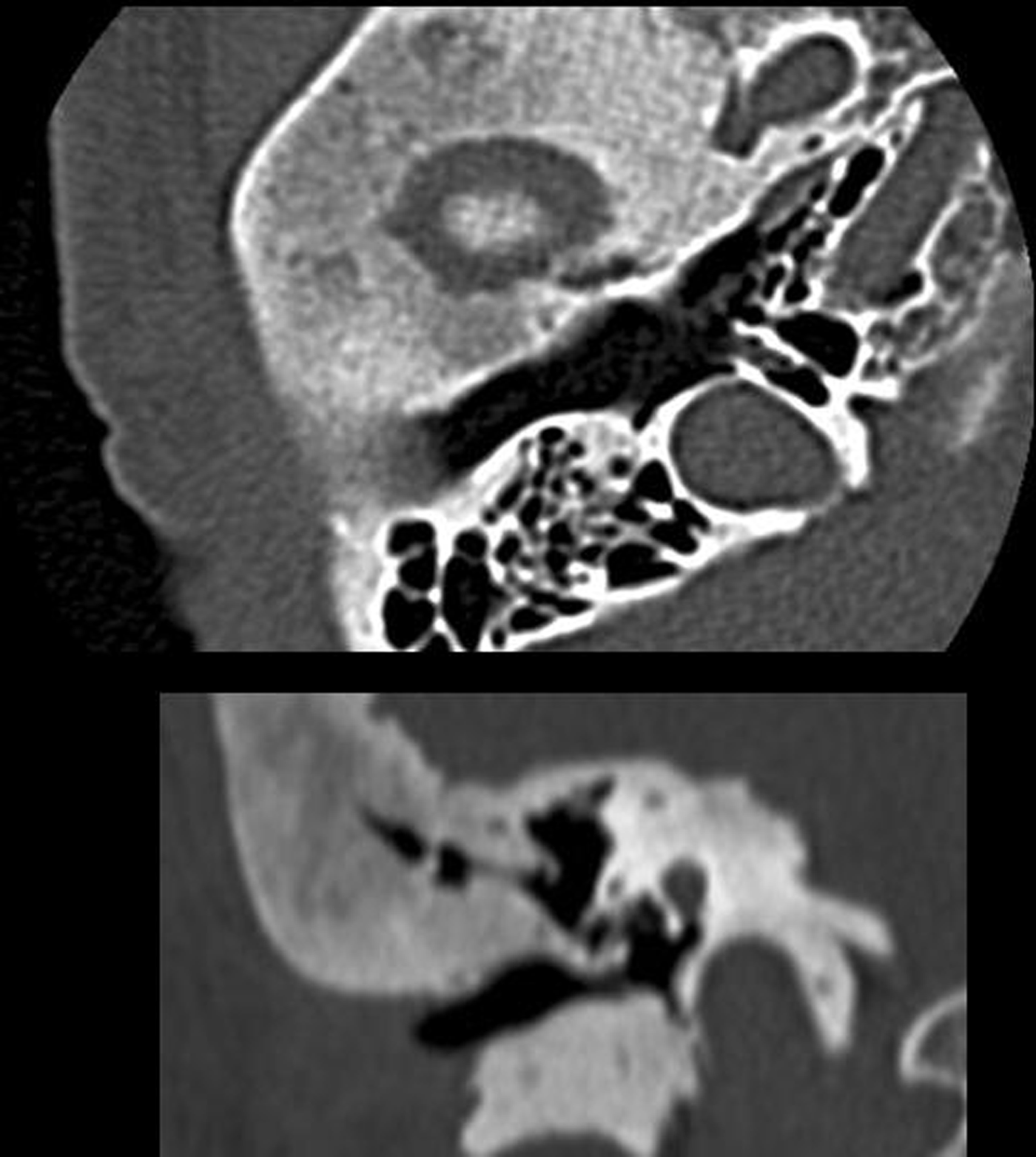
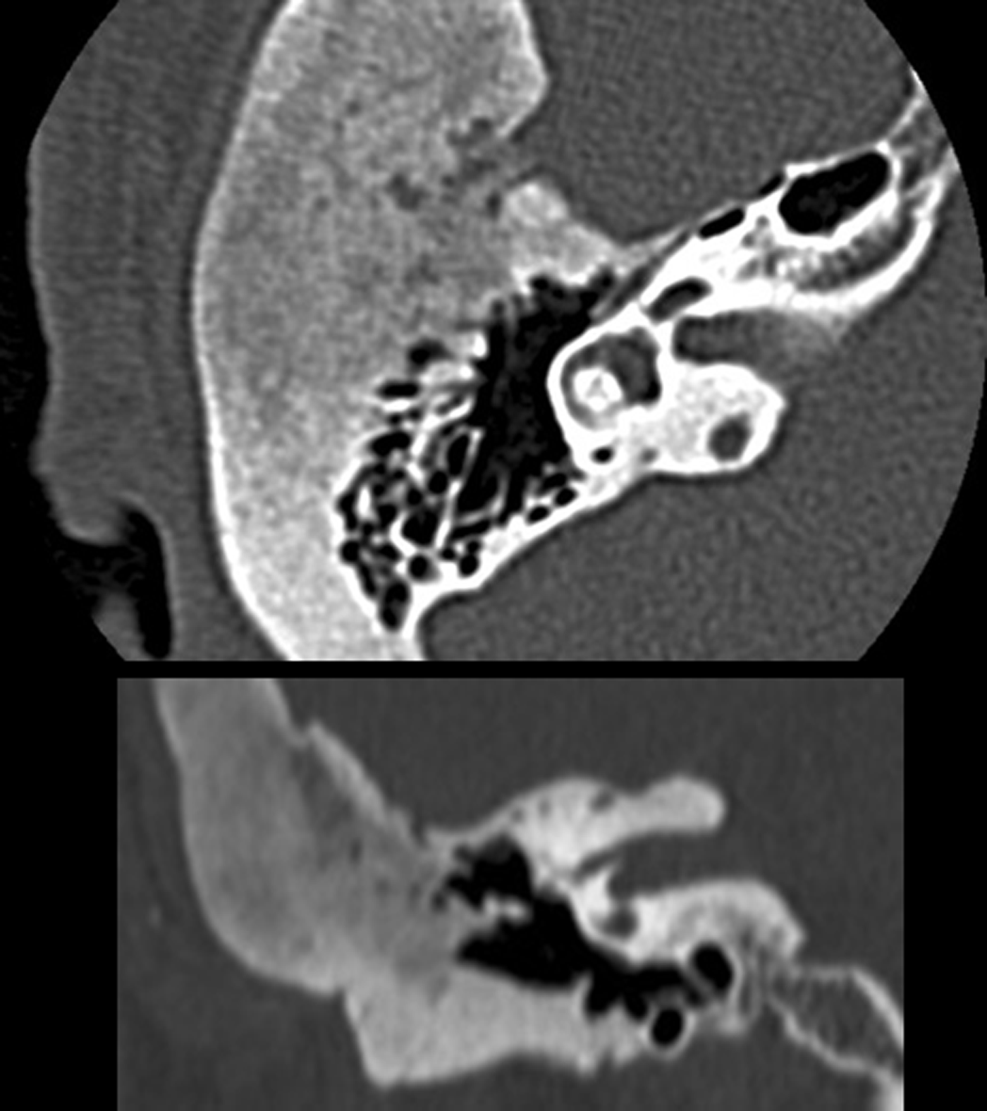
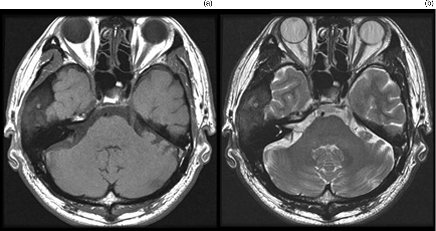
A CT scan of the right temporal bone demonstrated extensive thickening of the diploic and medullary spaces with a ground-glass appearance, moderately dense new bone and fibrous tissue (Figure 1). There was considerable expansion of the temporomandibular joint, but the patient did not have any functional complaints regarding mastication. The external auditory canal (Figure 2), middle ear, ossicular chain, cochlear capsule and internal auditory canal (Figure 3) appeared normal. Both the inner and outer cortices of the involved bones were intact, an important characteristic of fibrous dysplasia. Magnetic resonance imaging (MRI) showed a low-to-intermediate signal on T1-weighted images and a heterogeneous signal on T2-weighted images (Figure 4).
Axial computed tomography scan demonstrating a massive fibrous dysplasia in the right temporal bone. The overlying cortical bone remained intact.

Computed tomography scan of the right temporal bone demonstrating no narrowing of the external auditory canal. There is expansion of the disease into the temporomandibular joint. Upper = axial image; Lower = coronal image
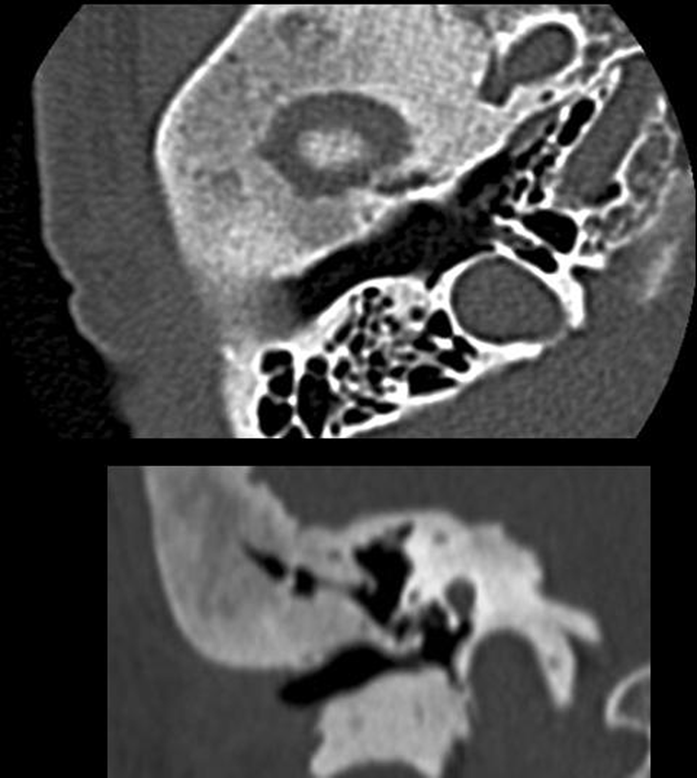
Computed tomography scan of the right temporal bone demonstrating no narrowing of the internal auditory canal. Upper = axial image; Lower = coronal image
Magnetic resonance imaging. (a) T1-weighted image. (b) T2-weighted image.
His fainting and headache disappeared the following day and there have not been any further complaints. Therefore, surgical exploration has not been performed.
Discussion
Fibrous dysplasia is a relatively uncommon condition of the bone and rarely affects the temporal bone. It is classified into three subgroups: (1) monostotic fibrous dysplasia – involvement of a single bone (70 per cent); (2) polyostotic fibrous dysplasia – involvement of multiple bones (27 per cent); and (3) McCune–Albright syndrome – bony involvement associated with endocrinopathies and skin hyperpigmentation (3 per cent).Reference Nager, Kennedy and Kopstein2 Craniomaxillofacial bony involvement occurs in only 10 per cent of monostotic fibrous dysplasia cases.Reference Windolz3 The maxilla and mandible are the most frequently affected craniomaxillofacial bones. The temporal bone is affected in only 18 per cent of those with skull involvement.Reference Magerian, Sofferman, McKenna, Eavey and Nadol4, Reference Morrissey, Talbot and Schleuning5
The mean age of occurrence of the three types is 14, 11 and 8 years old for the monostotic form, polyostotic form and McCune–Albright syndrome, respectively.Reference Van Horn, Dahlin and Bickel6 Yaunus and HaleemReference Yaunus and Haleem7 reported an infant case in a nine months old with an absent kidney. Temporal bone involvement with fibrous dysplasia is twice as common in males as it is in females.Reference Nager, Kennedy and Kopstein2 This disease has a racial predominance, in that Caucasians account for >80 per cent of all cases, African-Americans 2 per cent and Asians only 1 per cent.Reference Magerian, Sofferman, McKenna, Eavey and Nadol4
Pathologically, fibrous dysplasia appears as a poorly localised lesion replacing normal bone. The tissue contains disorganised bony trabeculae and spindle cells surrounded by a fibrous matrix.Reference Magerian, Sofferman, McKenna, Eavey and Nadol4, Reference Yaunus and Haleem7 Theories of the aetiology of this disease include aberrant differentiation of the mesenchyme during bone formation, an arrest of bone at the immature woven stage or a disturbance in cancellous bone maintenance.Reference Nager, Kennedy and Kopstein2 The bony cortex is expanded and thinned, but usually remains intact, maintaining a smooth contour. There is no true capsule, and the margin between normal and abnormal bone is indistinct.
Barrionuevo et al. Reference Barrionuevo, Marcallo, Coelho, Cruz, Mocellin and Patrocinio8 classified fibrous dysplasia of the temporal bone into the following three stages according to the progression of the disease: stage 1, latent phase; stage 2, symptomatic phase; and stage 3, complication phase. Although our case was stage 1, almost all cases of fibrous dysplasia of the temporal bone in the 77 surveyed cases were in stage 3.Reference Abe, Suzuki, Ueyama, Ina and Mogi9 The most common presenting symptom is deafness. Other symptoms include post-aural or temporal swelling, otorrhoea, otalgia and pulsatile tinnitus. Abnormal physical findings are unilateral conductive or SNHL, stenosis of the ear canal including external auditory canal massReference Wang and Chen10 and cholesteatoma. Cholesteatoma is seen in 40 per cent of cases and usually originates from the external canal wall.Reference Morrissey, Talbot and Schleuning5, Reference Tochino, Sunami and Yamane11 Sensorineural hearing loss, resulting from involvement of the internal auditory canal, is seen in 17 per cent of patients.Reference Magerian, Sofferman, McKenna, Eavey and Nadol4 McCall et al. Reference McCall, Curtin and McKenna12 reported posterior semi-circular canal dehiscence arising from cystic degeneration of fibrous dysplasia. Facial nerve involvement is seen in 10 per cent of these patients.Reference Morrissey, Talbot and Schleuning5, Reference Sakamoto, Hayashida and Sugasawa13, Reference Reddy, Vinayak, Jefferis and Grieve14 Although not common, function of all cranial nerves can be affected.Reference Nager, Kennedy and Kopstein2 The case presented here showed expansion of the disease into the temporomandibular joint, but did not show any symptoms. If displacement of the temporomandibular joint occurs, the patient may complain of dental malocclusion or temporomandibular joint dysfunction.Reference Pouwels and Cremers15
The radiographic appearance varies with the ratio of fibrous and osseous tissue in the affected bone, ranging from radiolucency to radiopacity. Three radiographic patterns have been identified: pagetoid, sclerotic and cystic.Reference Magerian, Sofferman, McKenna, Eavey and Nadol4, Reference Reddy, Vinayak, Jefferis and Grieve14, Reference Lambert and Brackmann16 The pagetoid pattern seen in 56 per cent of cases is typified by a mixture of dense and radiolucent fibrous areas with bone expansion. Homogeneously dense areas with bone expansion are typical of the sclerotic pattern seen in 23 per cent of radiographic studies. The cystic pattern shows spheric or oval lucent regions with a dense boundary seen in 21 per cent of cases. Our case showed the most common pagetoid pattern, which is characterised by a ground-glass appearance on CT imaging. Magnetic resonance imaging, in general, does not give as much information as CT.Reference Chee and Chen17 It exhibits a low-to-intermediate signal on T1-weighted images and a heterogeneous signal on T2-weighted images. Post-gadolinium T1- and T2-weighted imaging shows moderate to marked enhancement.Reference Casselman, De Jonge, Neyt, De Clercq and D'Hont18 Signal intensity and the degree of enhancement depend on the relative proportions of bony trabeculae, cellularity, collagen and cystic changes.
• This report presents a patient with asymptomatic fibrous dysplasia of the temporal bone
• Diagnosis was based on computed tomography and magnetic resonance imaging
• Surgical exploration and treatment was not performed due to the patient's lack of complaints
• Treatment should be based on patient symptoms and disease progression
Treatment and definitive diagnosis rely on surgical debulking and biopsy. The decision to treat depends on the degree of cosmetic or functional impairment. Fibrous dysplasia is a benign condition with a good prognosis. As fibrous dysplasia is likely to become quiescent, surgical intervention should be as conservative as possible.Reference Morrissey, Talbot and Schleuning5, Reference Yaunus and Haleem7, Reference Reddy, Vinayak, Jefferis and Grieve14 The disease process usually stops at puberty.Reference Yaunus and Haleem7 However, when an aggressive or unpredictable progression occurs, including cases involving inner ear or skull base structures, appropriate surgical intervention should be considered. Despite some instances of recurrence, surgical treatment has revealed relatively good outcomes.Reference Kim, Song, Choi, Lee, Oh and Chang19, Reference Yang, Chen, Zheng, Xu, Zhang and Xiong20 Radiation therapy should be avoided,Reference Morrissey, Talbot and Schleuning5, Reference Reddy, Vinayak, Jefferis and Grieve14, Reference Yagoda and Selesnick21 because past experience shows an increase in the incidence of malignant transformation to 44 per cent after radiation treatment.Reference Slow, Stern and Friedman22, Reference Kaushik, Smoker and Frable23 Spontaneous malignant transformation is extremely rare (0.5 per cent);Reference Lambert and Brackmann16 however, malignant change is indicated by pain, aggressive growth and elevation of serum alkaline phosphatase.Reference Schwartz and Alpert24
Conclusion
This report presents a patient with asymptomatic fibrous dysplasia of the temporal bone. Diagnosis was based on CT and MRI. Surgical exploration and treatment was not performed due to the patient having no complaints.