Introduction
The main functions of the Eustachian tube are ventilation, protection and clearance of the middle ear. Middle-ear ventilation occurs at each deglutition by the action of paratubal muscles, leading to air pressure equilibration with atmospheric pressure. A role for Eustachian tube dysfunction in otitis media has been extensively investigated; currently evidence suggests that otitis media pathogenesis is strictly related to tubal dysfunction, leading to reduced ventilation and increased negative pressure in the middle ear.Reference Seibert and Danner 1 , Reference Corbeel 2
The tensor and levator veli palatini are the most important paratubal muscles in humans.Reference Teixeira, Banks, Swarts, Alper and Doyle 3 It is clear that the tensor veli palatine muscle, arising from the perichondrium of the lateral lamina of the tubal cartilage and the lateral membranous wall of the Eustachian tube, is the only muscle to actively open the Eustachian tube and promote ventilation of the middle ear.Reference Bluestone and Doyle 4 , Reference Honjo, Ushiro, Haji, Nozoe and Matsui 5 Eustachian tube function seems to be influenced by its physical composition (i.e. the distribution of elastic fibres), mechanical characteristics and geometry (the muscle–Eustachian tube vector relationship).Reference Doyle, Mandel, Seroky, Swarts and Casselbrant 6 , Reference Dinç, Damar, Uğur, Öz, Eliçora and Bişkin 7 This system improves with age and is down-regulated during concomitant inflammatory nasopharyngeal exacerbations.Reference Swarts, Casselbrant, Teixeira, Mandel, Richert and Banks 8 , Reference Bluestone 9
The present authors previously reported a qualitative and quantitative analysis of the tensor and levator veli palatini muscle fibres in animals in which otitis media was experimentally induced by obstructing the nasal cavities.Reference Scarano, Fetoni, Picciotti, Cadoni, Galli and Paludetti 10 In these animals, the number of tensor veli palatini fibres progressively decreased after nasal obstruction. These findings suggest that a functional muscle may reduce tubal dysfunction and consequently otitis media chronicity and recurrence. Eustachian tube rehabilitation using a combination of non-invasive methods such as improved nasal hygiene and breathing, muscle strengthening exercises and auto-insufflation can improve Eustachian tube opening.Reference D'Alatri, Picciotti, Marchese and Fiorita 11 , Reference Gersdorff, Cambier and Huysbrechts-Forester 12 Auto-inflation was first described by Valsalva in 1704 and Politzer in 1863 using a catheter and has a beneficial effect in otitis media treatment; it should be considered a first-line treatment (before planning antibiotic therapy or surgery).Reference Stangerup 13 , Reference Arick and Silman 14
Several attempts have been made to develop a reliable test to record Eustachian tube function. Recently, the current authors proposed a new technique for paratubal electromyography (EMG) using a surface, non-invasive electrode applied transnasally under nasopharyngoscopic guidance to record tensor veli palatini muscle activity during swallowing.Reference Picciotti, Rigante, Della Marca, Restuccia and Scarano 15
In the present study, this technique was used in otitis media with effusion (OME) patients to confirm that Eustachian tube muscle dysfunction is involved in OME pathogenesis. In particular, electronic recordings of the tensor veli palatini muscle on both sides were compared with data from normal healthy participants. Moreover, muscle activity before and after Eustachian tube rehabilitation is described.
Materials and methods
Twenty otitis media with effusion (OME) patients (10 women and 10 men) with a mean age of 42.5 ± 13.6 years were enrolled into the study and treated with Eustachian tube rehabilitation. Fourteen patients were affected by unilateral OME (unilateral OME group) and 6 by bilateral OME (bilateral OME group). A control group of 10 healthy volunteers (5 men and 5 women), aged between 27 and 37 years and without any otological or nasal pathology was also enrolled.
All patients underwent microtoscopy, a physical examination of the palate and nasopharynx, audiometric tests, tympanography, pressure/swallowing Eustachian tube function tests, the Toynbee test, the Valsalva manoeuvre and paratubal EMG. Electrophysiology of the tensor veli palatini was measured, as previously described.Reference Picciotti, Rigante, Della Marca, Restuccia and Scarano 15 For this, a metallic nasopharyngeal electrode was introduced through the nasal cavity and placed upon the pharyngeal ostium of the Eustachian tube, approximately 3 mm antero-inferiorly to the salpingo-palatine fold, under endoscopic guidance. The nasopharyngeal electrode, consisting of a Teflon-isolated steel wire with a silver ball tip inserted into a steel catheter electrode, measured tensor veli palatini muscle activity.Reference Restuccia, Di Lazzaro, Valeriani, Mariotti, Torrioli and Tonali 16 The reference electrode, consisting of a gold surface lead filled with electrolyte, was placed at the left mastoid and fixed with collodium. Impedance was less than 5 kOhm. The EMG signal was acquired using a Micromed System 98 digital polygraph (Mogliano Veneto, Italy), with a sampling rate of 1024 Hz, a high pass filter of 10 Hz, a low pass filter of 500 Hz and a gain of 5 μV per mm. During the recordings, participants were asked to swallow and the electrode was moved until the maximal contraction was recorded. After the first recording, the electrode was moved within the same nasal cavity to find the contralateral pharyngeal ostium of the Eustachian tube. All bilateral electrical activities including motor unit potentials, amplitude, duration phase, interference and swallowing contraction EMG activities were recorded, but only the most important (amplitude and duration phase) were analysed.
Patients in the bilateral OME group underwent Eustachian tube rehabilitation, comprising 12 therapy sessions with an average length of 30 minutes: the first 8 sessions were twice weekly and the last 4 were 15 days apart. In these, naso-diaphragmatic breathing exercises were performed to synchronise nasal and diaphragmatic breathing, and patients acquired an awareness of nostril dilatation via nasal valve exercises. Increased tubal opening was achieved via swallowing and yawning exercises. Moreover, the velopharyngeal sphincter was activated via exercises that involved moving the tongue, soft palate and jaws (separately or in coordination). The Valsalva and other passive auto-insufflation manoeuvres were also used. Finally, the regular use of chewing gum was encouraged to activate jaw movements and increase salivary flow, the swallowing rate, and the rate of paratubal muscle activation and tubal opening.Reference Kapila, Dodds, Helm and Hogan 17
In the unilateral OME group, EMG data for the affected side were compared with those for the contralateral side and normal controls. In the bilateral OME group, EMG data obtained before Eustachian tube rehabilitation were compared with data obtained after therapy and with data for controls.
All patients were informed about their disease and treatment options, and counselled about the potential risks and expectations of the procedure; all gave consent. All procedures were performed with the approval of the local ethics committee in accordance with the Helsinki Declaration of 1975, as revised in 2008.
Statistical analysis was performed using paired Student's t-tests, with statistical significance set at a p value of less than 0.05.
Results
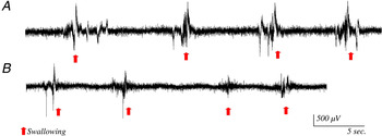
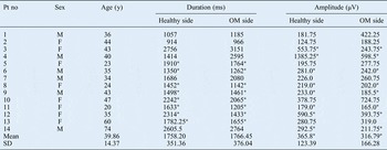
Electromyography results for individual patients are shown in Tables I and II. In the unilateral otitis media with effusion (OME) group, the duration phase and/or amplitude were lower in the healthy side than in the affected side in 11 patients (78.5 per cent): 5 patients (6, 8, 9, 11 and 12; 35.7 per cent) had both duration phase and amplitude impairment in the affected side (patient 6 data is shown in Figure 1); 3 (patients 5, 10 and 13; 21.4 per cent) showed only duration phase impairment; and 3 (patients 3, 4 and 14; 21.4 per cent) showed only amplitude impairment. Electromyography data for the other three patients (1, 2 and 7; 21.4 per cent) showed no impairment (Table I).
Electromyography recording of patient 6, showing impairment of both the duration phase and amplitude in the side affected by otitis media (OM). A = healthy side; B = OM side

Electromyography results of unilateral otitis media patients

*Reduced duration phase and/or amplitude. Pt = patient; no = number; y = years; OM = otitis media; M = male; F = female; SD = standard deviation
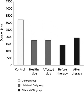
The EMG duration phase was reduced in affected ears compared with unaffected ears in eight patients (57.1 per cent), but there was no difference in the mean values for the two sides (p > 0.05). However, the mean duration phase in the normal control group (3215 ± 672 ms; data not shown) was significantly different from that of both the unaffected and OME sides (p < 0.001; Figure 2).
Graph comparing the duration phase in the control, and unilateral otitis media (OM) groups and in the bilateral OM group before and after Eustachian tube rehabilitation therapy. Data are expressed as the mean ± standard deviation.

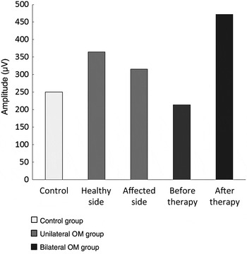
Similarly, the EMG amplitude was reduced in the affected ears compared with unaffected ears in eight patients, but there was no difference in the mean values for the two sides (p > 0.05). In contrast, the mean amplitude for the normal control group (250.1 ± 12 μV; data not shown) was not significantly different from that of the unaffected and affected sides (p > 0.05; Figure 3).
Graph comparing the amplitude in the control and unilateral otitis media (OM) groups and in the bilateral OM group before and after Eustachian tube rehabilitation therapy. Data are expressed as the mean ± standard deviation.

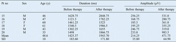
In the bilateral OME group, the EMG duration phase and amplitude were impaired both before and after Eustachian tube rehabilitation in all patients. Patient 15 showed the greatest change in duration phase (1220 ms) and patient 20 showed the greatest change in amplitude (750 μV). In contrast, patient 17 showed the least change in duration phase (61.75 ms) and patient 19 showed the least change in amplitude (72.25 μV; Table II).
Electromyography results of bilateral chronic otitis media with effusion

Pt = patient; no = number; y = years; M = male; F = female; SD = standard deviation
There was no significant difference in the mean duration phase before and after Eustachian tube rehabilitation (p > 0.05), but there was a significant difference in the mean amplitude before and after Eustachian tube rehabilitation (p < 0.05). Finally, the mean duration phase and mean amplitude before and after Eustachian tube rehabilitation were significantly different from equivalent data in the normal control group (3215 ± 672 ms and 250.1 ± 12 μV, respectively; p < 0.001; Figures 2 and 3).
Discussion
Although several factors involved in otitis media pathogenesis have been described, the complete pathological process has not been defined. One theory is that Eustachian tube dysfunction is responsible for the development of middle-ear diseases. Clinical and experimental studies have been conducted to investigate a possible correlation between otitis media and tubal dysfunction, and several test protocols (with adequate specificity and sensitivity) have been used to diagnose tubal dysfunction and identify possible underlying causes.
Doyle first reported that inefficient Eustachian tube function leads to mucosal inflammation and middle-ear effusion in monkeys.Reference Doyle 18 Casselbrant et al. observed the development of otitis media following temporary paralysis of the tensor veli palatini muscle after botulinum toxin injection in an animal model.Reference Casselbrant, Cantekin, Dirkmaat, Doyle and Bluestone 19 Surgical clefting of the palate induced otitis media and permanent dysfunction of the tensor veli palatini muscles.Reference Doyle, Ingraham, Saad, Cantekin, Lim, Bluestone, Klein and Nelson 20 In addition, clinical investigations into cleft palate in children affected by Down's syndrome showed a high incidence of otitis media related to Eustachian tube dysfunction due to deformation of the craniobasal regions.Reference White, Doyle, Bluestone, Lim, Bluestone, Klein and Nelson 21 Studies using different techniques have demonstrated that decreased Eustachian tube muscle function coincides with the presence of otitis media in younger children.Reference Swarts, Casselbrant, Teixeira, Mandel, Richert and Banks 8 , Reference Bhat, Kumar, Nag and Hegde 22 , Reference Bylander, Tjernstrom, Ivarsson, Ingvarsson, Lim, Bluestone, Klein and Nelson 23
Moreover, adult participants with a childhood history of otitis media were proven to have poorer Eustachian tube function compared with those with no previous history of middle-ear disease.Reference Alper, Swarts, Singla, Banks and Doyle 24 These data account for poor constitutive Eustachian tube function which remains throughout adulthood.
The most frequently used methods for testing Eustachian tube function are manometry, pressure chamber testing, the forced response test (a relatively invasive protocol needing direct access to the middle ear through a tympanic perforation) and sonotubometry (a non-invasive test that monitors the sound level in the ear canals after sound is introduced into the nasopharynx).Reference Teixeira, Banks, Swarts, Alper and Doyle 3 , Reference Mandel, Swarts, Casselbrant, Tekely, Richert and Seroky 25
The current authors previously demonstrated in rats that otitis media was related to reduced paratubal muscle fibre numbers caused by nasal obstruction.Reference Scarano, Fetoni, Picciotti, Cadoni, Galli and Paludetti 10 The present study confirmed a role for Eustachian tube muscle dysfunction in otitis media with effusion (OME) pathogenesis. The action potential in the affected side was modified compared with the healthy side in 78.5 per cent of patients. In particular, 57.1 per cent of patients had a reduced duration phase in the affected side compared with the healthy side, with a significant difference between normal controls and affected patients. The pattern of differences in amplitude was similar, with 57.1 per cent of patients showing impairment on the affected side. Studies have suggested that an increase in the EMG parameters is related to neurological disorders, whereas a decrease may suggest myopathic damage.Reference De Bleecker 26 The reduction in duration phase and amplitude described in the present study demonstrates that modified tensor veli palatini activity may be related to myopathic dysfunction.
These findings contradict those of Sapci et al., who analysed the function of the tensor veli palatini muscle by EMG (using a needle introduced transnasally) in chronic middle-ear pathologies.Reference Sapci, Mercangoz, Evcimik, Karavus and Gozke 27 These authors found no significant differences between the affected and control sides and between affected patients and normal controls, concluding that myogenic defects in the Eustachian tube have a limited effect in otitis media.
Different techniques have been proposed for paratubal EMG, including a bipolar wire-hooked electrode inserted transorallyReference Hairston and Sauerland 28 ; a unipolar needle electrode inserted transorallyReference Kamere 29 ; a surface electrode applied to the soft palateReference Guindi, Payne and Higenbottan 30 ; a needle monopolar electrode inserted transnasallyReference Honjo, Kumazawa, Honda and Shimojo 31 ; and a concentric needle electrode applied transnasally under endoscopic guidance.Reference Su, Hsu and Chee 32 The present study used a surface electrode applied transnasally under nasopharyngoscopic guidance to record tensor veli palatini muscle activity during swallowing and objectively evaluate the function of this muscle. This method has several advantages: it is non-invasive; the electrode is easy to position; the procedure is not painful; and the endoscopy guide helps ensure correct positioning of the electrode. Therefore, the procedure is also suitable for use in children.
The present study also showed that tensor veli palatini muscle activity before and after Eustachian tube rehabilitation can be easily monitored by EMG. In fact, all patients who underwent Eustachian tube therapy showed improvement in the most important EMG parameters, duration phase and amplitude. Differences in the degree of improvement among patients was probably caused by inter-individual variability in signal characteristics.
A potential limitation of the study was the small number of patients, with high inter-individual variability and high SD values. This prevented the calculation of reliable cut-off values for ear disease based on duration and amplitude values. This preliminary study, however, unquestionably demonstrates that tensor veli palatini EMG parameters are significantly different between healthy individuals and otitis media patients. Good test reliability was indicated by the significant improvement in EMG parameters after Eustachian tube rehabilitation. Future studies will use a larger patient cohort to determine standardised criteria based on EMG amplitude and duration values for differentiating between healthy ears and those affected by Eustachian tube dysfunction.
-
• Otitis media pathogenesis is linked to Eustachian tube dysfunction via improper ventilation and increased negative middle-ear pressure
-
• Non-invasive Eustachian tube rehabilitation aims to improve Eustachian tube opening
-
• Paratubal electromyography using a non-invasive surface electrode was used to record tensor veli palatini muscle activity during swallowing in 20 otitis media patients
-
• In 78.5 per cent of patients, the duration phase and/or amplitude was reduced in the affected side
-
• All patients undergoing Eustachian tube rehabilitation had an impaired muscle action potential
Published literature and previous experimental data indicate that Eustachian tube muscle dysfunction plays an important role in the development of otitis media. Based on these findings, Eustachian tube rehabilitation may be considered crucial in OME therapy to ameliorate middle-ear ventilation and reduce the incidence of OME reappearance after surgery. Finally, EMG of the tensor veli palatini muscle with a transnasally applied surface electrode is a useful tool for evaluating Eustachian tube dysfunction and monitoring Eustachian tube rehabilitation.