Introduction
Diffusion tensor imaging (DTI) is a novel research approach to assess the amount and preferred direction of diffusion of water molecules in vivo (Basser et al. Reference Basser, Mattiello and LeBihan1994; Basser, Reference Basser1995). In the brain, water molecules tend to diffuse along the direction of the axonal fibres wrapped by myelin sheathes; thus, DTI serves as a proxy measure of the orientation and structure of white matter comprising millions of axonal fibres grouped together in myelinated sheaths (Basser, Reference Basser1995). Fractional anisotropy (FA) is a measure of the directional diffusion of water, values range from 0 (completely anisotropic or randomly directed) to 1 (completely isotropic or uni-directional). FA scores increase in healthy children with better reading and spelling skills (Deutsch et al. Reference Deutsch, Dougherty, Bammer, Siok, Gabrieli and Wandell2005) and cognitive performance (Schmithorst et al. Reference Schmithorst, Wilke, Dardzinski and Holland2005) and decrease in adults with multiple sclerosis (Horsfield et al. Reference Horsfield, Larsson, Jones and Gass1998), traumatic brain injury (Gupta et al. Reference Gupta, Saksena, Agarwal, Hasan, Husain, Gupta and Narayana2005) and normal and abnormal ageing (Moseley, Reference Moseley2002). This makes it a useful tool to detect and quantify dynamic changes in white matter microstructure during neurodevelopment and degeneration of the brain. Since schizophrenia can be regarded as a disorder of neurodevelopment (Bullmore et al. Reference Bullmore, Frangou and Murray1997; Rapoport et al. Reference Rapoport, Addington, Frangou and Psych2005), DTI presents a safe and convenient tool to study how brain connectivity in patients differs from healthy individuals.
Using DTI, our group was the first to report widespread white matter disruption in individuals newly diagnosed with schizophrenia prior to treatment with antipsychotic medication (Cheung et al. Reference Cheung, Cheung, McAlonan, Deng, Wong, Yip, Tai, Khong, Sham and Chua2008). We found that a range of white matter FA decreases adjacent to frontotemporal and striatolimbic regions in patients relative to healthy controls. Almost without exception, we and other groups have reported lower FA in patients compared with healthy controls (Kanaan et al. Reference Kanaan, Barker, Brammer, Giampietro, Shergill, Woolley, Picchioni, Toulopoulou and McGuire2009). Most often implicated are the prefrontal and temporal lobes (Ardekani et al. Reference Ardekani, Nierenberg, Hoptman, Javitt and Lim2003; Szeszko et al. Reference Szeszko, Ardekani, Ashtari, Kumra, Robinson, Sevy, Gunduz-Bruce, Malhotra, Kane, Bilder and Lim2005; Ellison-Wright & Bullmore, Reference Ellison-Wright and Bullmore2009) and their interconnecting white matter tracts: uncinate fasciculus; cingulum bundle (Kubicki et al. Reference Kubicki, Westin, Nestor, Wible, Frumin, Maier, Kikinis, Jolesz, McCarley and Shenton2003, Reference Kubicki, Park, Westin, Nestor, Mulkern, Maier, Niznikiewicz, Connor, Levitt, Frumin, Kikinis, Jolesz, McCarley and Shenton2005; Sun et al. Reference Sun, Wang, Cui, Breeze, Du, Wang, Cong, Zhang, Li, Hong and Zhang2003; Hubl et al. Reference Hubl, Koenig, Strik, Federspiel, Kreis, Boesch, Maier, Schroth, Lovblad and Dierks2004; Zetzsche et al. Reference Zetzsche, Preuss, Frodl, Watz, Schmitt, Koutsouleris, Born, Reiser, Möller and Meisenzahl2007); arcuate fasciculus (Hubl et al. Reference Hubl, Koenig, Strik, Federspiel, Kreis, Boesch, Maier, Schroth, Lovblad and Dierks2004; Kubicki et al. Reference Kubicki, Park, Westin, Nestor, Mulkern, Maier, Niznikiewicz, Connor, Levitt, Frumin, Kikinis, Jolesz, McCarley and Shenton2005); inferior longitudinal fasciulus (Ashtori et al. Reference Ashtori, Cottone, Ardekani, Cervellione, Szeszko, Wu, Chen and Kumra2007) (for a review, see Kanaan et al. Reference Kanaan, Kim, Kaufmann, Pearlson, Barker and McGuire2005, Reference Kanaan, Barker, Brammer, Giampietro, Shergill, Woolley, Picchioni, Toulopoulou and McGuire2009; Kubicki et al. Reference Kubicki, McCarley, Westin, Park, Maier, Kikinis, Jolesz and Shenton2007; Kyriakopoulos et al. Reference Kyriakopoulos, Bargiotas, Barker and Frangou2008). We also reported lower FA in the splenium of the corpus callosum (Cheung et al. Reference Cheung, Cheung, McAlonan, Deng, Wong, Yip, Tai, Khong, Sham and Chua2008) and previous groups have also described reduced diffusivity in this principal interhemispheric tract (Foong et al. Reference Foong, Maier, Clark, Barker, Miller and Ron2000, Reference Foong, Symms, Barker, Maier, Miller and Ron2002; Ardekani et al. Reference Ardekani, Nierenberg, Hoptman, Javitt and Lim2003; Hubl et al. Reference Hubl, Koenig, Strik, Federspiel, Kreis, Boesch, Maier, Schroth, Lovblad and Dierks2004; Kalus et al. Reference Kalus, Slotboom, Gallinat, Federspiel, Gralla, Remonda, Strik, Schroth and Kiefer2005) as well as in the striatum (Hubl et al. Reference Hubl, Koenig, Strik, Federspiel, Kreis, Boesch, Maier, Schroth, Lovblad and Dierks2004; Kubicki et al. Reference Kubicki, Park, Westin, Nestor, Mulkern, Maier, Niznikiewicz, Connor, Levitt, Frumin, Kikinis, Jolesz, McCarley and Shenton2005, Szeszko et al. Reference Szeszko, Ardekani, Ashtari, Kumra, Robinson, Sevy, Gunduz-Bruce, Malhotra, Kane, Bilder and Lim2005) and the hippocampal/parahippocampal region (Ardekani et al. Reference Ardekani, Nierenberg, Hoptman, Javitt and Lim2003; Kalus et al. Reference Kalus, Buri, Slotboom, Gralla, Remonda, Dierks, Strik, Schroth and Kiefer2004, Reference Kalus, Slotboom, Gallinat, Federspiel, Gralla, Remonda, Strik, Schroth and Kiefer2005).
Few studies have examined the relationship between white matter connectivity and symptomatology. Hubl et al. (Reference Hubl, Koenig, Strik, Federspiel, Kreis, Boesch, Maier, Schroth, Lovblad and Dierks2004) first explored this by comparing 13 chronically auditorily hallucinating patients with 13 non-auditorily hallucinating patients and 13 healthy controls. They reported significantly higher FA scores in the hallucinators versus the other two groups in the lateral parts of the temporo-parietal section of the arcuate fasciculus and in parts of the anterior corpus callosum. The lateral part of the arcuate fasciculus links Broca's area in the frontal lobe to Wernicke's area in the temporal lobe and is disrupted in aphasia. The hallucinating group also had greater FA than the non-hallucinating group in the left cingulum bundle, which may represent aberrant emotional circuitry. Others have failed to replicate these results. Seok and co-workers, in a group of likewise intractably hallucinated patients as compared with non-hallucinators and healthy controls, found only a positive correlation between severity of auditory hallucinations and FA score in the left frontal part of the superior longitudinal fasciculus (Seok et al. Reference Seok, Park, Chun, Lee, Cho, Kwon and Kim2007). Fujiwara and co-workers also used DTI based on a region-of-interest analysis restricted to the anterior and posterior cingulate bundles and found that positive symptoms in chronically psychotic patients were positively correlated with FA scores in the posterior cingulate bundle (Fujiwara et al. Reference Fujiwara, Namiki, Hirao, Miyata, Shimizu, Fukuyama, Sawamoto, Hayashi and Murai2007). In contrast, Skelly and co-workers used voxel-based DTI to show lower FA in 25 patients with chronic schizophrenia compared with 25 healthy controls. They reported that positive symptoms were inversely correlated with FA scores in diverse regions, including the left uncinate fasciculus, right sagittal striatum and left superior longitudinal fasciculus (Skelly et al. Reference Skelly, Calhoun, Meda, Kim, Mathalon and Pearlson2008).
Thus, there is discrepancy in the literature and it may be useful to clarify the direction of the relationship between white matter FA and symptom severity using a first-episode sample. A handful of DTI studies have recruited such patients (Price et al. Reference Price, Cercignani, Parker, Altmann, Barnes, Barker, Joyce and Ron2007a, b; Cheung et al. Reference Cheung, Cheung, McAlonan, Deng, Wong, Yip, Tai, Khong, Sham and Chua2008; Friedman et al. Reference Friedman, Tang, Carpenter, Buchsbaum, Schmeidler, Flanagan, Golembo, Kanellopoulou, Ng, Hof, Harvey, Tsopelas, Stewart and Davis2008; Peters et al. Reference Peters, de Haan, Dekker, Blaas, Becker, Dingemans, Akkerman, Majoie, van Amelsvoort, den Heeten and Linszen2008) and while there have been negative findings (Friedman et al. Reference Friedman, Tang, Carpenter, Buchsbaum, Schmeidler, Flanagan, Golembo, Kanellopoulou, Ng, Hof, Harvey, Tsopelas, Stewart and Davis2008; Peters et al. Reference Peters, de Haan, Dekker, Blaas, Becker, Dingemans, Akkerman, Majoie, van Amelsvoort, den Heeten and Linszen2008), others noted deep frontotemporal white matter connections (Kyriakopoulos et al. Reference Kyriakopoulos, Bargiotas, Barker and Frangou2008), especially the inferior fronto-occipital fasciculus (Ellison-Wright & Bullmore, Reference Ellison-Wright and Bullmore2009), uncinate fasciculus (Price et al. 2007 b) as well as callosum (Price et al. 2007 a;Kyriakopoulos & Frangou, Reference Kyriakopoulos and Frangou2009) and cerebellar tracts (Kyriakopoulos & Frangou, Reference Kyriakopoulos and Frangou2009). These findings appear weaker than those described above in chronic populations, in which dose-dependent FA increase may have contributed (Okugawa et al. Reference Okugawa, Nobuhara, Minami, Tamagaki, Takase, Sugimoto, Sawada and Kinoshita2004; Shergill et al. Reference Shergill, Kanaan, Chitnis, O'Daly, Jones, Frangou, Williams, Howard, Barker, Murray and McGuire2007). The tendency to step up medication afflicts chronically relapsing samples and this in turn can confound the interpretation of results (Kanaan et al. Reference Kanaan, Kim, Kaufmann, Pearlson, Barker and McGuire2005; Kyriakopoulos et al. Reference Kyriakopoulos, Bargiotas, Barker and Frangou2008). In addition, the issue of sample size and heterogeneity in data processing (Zetzsche et al. Reference Zetzsche, Preuss, Frodl, Watz, Schmitt, Koutsouleris, Born, Reiser, Möller and Meisenzahl2007; Ellison-Wright & Bullmore, Reference Ellison-Wright and Bullmore2009) may have contributed to inconsistencies across studies. For example, antipsychotic medication can increase basal ganglia volume (Chakos et al. Reference Chakos, Lieberman, Bilder, Borenstein, Lerner, Bogerts, Wu, Kinon and Ashtari1994; Sigmundsson et al. Reference Sigmundsson, Suckling, Maier, Williams, Bullmore, Greenwood, Fukuda, Ron and Toone2001; Chua et al. Reference Chua, Deng, Chen, Law, Chiu, Cheung, Wong, Lienenkaëmper, Cheung, Suckling and McAlonan2008; Deng et al. Reference Deng, McAlonan, Cheung, Chiu, Law, Cheung, Sham, Chen and Chua2009; Leung et al. Reference Leung, Cheung, Yu, Yip, Sham, Li, Chua and McAlonan2009) and even just a few days of antipsychotic treatment can modulate FA (Kanaan et al. Reference Kanaan, Barker, Brammer, Giampietro, Shergill, Woolley, Picchioni, Toulopoulou and McGuire2009). Therefore, we conceived a study to address the role of symptoms on white matter organization in a voxel-by-voxel basis in patients presenting for the first time with schizophrenia, who had never previously been exposed to antipsychotic medication. The aim of the study is to address the issues of chronicity and medication exposure as potential confounders. We sought to limit their effect by ensuring that our sample consists of individuals newly diagnosed with schizophrenia presenting in their first episode of illness and all neuroleptic-naive.
Method
Subjects
All subjects recruited were aged between 18 and 45 years. They had 6–18 years of education, were Chinese and had given fully informed written consent to participate in a study approved by the Hong Kong West Cluster Institutional Review Board of the hospital. Subjects were excluded if they had a history of neurological illness (such as epilepsy, strokes or meningitis), head injury that required hospitalization, persistent headaches, genetic disorder (such as tuberous sclerosis, fragile X syndrome), electroconvulsive therapy, history of drug and/or alcohol abuse, hyperactivity, mental retardation, suspension from school for >6 months. They were also excluded if they had taken medication for any chronic medical condition in the previous week.
Patients were recruited from the teaching hospital of The University of Hong Kong. Inclusion criteria were no previous antipsychotic treatment, first experience of psychotic symptoms (i.e. hallucinations, and/or delusions and/or thought disorder), no significant mood/organic disorder, with decline in daily functioning and fulfilling criteria for a diagnosis of DSM-IV schizophrenia (APA, 1994) after assessments by specialist clinician investigators in psychiatry (C.P.Y.C., C.W.L.). Healthy controls from the local community were recruited by advertisement.
Patients were screened on the first day of presentation to hospital and gave full informed written consent to participate. Symptoms were rated by a specialist psychiatrist with clinical responsibility for the care of the patients using the Positive and Negative Syndrome Scale (PANSS; Kay et al. Reference Kay, Fiszbein and Opler1987, Reference Kay, Opler and Lindenmayer1988), with intra-class correlation coefficients 0.73–0.83 (Chen et al. Reference Chen, Hui, Dunn, Miao, Yeung, Wong, Chan and Tang2005) based on a 30-item scale with positive, negative and general psychopathology items demonstrating high reliability and validity (Kay et al. Reference Kay, Opler and Lindenmayer1988; Kay, Reference Kay1990). A magnetic resonance imaging (MRI) scan was performed according to the protocol below before initiating antipsychotic medication.
MRI data acquisition
DTI and T2/PD datasets were acquired by GE Signa 1.5 T system (General Electric, USA). A 5-min DTI sequence followed the protocol of Leung et al. (Reference Leung, Ooi, Kwong, Chan, Cao and Khong2004) and comprised single-shot spin-echo echo-planar imaging with TR=10 000 ms, TE=100 ms, acquisition matrix=128×128, and field of view=28 cm, slice thickness of 5- with 1.5 mm gap. In total, 25 volumes had diffusion-sensitizing gradient encoding (Basser et al. Reference Basser, Mattiello and LeBihan1994; Basser, Reference Basser1995) applied along 25 directions with a diffusion-weighted factor b=1200 s/mm2 and one volume, B0, without a diffusion gradient, b=0 s/mm2 (see Cheung et al. Reference Cheung, Cheung, McAlonan, Deng, Wong, Yip, Tai, Khong, Sham and Chua2008). A dual-echo fast spin echo sequence provided T2/PD datasets aligned to AC-PC across the whole brain, 3 mm thick, Nex=0, TR 5–6 s, TE 20/80 ms, matrix 256×192 (see McAlonan et al. Reference McAlonan, Cheung, Cheung, Suckling, Lam, Tai, Yip, Murphy and Chua2005).
Imaging processing and analysis
Image pre-processing was performed using SPM2 (Wellcome Department of Cognitive Neurology, Institute of Neurology, UK) and the Diffusion II toolbox (http://sourceforge.net/projects/spmtools) running in MATLAB 6.5 (The MathWorks, Inc., USA). The DTI pre-processing sequence was based upon the optimized voxel-based morphometric approach of Good et al. (Reference Good, Johnsrude, Ashburner, Henson, Friston and Frackowiak2001) and Cheung et al. (Reference Cheung, Cheung, McAlonan, Deng, Wong, Yip, Tai, Khong, Sham and Chua2008).
To examine the relationship between symptoms and FA, whole brain FA maps of the patient group were correlated separately with positive and negative PANSS scores in a voxel-by-voxel manner using a standard linear regression model (Genovese et al. Reference Genovese, Lazar and Nichols2002) with results thresholded at a false discovery rate (FDR) of p<0.05 (two tailed, corrected). As in our previous study (Cheung et al. Reference Cheung, Cheung, McAlonan, Deng, Wong, Yip, Tai, Khong, Sham and Chua2008), we used the FDR threshold of p<0.05 (two tailed, corrected), where FDR is the proportion of false positives (incorrect rejections of the null hypothesis) among those tests for which the null hypothesis is rejected (Genovese et al. Reference Genovese, Lazar and Nichols2002). We relied upon the FDR method to control for multiple comparisons leading to type 1 error under the null hypothesis (Bullmore et al. Reference Bullmore, Suckling, Overmeyer, Rabe-Hesketh, Taylor and Brammer1999) as opposed to the Bonferroni correction, which is regarded as overly strict, where neighbouring pixels are interdependent (Genovese et al. Reference Genovese, Lazar and Nichols2002). All MNI coordinates were converted to Talairach using a standard non-linear algorithm (Brett, Reference Brett2002).
Results
Subjects with schizophrenia (n=34) had a mean age of 25.4 (s.d.=7.5) years. There were 17 females, who were all right-handed. They had a PANSS positive symptomatology mean score of 19.9 (s.d.=4.2), PANSS negative symptomatology mean score of 15.0 (s.d.=7.2). The healthy controls had a mean age of 27.6 (s.d.=8.5) years and did not differ significantly from the patients (p>0.03), 15 were female and all were right-handed and did not differ significantly with the patients (p=0.9). In this study, all subjects were right-handed and are extended from a previous sample (25 patients, 26 healthy controls) from Cheung et al. (Reference Cheung, Cheung, McAlonan, Deng, Wong, Yip, Tai, Khong, Sham and Chua2008).
Voxel-wise correlation of FA scores and PANSS
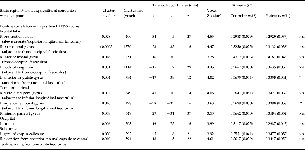
In total, 34 never-medicated patients with first-episode schizophrenia and 32 healthy controls successfully underwent a MRI scan. Altogether, 31 patients with never-medicated first-episode schizophrenia who had PANSS data for positive and negative PANSS scores were entered into the analysis. We then compared all 34 patients with the 32 healthy controls on mean FA values for 11 clusters that showed significant correlation with PANSS scores (Table 1).
White matter fractional anisotropy (FA) differences in patients compared with healthy controls

PANSS, Positive and Negative Syndrome Scale; R, right; L, left; n.s., non-significant group difference in mean FA.
a Brain regional labels are suggested for guidance only.
b p<0.05 (false discovery rate-corrected).
* Significant group difference in mean FA; p<0.05, uncorrected.
** Significant group difference in mean FA; p<0.005, uncorrected.
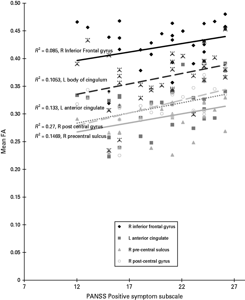
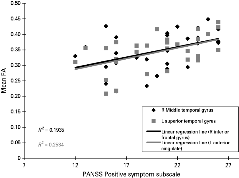
We found that FA values in 11 frontal (Figs 1 and 2) and temporal-limbic (Figs 1 and 3) clusters were positively associated with high positive symptom scores [voxel level p<0.05 (FDR-corrected), cluster size 100, cluster level p<0.05 (uncorrected)]. Notwithstanding this, for all 11 clusters, the FA values were lower in patients than in healthy controls, significantly so around the left anterior cingulate and superior temporal regions (see Table 1). Thus, the closer FA values in the patient group were to control measures, the greater the number of positive symptoms. There were no brain regions showing negative correlation with positive PANSS. There were also no brain regions showing any correlation with negative PANSS.
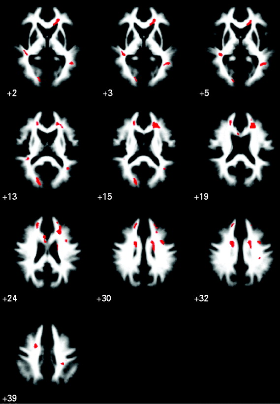
Functional anisotropy (FA) score correlates of positive symptoms in schizophrenia. Red clusters show significant positive correlations of white matter FA with positive symptom scores in never-medicated patients with first-episode schizophrenia (n=31). There were no significant negative correlations of white matter FA with negative symptom scores. The maps are orientated with the right side of the brain shown on the right side of the panel. The z coordinate for each axial slice in the standard space of Talairach and Tournoux is given in mm.

Scatter plot of the relationship between functional anisotropy (FA) of significant frontal clusters with positive symptom subscale on the Positive and Negative Syndrome Scale (PANSS). A best-fitted least square line was plot for each of the five frontal areas and their corresponding R 2 given next to each of the best-fitted lines.

Scatter plot of the relationship between functional anisotropy (FA) of significant temporal clusters with positive symptom subscale on the Positive and Negative Syndrome Scale (PANSS). A best-fitted least square line was plotted for each of the five frontal areas and their corresponding R 2 given next to each of the best-fitted lines.

Discussion
Our principal finding is that in antipsychotic-naive patients newly diagnosed with schizophrenia, positive symptoms showed a significant positive correlation with widespread regions, including fronto-occipital and temporo-limbic white matter FA scores. Specifically, these regions included the right frontal lobe, left anterior cingulate gyrus, left body of cingulum, left superior temporal gyrus, right middle temporal gyrus, right inferior parietal gyrus, left cuneus, left genu of corpus callosum, right extension of posterior internal capsule. However, in all of these regions FA scores were lower in patients than healthy controls. Our present study extends our previous DTI work on never-medicated patients in their first episode of schizophrenia (Cheung et al. Reference Cheung, Cheung, McAlonan, Deng, Wong, Yip, Tai, Khong, Sham and Chua2008), indicating that patients with FA values approaching control levels in fronto-occipital and temporo-limbic tracts have the greatest number of positive symptoms.
In our previous study (Cheung et al. Reference Cheung, Cheung, McAlonan, Deng, Wong, Yip, Tai, Khong, Sham and Chua2008), we reported decreased FA in patients compared with healthy controls in a widespread pattern, which included the fronto-occipital and temporo-limbic regions corresponding to the left fronto-occipital fasciculus and inferior longitudinal fasciculus, white matter adjacent to right precuneus, splenium of corpus callosum, right posterior limb of internal capsule, white matter adjacent to right substantia nigra and left cerebral peduncle. The current study found that, despite lower FA in the patient group relative to controls, there was a positive correlation between positive symptoms and FA encompassing a widespread network including fronto-occipital and temporo-limbic pathways. This result may be considered rather counter-intuitive in that more florid positive symptoms of schizophrenia are associated with white matter FA values very similar to control levels. We note that FA relatively increased with positive symptoms even though, overall, the absolute FA scores were all lower in the group with schizophrenia.
Widespread cortical thinning has been described both in first-episode schizophrenia in the frontopolar, cingulate and occipital regions (Narr et al. Reference Narr, Toga, Szeszko, Thompson, Wood, Robinson, Sevy, Wang, Schrock and Bilder2005). The same group had earlier reported that very early onset individuals followed up over 5 years showed striking grey matter loss originating in the parietal cortex dynamically moving to temporal then frontal cortices (Thompson et al. Reference Thompson, Vidal, Giedd, Gochman, Blumental, Nicholson, Toga and Rappaport2001). They noted that this grey matter loss correlated with psychotic symptom severity as well as sensorimotor (including visuo-spatial and auditory functions) and executive function impairments (Thompson et al. Reference Thompson, Vidal, Giedd, Gochman, Blumental, Nicholson, Toga and Rappaport2001). Cortical thinning is believed to reflect underlying reduced neuropil and neuronal size with alterations in synaptic, dendritic and axonal organization (Harrison, Reference Harrison1999) and, if progressive, may imply that dyconnectivity gets worse over time. Hence, our data in newly diagnosed and never-medicated subjects with schizophrenia support the position that there is already widespread cortical dysconnectivity with lower FA scores in patients compared with controls across the board.
When we examined the overlap in spatial distribution of ‘signature’ between our previous study (Cheung et al. Reference Cheung, Cheung, McAlonan, Deng, Wong, Yip, Tai, Khong, Sham and Chua2008) and the current one, we noted that three regions in the present study overlapped with the previous one, in which seven regions had been reported. All showed significantly reduced FA in patients as compared with controls. These three regions in the present study are left anterior cingulate gyrus (anterior to fronto-occipital fasciculus), left superior temporal gyrus (adjacent to inferior longitudinal fasciculus) and right extension from posterior limb of internal capsule. Since these regions are dopamine-rich, we believe that they resonate with the propensity for antipsychotics to modulate positive symptoms and may be evidence of state- rather than trait-related dysconnectivity. Hubl et al. (Reference Hubl, Koenig, Strik, Federspiel, Kreis, Boesch, Maier, Schroth, Lovblad and Dierks2004) had reported higher white matter directionality in the temporo-parietal region in auditorily hallucinated patients versus non-auditorily hallucinated controls. Other studies have also reported findings in this direction (Fujiwara et al. Reference Fujiwara, Namiki, Hirao, Miyata, Shimizu, Fukuyama, Sawamoto, Hayashi and Murai2007; Seok et al. Reference Seok, Park, Chun, Lee, Cho, Kwon and Kim2007), although they are not directly comparable to ours since all assessed chronically ill patients with persistent symptoms and possibly lengthy exposure to antipsychotics. Our findings are in keeping with those from three recent DTI studies, which have also reported a positive correlation between positive symptoms and FA. Delusions/hallucinations positively correlated with FA in the left inferior fronto-occipital fasciculus (Szeszko et al. Reference Szeszko, Robinson, Ashtari, Vogel, Betensky, Sevy, Ardekani, Lencz, Malhotra, McCormack, Miller, Lim, Gunduz-Bruce, Kane and Bilder2008). A positive correlation was also noted between delusions of passivity and FA in the frontocingulate region (Sim et al. Reference Sim, Yang, Loh, Poon, Sitoh, Verma, Keefe, Collinson, Chong, Heckers, Nowinski and Pantelis2009) and between auditory hallucinations with FA in the arcuate/superior longitudinal fasciculi and anterior cingulum (Shergill et al. Reference Shergill, Kanaan, Chitnis, O'Daly, Jones, Frangou, Williams, Howard, Barker, Murray and McGuire2007). These sites of white matter dysconnectivity are extensive (for review, see Catani et al. Reference Catani, Howard, Pajevic and Jones2002), comprising the inferior fronto-occipital tract (connects the frontal cortex with posterior temporal and occipital cortex), the superior longitudinal fasculus (links the frontal with parietal and temporal cortices, the cingulum (joins the inferior temporal pole with the subrostral frontal lobe, cingulate gyrus and genu of corpus callosum). We believe that this myriad of white matter deficits is in keeping with evidence in the literature of established cortical thinning in first-episode, minimally treated (Narr et al. Reference Narr, Toga, Szeszko, Thompson, Wood, Robinson, Sevy, Wang, Schrock and Bilder2005), as well as very early onset, schizophrenia (Thompson et al. Reference Thompson, Vidal, Giedd, Gochman, Blumental, Nicholson, Toga and Rappaport2001). Conceivably, some discrepancy in the extant literature may arise from sample characteristics, such as effects of medication and chronicity, since these are difficult to disentangle from pathology. For example, of the three studies cited above, the Szeszko group included the youngest patients (mean age=25 years), who were newly diagnosed but many already in receipt of medication. They noted that medication effects are still not well understood but that FA scores inversely correlated with the duration of antipsychotic treatment in the left superior longitudinal fasciculus (Szeszko et al. Reference Szeszko, Robinson, Ashtari, Vogel, Betensky, Sevy, Ardekani, Lencz, Malhotra, McCormack, Miller, Lim, Gunduz-Bruce, Kane and Bilder2008). Our study was conducted with neuroleptic-naive individuals and our findings, taken with these recent studies, argue for the presence of frontotemporo-limbic dysconnectivity. This parsimonious interpretation is also propounded in a recent meta-analysis by Ellison-Wright & Bullmore (Reference Ellison-Wright and Bullmore2009). However, for patients on treatment it is worth bearing in mind the possible contribution from neuroleptic treatment, since there is a duration- (Szeszko et al. Reference Szeszko, Robinson, Ashtari, Vogel, Betensky, Sevy, Ardekani, Lencz, Malhotra, McCormack, Miller, Lim, Gunduz-Bruce, Kane and Bilder2008) and dosage- (Okugawa et al. Reference Okugawa, Nobuhara, Minami, Tamagaki, Takase, Sugimoto, Sawada and Kinoshita2004) dependent relationship between antipsychotic treatment and FA score. Our finding that positive symptoms are positively correlated with FA appears at odds with the study by Skelly et al. (Reference Skelly, Calhoun, Meda, Kim, Mathalon and Pearlson2008). The latter group reported that chronically treated patients (mean age=34 years) had lower brain FA in association tracts (including left superior longitudinal fasciculus), which inversely correlated with positive symptoms. This discrepancy with our own results deserves to be carefully addressed. One possibility is that younger patients with minimal or no exposure to medication experience FA lowering further over time, i.e. an inverse relationship with positive symptoms may emerge as the illness progresses or as a consequence of medication exposure (Szeszko et al. Reference Szeszko, Robinson, Ashtari, Vogel, Betensky, Sevy, Ardekani, Lencz, Malhotra, McCormack, Miller, Lim, Gunduz-Bruce, Kane and Bilder2008). However, as our study was cross-sectional, we anticipate that a future longitudinal follow-up of our patients would be an ideal approach to try to reconcile results from samples of patients at different stages of illness.
Our cross-sectional analysis cannot speak to ‘causal’ mechanisms but does prompt some interpretation. Mild variation in FA may not reflect serious pathology in neurotypical individuals but here potentially reveals vulnerability in those with psychosis. For example, positive symptoms could be generated via aberrant activation of white matter pathways, which remain reasonably ‘intact’. Classically, symptoms in schizophrenia aggregate into distinct symptom complexes, suggesting that a distinct neural substrate can be investigated by neuroimaging (Buchanan & Carpenter, Reference Buchanan and Carpenter1997). ‘Positive’ symptoms (hallucinations, delusions, thought disorder) are thought to reflect ‘state’ and are more treatment-responsive (Buchanan & Carpenter, Reference Buchanan and Carpenter1997), ‘negative’ symptoms represent ‘trait’ (Buchanan & Carpenter, Reference Buchanan and Carpenter1997) and are regarded as having a likely brain structural basis (Crow, Reference Crow1985). Persistently untreated positive (Flaum et al. Reference Flaum, O'Leary, Swayze, Miller, Arndt and Andreasen1995; Sumich et al. Reference Sumich, Chitnis, Fannon, O'Ceallaigh, Doku, Faldrowicz and Sharma2005) or negative (Chua et al. Reference Chua, Wright, Poline, Liddle, Murray, Frackowiak, Friston and McGuire1997; Wible et al. Reference Wible, Anderson, Shenton, Kricun, Hirayasu, Tanaka, Levitt, O'Donnell, Kikinis, Jolesz and McCarley2001) symptoms may well increase the likelihood of brain structural abnormality but, at first episode, the extent of white matter pathophysiology is still limited.
We cautiously suggest that our results indicate that such limited brain structural pathology at first presentation might provide a substrate for expression of positive symptoms, characteristic of the early phase of illness. There is some evidence to support this in the literature. For example, the duration of untreated illness is associated with worse prognosis (Barnes et al. Reference Barnes, Leeson, Mutsatsa, Watt, Hutton and Joyce2008) and more extensive brain structural differences (Lappin et al. Reference Lappin, Morgan, Morgan, Hutchison, Chitnis, Suckling, Fearon, McGuire, Jones, Leff, Murray and Dazzan2006). Positive symptoms are less prominent over time (Arndt et al. Reference Arndt, Andreasen, Flaum, Miller and Nopoulos1995) and, with each relapse, prognosis worsens and treatment dosages often escalate (Lieberman et al. Reference Lieberman, Jody, Geisler, Alvir, Loebel, Szymanski, Woerner and Borenstein1993). Our data concur with others (Hubl et al. Reference Hubl, Koenig, Strik, Federspiel, Kreis, Boesch, Maier, Schroth, Lovblad and Dierks2004; Fujiwara et al. Reference Fujiwara, Namiki, Hirao, Miyata, Shimizu, Fukuyama, Sawamoto, Hayashi and Murai2007; Seok et al. Reference Seok, Park, Chun, Lee, Cho, Kwon and Kim2007), which imply that disease progression and symptomatology are related to changes in the organization of white matter circuitry. The corollary of our finding of a positive correlation between FA scores and positive symptoms in the left fronto-occipital and temporo-limbic tracts is that, over time (as symptoms abate with treatment), FA may decline and this can represent a marker for treatment or prognosis. Addressing this question in a longitudinal follow-up study may help to determine the extent to which positive and negative symptoms reflect white matter pathology and whether FA can serve as a marker for treatment or prognosis. Perhaps one way forward would be to evaluate future treatment outcome based on symptoms and/or functioning at 1 year or 2, and then to enter this as covariate into the baseline scan in order to determine if baseline FA score may be used to help predict future outcome.
The brain pathology in schizophrenia is suggested to arise from dysregulation of the normal dendritic organization that accompanies a critical surge in white matter growth from adolescence to early adulthood (Sowell et al. Reference Sowell, Thompson, Holmes, Jernigan and Toga1999), when prodrome symptoms typically begin. Harrison (Reference Harrison1999) has argued cogently that key susceptibility genes (such as catechol-O-methyltransferase, neuregulin, dysbindin) converge toward the risk of schizophrenia by their role in synaptic plasticity/function (Frankle et al. Reference Frankle, Lerma and Laruelle2003) so as to impair higher order information processing. In other words, this disorder appears, in its fundamental form, to be one of impaired stabilization and development of cortical micro-circuitry (Harrison, Reference Harrison1999; Winterer and Weinberger, Reference Winterer and Weinberger2004). Consequently, the directionality of neuroimaging findings in schizophrenia may be quite difficult to predict, but DTI evidence for disruption of white matter connections linked to a multiplicity of symptoms fits with asynchronization of neural circuitry during neurodevelopment, resulting in synaptic misconnections (Andreasen et al. Reference Andreasen, Nopoulos, O'Leary, Miller, Wassink and Flaum1999; Frankle et al. Reference Frankle, Lerma and Laruelle2003; Whitefield-Gabrieli et al. Reference Whitefield-Gabrieli, Thermenos, Milanovic, Tsuang, Faraone, McCarley, Shenton, Green, Nieto-Castanon, LaViolette, Wojcik, Gabrieli and Seidman2009).
Strengths and weaknesses of study
The study involved a never-medicated, first-episode group of patients with schizophrenia and aimed to investigate how their symptoms are related to system of white matter connectivity. It employed a voxel-based approach not constrained by prior hypotheses. Patients with schizophrenia were carefully diagnosed and ethnically homogeneous and well balanced with the healthy controls. Although missing data meant that we were not able to enter the duration of untreated psychosis as a covariate to explore the possibility of a relationship between duration of psychosis and FA score, we observed that the duration of psychosis for many subjects was approximately 1 year. This is a potential limitation of the study, together with the DTI sequence used, which could be improved in power with thinner slices and a more powerful magnet, which, fortunately, will become accessible to our patients for future studies.
A between-group analysis followed by exploration of symptom correlates of regions showing significant differences would arguably have been the statistically more powerful and conventional approach. However, we previously published cross-sectional data on a sample including many of the patients and controls from the present study (Cheung et al. Reference Cheung, Cheung, McAlonan, Deng, Wong, Yip, Tai, Khong, Sham and Chua2008). We considered that the regions uncovered in a cross-sectional study might encompass aspects of the illness not exclusively pertaining to symptoms, such as predisposition/vulnerability to disorder. Therefore, since the main thrust of the current study was to examine the symptom–FA interaction, we decided to perform whole-brain correlational analysis. As discussed earlier, the FA correlates of positive symptoms coincided with fewer than half the regions of group difference identified in the cross-sectional analysis. We interpret this finding as pointing towards a difference in the white matter pathways contributing to more ‘static’ disease traits responsible for group differences and those that are involved in generation of positive symptoms.
In conclusion, fronto-occipital and temporo-limbic white matter connectivity positively correlates with positive symptom scores in first-episode schizophrenia. These effects on white matter connections are not influenced by pharmacotherapy and therefore appear to be disease-related. Future work will focus on mapping the progression of illness using FA, together with other neuroimaging markers, to investigate how the administration of antipsychotic agents can modify brain structure and symptoms.
Acknowledgements
This work was supported by a research grant to Dr S. E. Chua and Dr E. Y. H. Chen from the CRCG, The University of Hong Kong.
Declaration of Interest
None.