Congenital cardiac disease is a frequent manifestation in Noonan’s syndrome. The most common lesion, pulmonary stenosis, affects 57% of these patients,Reference Prendiville, Guvreau and Tworog-Dube 1 of whom 47% may require either balloon pulmonary valvoplasty or surgical valvotomy.Reference Prendiville, Guvreau and Tworog-Dube 1 The subsequent re-intervention rate is high in comparison with non-syndromic patients (65 versus 16%).Reference Prendiville, Guvreau and Tworog-Dube 1 , Reference Rao, Galal and Patnana 4 The reasons for this are not well understood, although may relate to their immediate response to valvoplasty or the nature of their valves.
Masura et alReference Masura, Burch and Deanfield 2 , in a 5-year follow-up study of patients with congenital pulmonary stenosis, who underwent balloon pulmonary valvoplasty, found that 80% of patients with Noonan’s syndrome had a sub-optimal response, defined as an immediate post-intervention pulmonary valve gradient >20 mmHg, compared with 15% for those with non-syndromic pulmonary stenosis. However, on follow-up, the pulmonary valve gradient of the patients with Noonan’s syndrome then reduced consistently over 5 years.Reference Masura, Burch and Deanfield 2 Considering this, the immediate response to balloon intervention does not appear to predict long-term outcome. McCrindleReference McCrindle 5 reported that dysplastic pulmonary valves, commonly associated with Noonan’s syndrome, were an independent risk factor for a poor response to balloon valvoplasty.
Clearly, the high re-intervention rate necessitates an analysis of contributing risk factors. Apart from anatomical considerations and the immediate haemodynamic procedural outcome, we hypothetised that more information could be gained by analysing the trajectory of echocardiographically derived valve gradients over time.
Two separate hypotheses were assessed:
∙ success can be predicted on the basis of age at procedure, pulmonary valve annulus, pulmonary valve gradient, and pulmonary valve gradient decrease immediately after procedure; and
∙ the likelihood of failure can be predicted from the trajectory of pulmonary valve gradient over time after pulmonary balloon valvoplasty.
Methods
All patients with Noonan’s syndrome, who underwent balloon pulmonary valvoplasty between 1 January, 1995 and 31 December, 2014, were identified from the departmental database (Heartsuite, Systeria, Glasgow, United Kingdom). Patients were excluded if they underwent surgery as a primary procedure; were over 18 years of age at the time of intervention; or did not have Noonan’s syndrome. Data collected included age at procedure; pulmonary valve morphology (dysplastic was defined as thickened immobile leaflets); the presence of supravalvar stenosis; the pre-procedure pulmonary valve annulus size measured echocardiographically and/or on angiogram; echocardiographic transpulmonary valve gradient, measured as peak systolic gradient, before procedure, immediately after procedure, and at follow-up appointments, documented pulmonary regurgitation before procedure, after procedure, and at follow-up, graded mild, moderate, and severe, invasive pulmonary artery pressure before and after procedure, the type of balloon, and the largest balloon size (including balloon/valve annulus ratio) used during the procedure. Data were obtained from electronic and paper notes. The decision for further intervention was based on a rise in the pulmonary valve gradient or at the clinical discretion of the cardiologist after multidisciplinary discussion.
End points were defined as progression to surgical intervention, and the last outpatient follow-up appointment attended.
The patients were divided into two groups. Success was defined as patients who did not require further intervention after initial balloon pulmonary valvoplasty and failures defined as patients who did. Follow-up for the failure group was defined as the time from the initial procedure to the second intervention.
Statistics
Data are expressed as mean, standard deviation, and range, as appropriate. Intergroup comparisons used the unpaired t-test or Fisher’s exact test, as appropriate. No adjustments were made for multiple testing. Trajectories were modelled using generalised estimating equations – using a Gaussian distribution and an exchangeable correlation structure – with non-linearities being explored with the use of multivariable fractional polynomials. The statistical program used was Stata v13.1 (StataCorp, Texas, United States of America). A p-value <0.05 was considered statistically significant.
Results
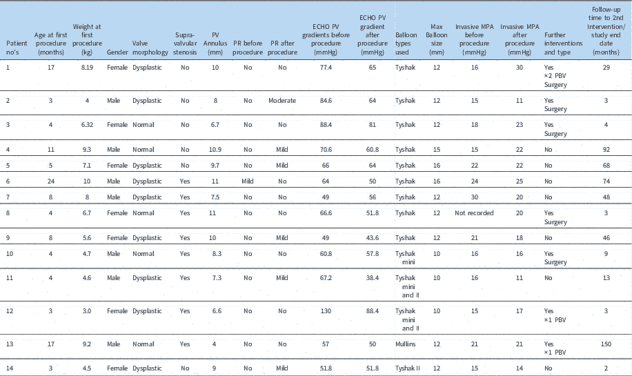
We identified 303 balloon pulmonary valvoplasty procedures during the study period, of which 14 were performed on patients with Noonan’s syndrome under 18 years of age. Seven of these underwent repeat interventions. Three had repeat valvoplasties, three surgical valvotomies, and one underwent two further pulmonary valvoplasties before a surgical valvotomy. The decisions for re-interventions were equally distributed over the decades of the study. One initial intervention was performed in the 1990s, nine in the 2000s, and four in the 2010s. One re-intervention was performed in the 1990s, three in the 2000s, and three in the 2010s. Individual patient data are depicted in Table 1.
Individual patient data.

BPV=balloon pulmonary valvoplasty; ECHO=on echocardiography; MPA=main pulmonary artery; PR=pulmonary regurgitation; PV=pulmonary valve
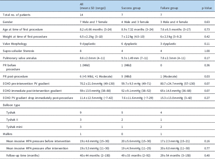
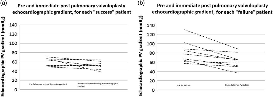
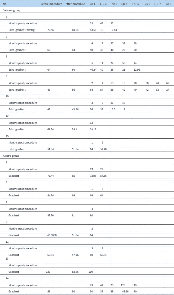
Re-interventions were carried out between 2.5 and 150.0 months, with a median of 3.3 months – failure group. The success group had a median follow-up of 68.2 months, ranging from 1.5 to 91.9 months. There were no significant differences between the groups regarding pre-procedural characteristics or in the initial echocardiographic pulmonary valve gradient immediately after procedure (Table 2). The failure group patients did not show appreciable differences compared with the success group patients in either absolute Doppler-derived gradient immediately after procedure (65 versus 52 mmHg, p=0.06) or change in gradient (−15 versus −8 mmHg, p=0.27) (Fig 1). In all but two patients the decision to re-intervene was taken within 10 months after the initial procedure.
Echocardiographically derived gradient across the pulmonary valve (PV) before and immediately after intervention. Pre and immediate post-pulmonary valvuloplasty echocardiographic gradient for each “success” ( a ) and “failure” ( b ) patient.

Demographics and outcome data.

ECHO=on echocardiography; MPA=main pulmonary artery; PBV=pulmonary balloon valvoplasty; PR=pulmonary regurgitation; PV=pulmonary valve
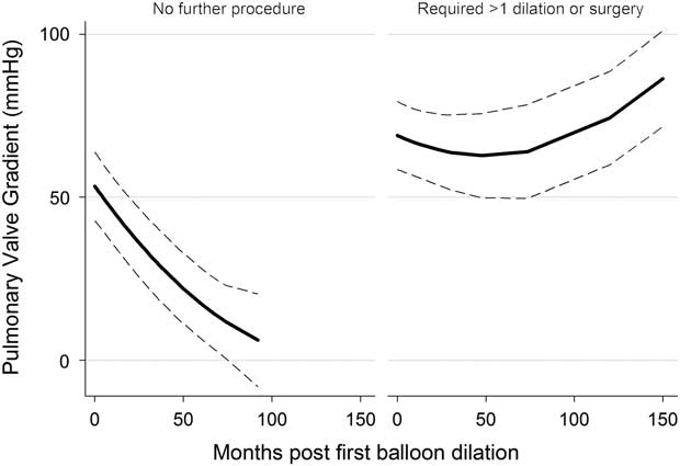
In comparison, the echocardiographic transpulmonary valve gradient trajectories on follow-up differed between the groups, as shown in Figure 2 and Table 3. The valve gradient trajectory of the success group had a continued downward slope with a mean reduction of approximately 8 mmHg/year for the first several years, tailing off to 4 mmHg/year after 5 years; this trajectory was significantly different from the failure group (p<0.02). Conversely, the failure group showed a more modest reduction at approximately 3 mmHg/year, which became static before increasing after 4 years, and was consistently higher than the success group (p<0.001). Valve annulus diameter was not a statistically significant risk factor for re-intervention.
Trajectories of Doppler-derived transvalvar pulmonary gradient.

Pulmonary valve gradients at follow-up.

Discussion
The immediate response in our patients to balloon pulmonary valvoplasty was a reduction in the mean transpulmonary gradient from 70±21 mmHg before procedure to 59±14 mmHg immediately after procedure. This contrasts with the published results of mainly non-syndromic patients, in whom the immediate post-procedure mean transpulmonary gradient decreased from 70±36 to 23±14mmHg.Reference McCrindle and Kan 3 In our patients, the re-intervention rate was 50%, compared with 16% reported in a long-term follow-up study of 85 mainly non-syndromic patientsReference Rao, Galal and Patnana 4 .
The balloon size used in the intervention was decided at the time of procedure to achieve the best possible result. Retrospectively, some of the choices appear quite large, including one three times the size of the pulmonary valve annulus.
It has been discussed that dysplastic valves rather than Noonan’s syndrome per se may contribute to sub-optimal immediate response to balloon pulmonary valvoplasty and subsequent higher rate of re-interventionReference McCrindle 5 , Reference Shaw, Kalidis and Crosby 6 . Interestingly, in our study population, two-thirds of the pulmonary valves were dysplastic, but only three out of the nine required re-intervention. Surprisingly, the presence of additional supravalvar stenosis did not seem to have an effect on the re-intervention rate (see Table 1).
By definition, none of our “success” patients needed any further intervention so far, although it could become necessary over time. Considering this, we noted that three-quarters of “failures” occurred within 30.3 months, time until re-intervention, whereas half of the success group had been followed up for ⩾68.2 months. We could not identify any pre-procedural factors predicting long-term success or failure. The pre-procedural transpulmonary valve gradient showed a statistically non-significant trend in predicting long-term outcome.
Although the decision for further intervention was made after discussion at a multidisciplinary meeting, a degree of clinician preference cannot be excluded. The presenting cardiologist was the initial interventionalist in two cases, but in one case after 10 years the patient was re-discussed after the attending cardiologist changed. It may be that the cardiologists managing the failure group had a lower threshold for re-intervention than the success group. During the 20-year study period, only one patient with Noonan’s syndrome was referred for surgical pulmonary valvotomy in our department. A comparison of both techniques therefore did not seem appropriate. In a PubMed research, we were not able to find articles systematically reviewing surgical treatment of pulmonary valve stenosis in Noonan’s patients, only case reports. Hence, comparison of catheter and surgical treatment is currently not possible.
Masura et alReference Masura, Burch and Deanfield 2 showed a consistent reduction in transpulmonary valve gradient over a 5-year follow-up period in all patients with Noonan’s syndrome. In our study, the immediate post-interventional gradient or the acute reduction of gradient was not predictive regarding re-intervention, but increasing gradients at any time after the intervention were predictive for re-interventions. In some patients, the gradient remained static for a period of 4–5 years and then increased. Hence, the trajectory over time seems predictive of the need for re-intervention. It remains unknown whether waiting longer can avoid re-intervention.
Given the small number of patients, the study data and statistical analysis should be interpreted with caution. So far, we could not identify predisposing factors predicting the results. Comparable national-level studies have been discussed to provide more robust data and an improved evidence base for clinical decision-making.
Conclusion
Pulmonary stenosis in patients with Noonan’s syndrome requires intervention in around 50% of cases. The immediate response to primary balloon pulmonary valvoplasty is often sub-optimal and re-intervention rates are high when compared with non-syndromic patients. Patients with a steady reduction of gradient over time do not need re-intervention, whereas a static or increasing gradient predicted re-intervention.
Further studies with higher patient numbers are required to identify characteristics predisposing to further intervention.
Acknowledgement
The authors would like to thank Thomas Witter for his invaluable help with the data collection.
Financial Support
This research received no specific grant from any funding agency or commercial or not-for-profit sectors.
Conflicts of Interest
None.
Ethical Standards
The study was approved by the institutional audit committee in accordance with ethical standards.