Introduction
Pneumocephalus is a rare condition characterised by the accumulation of air or gas within the intracranial cavity. Andrews and Canalis reported 59 cases of otogenic pneumocephalus, caused by temporal bone fracture, tumour or chronic otitis media, with trauma being the most common cause.Reference Andrews and Canalis1 Kangsanarak et al. analysed 24 321 cases of otitis media, and reported that 87 patients had intracranial complications caused by chronic otitis media, including meningitis, brain abscess, and lateral or sigmoid sinus thrombosis; however, they did not report pneumocephalus as an intracranial complication.Reference Kangsanarak, Navacharoen, Fooanant and Ruckphaopunt2 Another study reported 32 patients with intracranial complications due to chronic purulent otitis media; however, pneumocephalus was not encountered.Reference Dubey, Larawin and Molumi3
Pneumocephalus is a rare intracranial complication.Reference Andrews and Canalis1, Reference Candan, Katelioğlu, Ceylan and Köksal4 It is even rarer to encounter pneumocephalus extending into the lateral ventricles as a result of brain abscess due to chronic otitis media.
Here, we report a patient with extensive pneumocephalus extending into the lateral ventricles, as a result of a brain abscess arising from a cholesteatoma-induced defect in the skull base.
Case report
A 70-year-old man presented to our out-patient clinic with right-sided otalgia, otorrhoea and progressive headaches. The headaches were aggravated by body movements. The patient was alert and well orientated, with no meningeal signs such as neck stiffness or Kernig's sign.
Otoscopic examination revealed bone destruction of the right postero-superior external auditory canal (scutal defect), along with the presence of keratinaceous debris and mucopurulent discharge.
An audiogram showed profound right-sided sensorineural hearing loss, with a pure tone average of 105 dB (averaged threshold at 0.5, 1, 2 and 4 kHz).
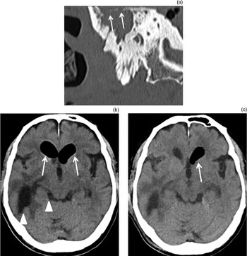
Computed tomography (CT) of the temporal bones revealed a soft tissue mass in the right attic and mastoid cavity. A tegmental defect (approximately 2 × 2 cm) was observed at the right mastoid antrum. A brain CT scan showed areas of pneumocephalus involving the temporal lobe and both lateral ventricles (Figure 1).
(a) Pre-operative, coronal computed tomography (CT) scan showing a soft tissue mass filling the right attic and mastoid cavity. Bony defects (arrows) are observed in the skull base at the mastoid cavity. (b) Pre-operative, axial CT scan showing expansion of the anterior part of both lateral ventricles due to air or gas accumulation (arrows). The brain abscess and the posterior part of both lateral ventricles are filled with cerebrospinal fluid (arrowheads). (c) Axial CT scan taken on post-operative day 1, showing that the pneumocephalic areas in both lateral ventricles are reduced (arrow) compared with pre-operative images.

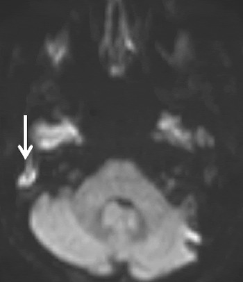
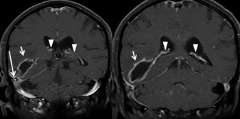
Magnetic resonance imaging (MRI) showed a high-intensity area, indicating cholesteatoma, in the right mastoid antrum (Figure 2). A T1-weighted, gadolinium-enhanced MRI scan showed a high intensity lesion indicative of an encapsulated abscess (Figure 3). The pneumocephalus in the lateral ventricles was directly connected to the mastoid cavity through the abscess and skull base bone defect (Figure 3).
Pre-operative, axial, diffusion-weighted magnetic resonance imaging scan, showing a high-intensity area (arrow) indicating cholesteatoma in the right mastoid antrum.

Pre-operative, coronal, T1-weighted, gadolinium-enhanced magnetic resonance imaging scan series showing an encapsulated abscess (short arrows) and part of the (gadolinium-enhanced) choroid plexus of both lateral ventricles (arrowheads). The pneumocephalus in the right lateral ventricle is directly connected to the skull base, close to the mastoid cavity, via the abscess (long arrow).

Culture results for otorrhoea samples revealed the presence of Stenotrophomonas maltophilia. Cerebrospinal fluid (CSF) was collected by lumbar puncture; CSF analysis showed an increased protein concentration (157 mg/dl), decreased glucose concentration (45 mg/dl) and elevated white blood cell count (815/mm3), suggesting bacterial meningitis.
The patient was administered ceftriaxone sodium hydrate and gentamicin for 3 days.
Radical mastoidectomy was then performed to remove the cholesteatoma. No liquorrhoea was observed from the roof of the mastoid cavity during surgery. The thick granulation tissue covering the tegmen in the mastoid antrum was pulsating, suggesting an area of tegmental defect. We extensively covered the roof of the mastoid cavity with temporal muscle fascia and a pedicled temporal muscle flap.
Post-operatively, piperacillin (2 g/day) was intravenously administered for one week.
On post-operative day 1, CT showed reduced pneumocephalus in the lateral ventricles (Figure 1). On post-operative day 6, CT and MRI revealed complete disappearance of the pneumocephalus from the lateral ventricles.
Discussion
We describe here a very rare case of extensive pneumocephalus extending into both lateral ventricles, arising from a brain abscess in a patient suffering from cholesteatoma. In our patient, although the fistula in the roof of the mastoid cavity was not directly identified during surgery, CT showed a decrease in the pneumocephalic area in the lateral ventricles on the day after surgical reconstruction of the roof of the mastoid cavity. On post-operative day 6, CT and MRI showed complete disappearance of the pneumocephalus in both lateral ventricles.
To the best of our knowledge, this is the first documented case of extensive pneumocephalus extending into the lateral ventricles from a brain abscess caused by a cholesteatoma-induced tegmen defect. These results suggest that the pneumocephalus occurred because of a cholesteatoma-induced tegmental defect at the skull base, indicating that this tegmental defect required urgent closure, following control of the intracranial infection.
Taguchi et al. have reported a patient with a gas-filled brain abscess occurring due to infection by an anaerobic bacillus.Reference Taguchi, Sato and Nakamura5 In our patient, microbiological investigation indicated bacillus infection but no gas production.
• This patient had extensive lateral ventricle pneumocephalus extending from a brain abscess caused by a cholesteatoma-induced skull base defect
• Pneumocephalus is an important complication with an atypical imaging appearance
• The patient's tegmental defect required urgent surgical repair, under appropriate antibiotic cover
Pneumocephalus development is considered to occur via two principle mechanisms – nose-blowing and Valsalva manoeuvre – both of which cause intermittent episodes of increased pressure within the middle ear.Reference Dubey, Jacob and Gandhi6 There are two types of Valsalva manoeuvre: (1) ‘nose-pinched’ Valsalva, whereby forceful exhalation is attempted against pinched nostrils, and (2) ‘glottic’ Valsalva, whereby positive pressure is applied against a closed glottis. In our patient, the mastoid cavity with the bone defect was not directly connected to the auditory tube. Therefore, it is unlikely that the nose-pinched Valsalva manoeuvre caused a pressure gradient in the mastoid cavity. However, a pressure gradient could have been created by the glottic Valsalva manoeuvre, which increases the intrathoracic pressure, thereby decreasing the jugular venous return and raising the intracranial pressure, and resulting in CSF leakage through any defect in the dura mater. In cases of pneumocephalus, air replaces the leaking CSF in the intracranial compartment.
Horowitz has reported a third mechanism: negative intracranial pressure induced by a change in head position from horizontal to vertical.Reference Horowitz7 The resulting negative pressure pulls in air, while a compensatory increase in CSF absorption creates space for the trapped air.
In our patient, air from the mastoid cavity might have entered the lateral ventricles through the abscess cavity. Since his headache was aggravated by body movements, it is possible that his otogenic pneumocephalus accumulated gradually as a result of daily bodily movements.