Introduction
According to studies in various animal models, meiotic differentiation is connected with the presence of the so-called ‘germ plasm’, a cytoplasmic substance whose formation and transformation are generally conserved during animal life cycles (Saffman and Lasko, Reference Saffman and Lasko1999). To date, it is known that germ plasm arises and is stored during oogenesis with the participation of the mitochondrial cloud, also known as Balbiani body (Kalt, Reference Kalt1973; Kloc et al., Reference Kloc, Dougherty, Bilinski, Chan, Brey, King, Patrick and Etkin2002; Saffman and Lasko, Reference Saffman and Lasko1999; Cox and Spradling, Reference Cox and Spradling2003; Chang et al., Reference Chang, Torres, Lewis, Mowry, Houliston and King2004; Wilk et al., Reference Wilk, Bilinski, Dougherty and Kloc2004). The germ plasm is called polar granules in Drosophila (Mahowald, Reference Mahowald1962; Liang et al., Reference Liang, Diehl-Jones and Lasko1994), germinal granules in Xenopus (Watanabe et al., Reference Watanabe, Itoh, Abe, Akizawa, Ikenishi and Furusawa1992; Kloc et al., Reference Kloc, Dougherty, Bilinski, Chan, Brey, King, Patrick and Etkin2002; Chang et al., Reference Chang, Torres, Lewis, Mowry, Houliston and King2004; Cuykendall and Houston, Reference Cuykendall and Houston2010), P granules in Caenorhabditis elegans (Strome and Wood, Reference Strome and Wood1983; Wolf et al., Reference Wolf, Priess and Hirsh1983). Despite the obvious link between mitochondrial cloud and germ plasm granules, the precise mechanism of their formation is still unknown.
Interesting data about the formation of germ plasm granules were obtained by transmission electron microscopy (TEM). The presence of mitochondrial ribosomal RNAs was documented in close vicinity to the forming polar granules of Drosophila late oocytes and early embryos (Kobayashi et al., Reference Kobayashi, Amikura and Okada1993, Reference Kobayashi, Amikura and Okada1994) as well as to the forming germinal granules of Xenopus oocytes (Kobayashi et al., Reference Kobayashi, Amikura and Okada1994, Reference Kobayashi, Amikura and Mukai1998; Kashikawa et al., Reference Kashikawa, Amikura and Kobayashi2001; Kloc et al., Reference Kloc, Bilinski, Chan and Etkin2001). Amikura et al. (Reference Amikura, Kashikawa, Nakamura and Kobayashi2001, Reference Amikura, Sato and Kobayashi2005) showed that not only mitochondrial ribosomes but also mitochondrial proteins exit mitochondria and reach polar granules in late oocytes and embryos of Drosophila. Reunov (Reference Reunov2004) suggested that mitochondria may contribute material during the formation of germ plasm granules in mouse oocytes, and Ninomiya and Ichinose (Reference Ninomiya and Ichinose2007) supported this hypothesis on the basis of their findings. It was documented that in mouse oocytes the mitochondrion-originated germinal body-like structures accumulate the germ plasm marker VASA (Reunov and Reunova, Reference Reunov and Reunova2015). It appears plausible that, also in other species, mitochondria can contribute to the formation of germ plasm granules, but further investigations are needed to clarify the process.
Being formed in oocytes/eggs/early embryos, and eventually incorporated into primordial germ cells (PGC), the germ plasm is segregated throughout embryogenesis and plays some role in meiotic differentiation of mitotically multiplying germline stem cells of the new generation (Kalt, Reference Kalt1973; Strome and Wood, Reference Strome and Wood1983; Williamson and Lehmann, Reference Williamson and Lehmann1996; Matova and Cooley, Reference Matova and Cooley2001; Carré et al., Reference Carré, Djediat and Sardet2002). However, the function of germ plasm during the shift from mitosis to meiosis is still poorly understood.
Germ plasm mechanisms related to the shift from mitosis to meiosis are of special interest for reproductive technologies. Although some progresses have been achieved with stem cell differentiation into gametes, the results obtained are far from establishing a reliable approach. The main obstacle is the lack of knowledge concerning cellular and subcellular mechanisms that provide viable meiosis triggering and progression. Therefore, studies of meiosis-induction mechanisms occurring inside the gonadic niche appear to be basic to aid in vitro and in vivo technologies (Kashir et al., Reference Kashir, Jones, Child, Williams and Coward2012; Medrano et al., Reference Medrano, Reijo Pera and Simon2013; Hendriks et al., Reference Hendriks, Dancet, van Pelt, Hamer and Repping2015; Moreno et al., Reference Moreno, Miguez-Forjan and Simon2015; Nikolic et al., Reference Nikolic, Volarevic, Armstrong, Lako and Stojkovic2016). It is noticeably that in the course of the ongoing experiments dedicated to meiotic determination of stem cells, no germ plasm element was considered. It seems timely to investigate this topic and find out whether the understanding of the germ plasm may be of help.
The ‘mitosis-to-meiosis’ phase has not been in the focus of germ plasm studies yet, and the rare data present in bibliography more often pertain spermatogenesis rather than oogenesis. Reunov et al. (Reference Reunov, Isaeva, Au and Wu2000) showed that the release of mitochondrial substance activated by germ plasm occurs prior to male meiosis. It was reported out that during male meiosis mitochondrial ribosomes translate nuclearly encoded mRNAs, although it is unclear if nuclear mRNAs penetrate into mitochondria, or mitochondrial ribosomes undergo translocation into the cytoplasm by some unknown mechanisms (Villegas et al., Reference Villegas, Araya, Bustos-Obregon and Burzio2002; Gur and Breitbart, Reference Gur and Breitbart2006, Reference Gur and Breitbart2008). Based on these findings, it might be suggested that in spermatogenesis the meiosis onset is accompanied by some kind of germ plasm activity that induces extrusion of mitochondrial substance containing mitochondrial ribosomes. In-depth analysis seems necessary to highlight the specificity of the germ plasm and mitochondria interaction during meiosis induction in spermatogenesis.
Regarding oogenesis, we did not find reports that clearly differentiate germ plasm that induces the mitosis-to-meiosis shift from the germ plasm that is formed de novo during oogenesis and is then transferred to the next generation. It seems that special research is necessary to find out what germ plasm is meiosis triggering in oogenesis, and some approach should be found to clarify this point. Assuming that spermatogenesis is not known as a system with germ plasm storage, having meiosis promoting germ plasm only, a comparison between oogenesis and spermatogenesis appears a logical way to find criteria for mitosis-to-meiosis-related germ plasm identification in oogenesis.
At a first glance, the comparison of oogenesis and spermatogenesis is an odd task. Oogenesis is peculiar in having meiotic division arrest to facilitate oocyte growth until prezygotic egg cell size. By contrast, in spermatogenesis, the meiotic differentiation directly forms relatively small, highly specialized cells. Therefore, although meiosis is a common feature for both processes, spermatogenesis and oogenesis represent two absolutely different morphologic systems. Nonetheless, it seemed rather likely that early female and male germline cells could be compared. The mitosis-to-meiosis shift occurs at the transformation of oogonia and spermatogonia to primary oocytes (oocytes I) and primary spermatocytes (spermatocytes I), respectively. According to some reports based on TEM data, oogonia and spermatogonia are ultrastructurally similar and have a nucleolus in their dispersed chromatin. Oocytes I and spermatocytes I are peculiar in chromatin condensation and appearance of distinctive synaptonemal complexes of synapsed chromosomes that occur when chromosomal crossing-over takes place at the zygotene–pachytene stage of meiosis (Eckelbarger, Reference Eckelbarger2005; Reunov et al., Reference Reunov, Yurchenko, Alexandrova and Radashevsky2009; Ables, Reference Ables2015). So, thanks to ultrastructural criteria allowing the recognition of premeiotic cells and zygotene–pachytene meiotic cells, the identification of germ plasm structures and the description of their transformation during the premeiotic/zygotene–pachytene stage is possible in both spermatogenesis and oogenesis. Noteworthy, it was so far unclear whether the premeiotic germ plasm activity may differ in the two sexes. This question needs to be addressed to understand the process, as reproductive technology may need such data to target sex-specific meiotic differentiation.
The aim of this study was to investigate: (1) germ plasm-related structure (GPRS) activity occurring during mitosis-to-meiosis shift; (2) sex-specific difference/similarity of GPRS activity occurring during mitosis-to-meiosis shift; and (3) GPRS interplay with mitochondrial material occurring during both meiosis triggering in both sexes, and storage phases during oogenesis. To reach these goals, we compared the mechanisms of GPRS transformation in oogenesis versus spermatogenesis using the Manila clam Ruditapes philippinarum as model species. The Manila clam undergoes an annual reproductive cycle of gametogenesis and spawning that alternates with a period of sexual rest. Both testes and ovaries contain a stock of primary mitotically multiplying germ cells that are located at the wall of acini (gonadic units) and periodically enter meiosis leading to gamete formation (Delgado and Perez-Camacho, Reference Delgado and Perez-Camacho2007; Milani et al. Reference Milani, Ghiselli, Maurizii and Passamonti2011, Reference Milani, Ghiselli, Pecci, Maurizii and Passamonti2015). Normally, each acinus contains germ cells at different stages of differentiation, making R. philippinarum gametogenesis a convenient model to study GPRS dynamics. Some preliminary analyses on clam GPRS were previously carried out on discrete steps of gametogenesis both with electron microscopy and immunostaining using light microscopy (Milani et al., Reference Milani, Ghiselli, Maurizii and Passamonti2011, Reference Milani, Ghiselli, Pecci, Maurizii and Passamonti2015, Reference Milani, Pecci, Ghiselli, Passamonti, Bettini, Franceschini and Maurizii2017). Here we investigated these structures in a processual perspective.
Materials and methods
Animal
Samples of the Manila clam Ruditapes philippinarum were collected in May 2016 from the Adriatic Sea (Goro, Ferrara, Italy). Upon arrival at the laboratory at the University of Bologna (Italy), the samples were prepared for successive analysis.
Histology
The ovaries were fixed in 2.5% glutaraldehyde in a 0.1 M cacodylate buffer and post-fixed in OsO4 (2%) in a 0.1 M cacodylate buffer. After washing in 0.1 M cacodylate buffer the tissues were dehydrated through an ethanol series. The material was then penetrated by an increasing series of LR White Resin diluted in alcohol. Finally, the tissues were embedded in LR White Resin. Semithin sections (0.5 μm) were sectioned with an Ultracut Leica UC6 ultramicrotome (Leica Microsystems, Germany) using a glass knife and placed onto slide. Sections were stained with methylene blue and examined under a light microscope.
Transmission electron microscopy
The ovaries and testes were fixed in 2.5% glutaraldehyde in a 0.1 M cacodylate buffer and post-fixed in OsO4 (2%) in a 0.1 M cacodylate buffer. After washing in 0.1 M cacodylate buffer the tissues were dehydrated through an ethanol series. The material was then penetrated by an increasing series of LR White Resin diluted in alcohol. Finally, the tissues were embedded in LR White Resin. Ultrathin sections (80 nm) were sectioned with an Ultracut Leica UC6 ultramicrotome (Leica Microsystems, Germany) using a diamond knife and placed onto copper grids coated with formvar film. Sections were stained with uranyl acetate and lead citrate solutions and examined using a Zeiss Libra 120 transmission electron microscope (Karl Zeiss Group, Germany).
Immunoelectron microscopy
Ovaries and testes were fixed in 4% paraformaldehyde and 0.5% glutaraldehyde in 0.1 M sodium cacodylate buffer (pH 7.4). After dehydrating the samples through increasing concentrations of ethanol, the pellets were embedded into LR White Resin. The blocks were sectioned with an Ultracut Leica UC6 ultramicrotome (Leica Microsystems, Germany) using a diamond knife. The thin sections were mounted on formvar-coated nickel grids. The grids were floated on drops of 1% BSA/0.01% Tween 20 in PBS for 1 h, then incubated for 1 h with primary antibodies specific for R. philippinarum: anti-VASA antibodies (in rabbit; Milani et al., Reference Milani, Ghiselli, Pecci, Maurizii and Passamonti2015) and anti-CYTB antibodies (in rabbit and in chicken; Milani et al., unpublished) diluted with PBS-0.05% Tween 20 (PBST) at 1:200 concentration. Next, the samples were washed in PBST and incubated for 2 h with a mixture of secondary antibody in PBST. The secondary antibodies were 12-nm colloidal gold-conjugated goat anti-rabbit IgG antibody (111-205-144, Jackson) and 18-nm colloidal gold-conjugated goat anti-chicken IgG antibody (703-215-155, Jackson) that were applied in 1:50 dilutions for 2 h at room temperature. For control sample preparation, primary antibodies were omitted and only secondary antibodies were used. The grids were washed three times in PBST, rinsed in distilled water, stained with uranyl acetate and lead citrate, and observed and photographed with a Zeiss Libra 120 transmission electron microscope (Karl Zeiss Group, Germany).
Quantitative analysis of GPRS during mitosis-to-meiosis phase
The identification of successive stages of meiotic cells was carried out accordingly to previous descriptions of animal gametogenesis (Eckelbarger, Reference Eckelbarger2005; Reunov et al., Reference Reunov, Yurchenko, Alexandrova and Radashevsky2009; Ables, Reference Ables2015). Oogonia and spermatogonia (premeiotic cells) are ultrastructurally similar and have a nucleolus in their dispersed chromatin. Oocytes I and spermatocytes I (cells that entered meiosis) are peculiar in chromatin condensation and in the appearance of distinctive synaptonemal complexes of synapsed chromosomes that occur when chromosomal crossing-over takes place at zygotene–pachytene stage of meiosis.
Three pieces of testis and three pieces of ovary were taken from each of three males and each of three females, and these pieces were embedded in resin blocks. From each of these 18 blocks (nine for females and nine for males), three sections were taken from different levels. Therefore, 54 sections were investigated (27 for females and 27 for males). At each section, the 10 premeiotic cells and 10 zygotene–pachytene cells were identified to give, in total, 270 cells of each type. Therefore, the GPRS were investigated in 540 female cells and 540 male cells, to give, in total, 1080 cells. The presence of such GPRS as the: (1) compact germ plasm granules; and (2) fragmented germ plasm granules were identified by electron microscopy and counted. The results were analysed using the Microsoft Excel program. All values were expressed as means with standard error of the mean (SEM). Percentages were calculated using Student’s t-test and *P <0.05, **P <0.001 were considered to be statistically significant.
Quantitative analysis of GPRS during storage phase
Three pieces of ovary were taken from each of three females, and these pieces were embedded in resin blocks. From each of these nine blocks, 30 sections were taken from different levels. Therefore, 270 sections were investigated. For each section, the presence of MO1–MO4, gradual stages of oocyte maturation were found. One oocyte of each stage was found and analysed at each section to give, in total, 270 oocytes. So, 1080 oocytes were investigated. Such GPRS as: (1) GSM (granular substance surrounded by mitochondria); (2) GSMgg (granular substance having initial germ plasm granule and surrounded by mitochondria); (3) GSgg (granular substance having germ plasm granule and lacked mitochondria). (4) GSggL (granular substance having larger germ plasm granule and lacked mitochondria); and (5) GG (germ plasm granule) were identified by electron microscopy and counted. The results were analysed using the Microsoft Excel program. All values were expressed as means with SEM. Percentages were calculated using Student’s t-test and *P <0.05, **P <0.01, #P <0.001 were considered to be statistically significant.
Results
GPRS in oogonia and primary oocytes
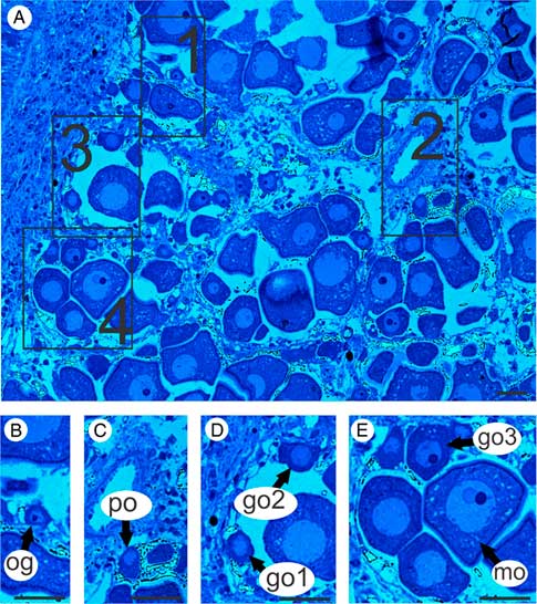
As it could be seen from the histology section, germ cells at different stages of oogenesis were present in the ovary (Fig. 1A). During our study we chose some areas containing developing stages of oogenesis (Fig. 1A) and investigated the cells according to their increasing size (Fig. 1B–E). We discriminated oogonia (average diameter 11 μm) and primary oocytes (average diameter 12 μm), growing oocytes 1 (average diameter 16 μm), growing oocytes 2 (average diameter 18 μm), growing oocyte 3 (average diameter 27 μm), as well as maturing oocytes (average diameter 65 μm) (Fig. 1B–E).
Oogenesis of the Manila clam Ruditapes philippinarum by light microscopy. Semisections were stained with methylene blue. (A) General view of the ovary. Note the squares 1–5 showing some oocytes corresponding to different growing oocyte stages. (B–E) Enlarged images of 1–5 squares, arranged in order of differentiation: oogonium (og), primary oocyte (po), growing oocyte 1 (go1), growing oocyte 2 (go2), growing oocyte 3 (go3) and mature oocyte (mo). Scale bars = 30 μm.

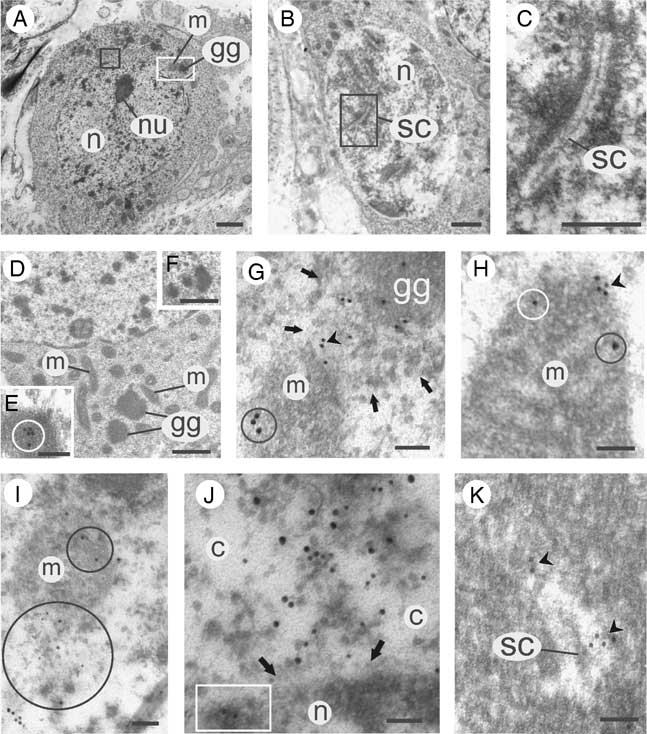
Oogonia are located along the acinus wall. By light microscopy and using serial sectioning allowing to pass through the whole cell, oogonia can be discriminated by the presence of a nucleolus in the nucleus (Fig. 1B), by contrast primary oocytes showed no nucleolus (Fig. 1C). The study by TEM confirmed that the oogonium nucleus is filled with granular chromatin and is peculiar in having a nucleolus (Fig. 2A). Primary oocyte chromatin appeared as elongated strands visible in a lighter background (Fig. 2B). Larger magnification enabled the discrimination of synaptonemal complexes, that allowed the recognition of the zygotene/pachytene stage of meiosis (Fig. 2B, C).
GPRS in prezygotene/pachytene oogenesis of the Manila clam Ruditapes philippinarum. (A) Oogonium with a clear nucleolus (nu) in the nucleus (n); m, mitochondria; gg, germ plasm granule. (B) Primary oocyte with synaptonemal complexes (sc) in the nucleus (n). (C) Magnification of the synaptonemal complex (sc) in the square in (B). (D) An oogonium with germ plasm granules (gg) surrounded by mitochondria (m). (E) A germ plasm granule that is labelled by gold particles (surrounded by circle) corresponding to VASA protein. (F) A fragmented germ plasm granule. (G) A part of germ plasm granule (gg) that is labelled by 12 nm colloidal gold particles corresponding to the VASA protein; note that VASA-positive periphery of granule is dispersed (shown by four arrows); also note that VASA (arrowhead) of dispersed substance is situated very close to a mitochondrion (m) that is labelled by 18 nm colloidal gold corresponding to CYTB (circle). (H) VASA (arrowhead) is located on the surface of a mitochondrion (m) and inside the organelle (light circle); note the intramitochondrial CYTB (dark circle). (I) Magnification of the white square in A, in which the mitochondria (m) are in contact with the germ plasm granule (gg); note the 18 nm colloidal gold particles (CYTB) both in the mitochondrial body (upper circle) and in its dispersed part that contain both the 18 nm gold particles (CYTB) and 12 nm particles (VASA) (bottom circle). (J) Magnification of the black square in (A); note the substance dispersed in the cytoplasm (c) that is marked with both the 18 nm gold particles (CYTB) and 12 nm particles (VASA); two arrows point the portion of the nucleus (n) that is in contact with this substance; note that 12 nm gold particles (VASA) are seen inside of the nucleus (square). (K) A synaptonemal complex (sc) labelled with 12 nm gold particles corresponding to VASA (arrowheads). Scale bars = 1 μm (A, B), 0.5 μm (C, D, F), 0.1 μm (E, G–K).

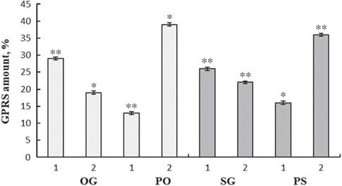
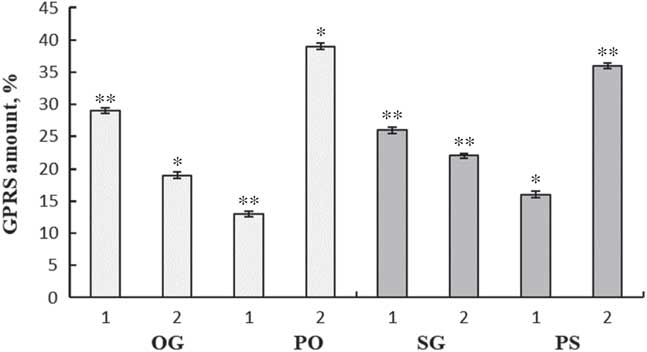
The cytoplasm of both oogonia and primary oocytes contained electron-dense granules that had a compact round shape, not surrounded by a membrane, and were located close to mitochondria (Fig. 2D). Using immunoelectron microscopy (iEM), despite VASA absence in the cytoplasm, these granules were typically VASA positive (Fig. 2E). So, these granules clearly corresponded to germ plasm granules. Some of these granules appeared fragmented (Fig. 2F). Each fragment normally had a diffused VASA-positive periphery (Fig. 2G). The compact and fragmented germ plasm granules were found both in the cytoplasm of oogonia and in primary oocytes. Quantification showed that compact germ plasm granules were more often found in the oogonia and fragmented germ plasm granules were more typical of primary oocytes (Fig. 3, on the left). VASA-positive, dispersed parts of germ plasm granules were often seen in close vicinity to mitochondria that are, as expected, labelled by anti-CYTB antibodies (Fig. 2G). VASA was also found inside some mitochondria (Fig. 2H). Higher magnification showed the presence of VASA and CYTB in the mitochondria that appeared to disassemble and emit material (Fig. 2I). At higher magnification, it was possible to see that a mixture of VASA and CYTB were regularly present in the cytoplasmic substance adjacent to the nucleus (Fig. 2J). Intranuclear VASA was frequently found in the chromatin (Fig. 2J) and synaptonemal complexes (Fig. 2K).
Dynamic of GPRS transformation in prezygotene/pachytene germ cells of the Manila clam Ruditapes philippinarum. Oogonia (og), primary oocytes (po), spermatogonia (sg), primary spermatocytes (ps). 1, compact germ plasm granules; 2, fragmented germ plasm granules. Each value is the mean ± SEM obtained from 270 cells of each germ cell type from three females and thee males. Values of GPRSs quantity are statistically different (*P <0.05, **P <0.001).
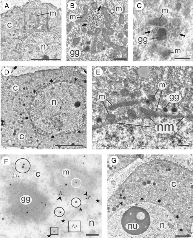
Growing oocyte 1 (Fig. 1D) had elongated mitochondria abundant in the cytoplasm and usually forming aggregates (Fig. 4A). Larger magnification enabled visualization of the mitochondria enclosed granular substance (Fig. 4B). Germ plasm granules were normally seen in the cytoplasm in close proximity to the granular substance (Fig. 4B), or inside this substance (Fig. 4C).
Post-zygotene/pachytene oogenesis of the Manila clam Ruditapes philippinarum. (A–C) growing oocyte 1 (go1). (A) Note the aggregation of mitochondria (m) in the cytoplasm (c); n, nucleus. (B) Magnification of the square in (A); mitochondria (m) surround an aggregate of granular substance (arrows) and the electron-dense germ plasm granules (gg) are located in the vicinity. (C) In some go1 the germ plasm granule (gg) can be found inside of the granular substance (arrows) that is surrounded by mitochondria (m). (D–F) Growing oocyte 2 (go2). (D) The cytoplasm (c) does not contain aggregations of mitochondria and granular substance as well as germ plasm granules, but there is a characteristic perinuclear layer (asterisks). (E) A magnification shows that the perinuclear layer consists of mitochondria (m) and germ plasm granules (gg) that are situated in close vicinity to the nuclear membrane (nm). (F) In the perinuclear layer the germ plasm granules (gg) are typically labelled with 12 nm gold particles (VASA) although VASA is also seen (white arrowhead) in the cytoplasm (c), at the periphery (black arrowheads) of the mitochondrion (m), inside (white circle) of the mitochondrion (m), VASA is also located (small square) in the nuclear membrane (stars) and inside (large square) the nucleus (n); the 18 nm gold particles (CYTB) are seen inside the mitochondrion (m), and also are found (larger black circle) in the cytoplasm (c) as well as in close vicinity (smaller black circles) to the nuclear membrane (stars). (G) Growing oocyte 3 (go3); note the absence of mitochondrial aggregations as well as any GPRS both in the cytoplasm (c) and perinuclear layer (asterisks); note the large nucleolus (nu) in the nucleus (n). Scale bars = 5 μm (A, D, G), 0.5 μm (B, C, E), 0.1 μm (F).
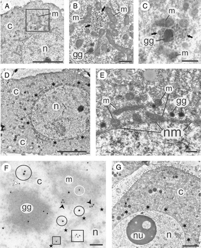
A characteristic feature of growing oocyte 2 (Fig. 1D) was a perinuclear layer (Fig. 4D). Higher magnification showed that this layer consisted of mitochondria and germ plasm granules that were adjacent to the nuclear membrane (Fig. 4E). Using iEM, VASA labelling, as expected, was found in the germ plasm granules and CYTB was found in mitochondria. Additionally, both CYTB and VASA were localized in the perinuclear area of the cytoplasm, and VASA was also present inside mitochondria and nucleus (Fig. 4F). No sign of granular substance was found at this stage.
Growing oocyte 3 had a characteristic large nucleolus in the nucleus (Fig. 1E). No granular substance was found despite the scrupulous examination of serial sections using TEM. The perinuclear area lacked the layer of germ plasm granules and mitochondria. Therefore, an absence of any GPRS was characteristic for this stage of oogenesis (Fig. 4G).
GPRS in maturing oocytes
Mature oocytes are the largest oocytes in the ovary (Fig. 1E). These oocytes also have developed a vitelline envelope (Fig. 5A–F). Using TEM, these oocyte GPRS presented five patterns. We shortly described these patterns and used the following abbreviations:
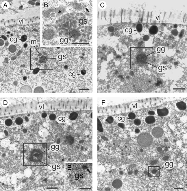
Pattern 1: granular substance surrounded by mitochondria – ‘GSM’ (Fig. 5A).
Pattern 2: granular substance surrounded by mitochondria and having central germ plasm granule – ‘GSMgg’ (Fig. 5B).
Pattern 3: granular substance that lacks a mitochondrial collar but has a central germ plasm granule that is surrounded by an electron-light cavity – ‘GSgg’ (Fig. 5C).
Pattern 4: similar to Pattern 3, but with a larger electron-light cavity that separates a germ plasm granule from the peripheral granular substance – ‘GSggL’ (Fig. 5D).
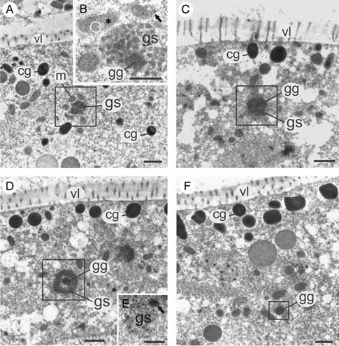
Pattern 5: single germ plasm granules – ‘GG’, that could be located near the granular substance (Fig. 5E), or without colocalization to this structure (Fig. 5F).
Variations of these GPRS patterns were at first observed in growing oocytes of stages go1 and go2 (Fig. 4A–C). The evaluation of these patterns in the successive steps of their arising turned out possible in maturing oocytes (MO) that could be classified according to ultrastructural criteria based on the extent of development their cortical layer:
The first stage (MO1) is characterized by the absence of cortical granules layer although some cortical granules are seen in the cytoplasm (Fig. 5A).
The second stage (MO2) shows cortical granules attached to the oocyte membrane (Fig. 5C).
The third stage (MO3) shows a complete monolayer of cortical granules (Fig. 5D).
The fourth stage (MO4) shows a more developed cortical layer, consisting in a double layer of cortical granules (Fig. 5F).
GPRS in oocytes of the Manila clam Ruditapes philippinarum. (A) Maturing oocyte MO1 has a few cortical granules (cg) that are distanced from the vitelline layer (vl); note GSM (square) that is a granular substance (gs) surrounded by mitochondria (m). (B) MO1 containing the GSMgg – a granular substance (gs) surrounded by mitochondria (asterisk) and having a central germ plasm granule (gg); note VASA (white circle) inside an intact mitochondrion (asterisk), CYTB (arrow) in the dispersed part of a disassembled mitochondrion, and also pay attention on the 12 nm (VASA) and 18 nm (CYTB) colloidal gold particles that are located in the granular substance (gs) as well as on the 18 nm (CYTB) colloidal gold particle that is located inside of the germ plasm granule (gg). (C) MO2 has cortical granules (cg) in the vicinity of the vitelline layer (vl); note the GSgg (square) with no mitochondria and consisting of granular substance (gs) and germ plasm granule (gg) that is separated from granular substance by a thin electron-light cavity. (D) MO3 has a monolayer of cortical granules (cg) located in the vicinity of the vitelline layer (vl); note the GSggL (square) that has no mitochondrial collar and has a characteristic larger electron-light cavity that separates increased germ plasm granule (gg) from the granular substance (gs). (E) MO3 that contains granular substance (gs) and a germ plasm granule (arrow) that is located at the periphery of the granular substance. (F) Mature oocytes MO4 is peculiar in having a double layer of the cortical granules (cg) close to the vitelline layer (vl); note a germ plasm granule (gg) that lacks granular substance (square). Scale bars = 0.5 μm.
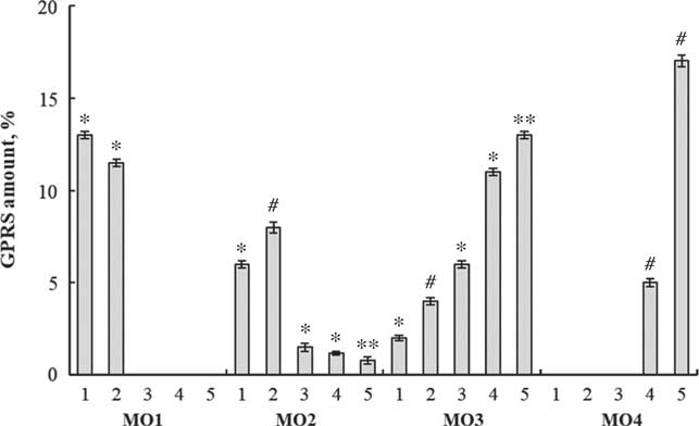
Using quantitative analysis, we tracked down the number of GPRS patterns that were detected for each stage of MO (Fig. 6). GSM and GSMgg were typical for MO1 with dominance of GSM, while the other three GPRS patterns were not found (Fig. 6). MO2 showed a substantial decrease of GSM and GSMgg quantity and the appearance of detectable GSgg, GSggL, and GG (Fig. 6). MO3 showed a further decrease of GSM and GSMgg, and an increase in GSgg, GSggL, and GG, with dominance of GG (Fig. 6). In MO4 we did not find GSM, GSMgg, and GSgg, but we observed GSggL and GG, with a dominance of GG (Fig. 6).
GPRS dynamic in maturing oocytes of the Manila clam Ruditapes philippinarum. MO1–MO4, gradual stages of oocyte maturation. 1, GSM (granular substance surrounded by mitochondria); 2, GSMgg (granular substance with initial germ plasm granule and surrounded by mitochondria); 3, GSgg (granular substance with germ plasm granule and without mitochondria). 4, GSggL (granular substance with larger germ plasm granule and without mitochondria); 5, GG (germinal granule). Each value is the mean ± SEM obtained from 270 cells of each germ cell types from three females. Values of GPRSs quantity are statistically different (*P <0.05, **P <0.01, #P <0.001).

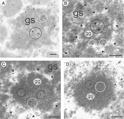
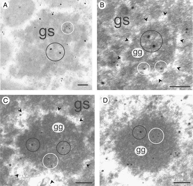
Using iEM, we investigated the localization of VASA and CYTB in GPRS of MO. Regarding to GSM, both VASA and CYTB were localized at the periphery and inside the granular substance (Fig. 7A, magnification of GSM showed in the square at Fig. 5A). GSMgg showed both VASA and CYTB in the granular substance and CYTB was also seen inside the GG (Fig. 5B). It is also noticeable that VASA was found inside intact mitochondria labelled with CYTB, and some mitochondria appeared disassembled, as some CYTB was found outside organelles with irregular shape (Fig. 5B). Both VASA and CYTB were detected in the granular substance and GG in the GSgg (Fig. 7B, magnification of GSgg showed in the square at Fig. 5C) and GSggL (Fig. 7C, magnification of GSggL showed in the square at Fig. 5D). Both proteins were also found at the periphery and inside the GG (Fig. 7D, magnification of GG showed in the square at Fig. 5F).
VASA and CYTB localization in GPRS in maturing oocytes of the Manila clam Ruditapes philippinarum. (A) Magnification of GSM in the square in Fig. 5(A); note that both the 12 nm (VASA) and 18 nm (CYTB) colloidal gold particles (showed by white and black circles, respectively) are present in the granular substance (gs). (B) Magnification of the GSgg in the square in Fig. 5(C); note the germ plasm granule (gg) that is separated from the granular substance (gs) by an electron-light cavity (arrowheads); note the 12 nm (VASA) located at the periphery of the germ plasm granule and 18 nm (CYTB) colloidal gold particles located inside of the germ plasm granule (shown by white and black circles, respectively). (C) Magnification of the GSggL in the square in Fig. 5(D); note the enlarged cavity (arrowheads) between the granular substance (gs) and the germ plasm granule (gg); note the 12 nm (VASA) and 18 nm (CYTB) colloidal gold particles (showed by white and black circles, respectively) located inside the germ plasm granule (gg). (D) Magnification of the GG in the square in Fig. 5(F); note the 12 nm (VASA) and 18 nm (CYTB) colloidal gold particles that are present around the germ plasm granule (gg); note that some VASA and CYTB are located inside the germ plasm granule (in white and black circles, respectively). Scale bars = 0.1 μm.
GPRS in spermatogenesis
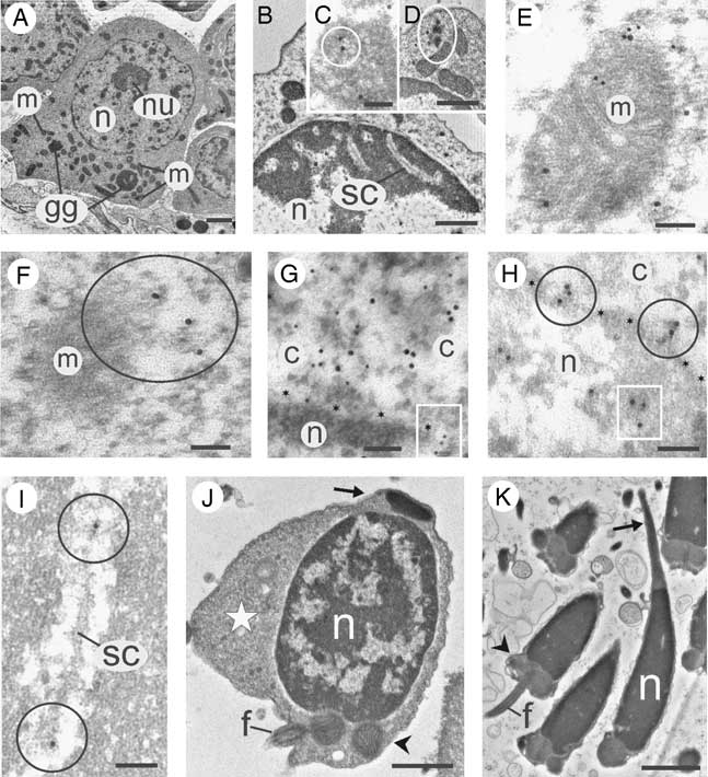
The early stages of spermatogenic cells entering meiosis (spermatogonia and primary spermatocytes) are the largest cells in testes and are situated near the acinus wall (Fig. 8A). Using serial sectioning and TEM, it was observed that the nuclei of spermatogonia had nucleoli and were filled with granular chromatin that is uniformly distributed across the nucleus (Fig. 8A). Instead, primary spermatocytes lacked a nucleolus in their nuclei and showed the chromatin arranged in elongated strands localized on a light background, similarly to that observed for oogonia. Synaptonemal complexes were visible in some strands (Fig. 8B). Both spermatogonia and primary spermatocytes showed electron-dense granules that are normally surrounded by mitochondria (Fig. 8A). iEM shows that these granules are positive for VASA protein (Fig. 8C). Therefore, these granules are considered GG. In some cells, these granules had no compact shape and appeared fragmented (Fig. 8D). Quantitative analysis showed that the amount of compact GG was higher in spermatogonia than in primary spermatocytes, whereas the fragmented form was more typical of primary spermatocytes (Fig. 3, on the right). It seems evident that VASA locate in the compact or dispersed forms of GG but is not translated in the cytoplasm of spermatogonia and primary spermatocytes.
GPRS in the spermatogenesis of the Manila clam Ruditapes philippinarum. (A) Spermatogonium with nucleolus (nu) in the nucleus (n); note germ plasm granules (gg) surrounded by mitochondria (m). (B) Primary spermatocyte with synaptonemal complex (sc) in the nucleus (n). (C) A germ plasm granule that is labelled by gold particles (surrounded by circle) corresponding to VASA protein. (D) Fragmented form of germ plasm granule in a spermatogonium (surrounded by oval). (E) A mitochondrion labelled with 18 nm colloidal gold particles (CYTB) and surrounded by 12 nm colloidal gold particles (VASA); also note intramitochondrial VASA and extramitochondrial CYTB. (F) 18 nm colloidal gold particles (CYTB) in the dispersed material (circle) of a mitochondrion (m). (G) Mitochondrial material that is dispersed in the cytoplasm (c) and is marked with both the 18 nm gold particles (CYTB) and 12 nm particles (VASA); note 12 nm particles (square) located in the nuclear membrane (showed by the asterisks). (H) The colloidal gold particles corresponding to VASA (circles) that are present on the nuclear membrane (asterisks) and inside (square) the nucleus (n); c, cytoplasm. (I) A synaptonemal complex (sc) labelled with colloidal gold particles corresponding to VASA (circles). (J) Spermatid with large amount of cytoplasm (star), acrosomal vesicle (arrow), nucleus (n), mitochondrial area (arrowhead) and flagellum (f); note that GPRS and intranuclear localization of VASA were not found in spermatids. (K) Spermatozoon with acrosome (arrow), nucleus (n), mitochondrial area (arrowhead) and flagellum (f); note that GPRS and intranuclear localization of VASA were not found in spermatozoa. Scale bars = 1 μm (A, J, K), 0.5 μm (B, D), 0.1 μm (C, E–I).
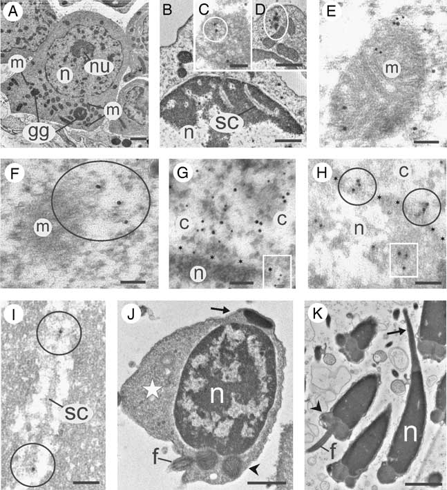
iEM shows that VASA-positive fragments of GG were located in the vicinity of mitochondria. Some VASA-positive substances were found in direct contact with mitochondria and inside the mitochondria (Fig. 8E). CYTB was also found in the cytoplasm adjacent to mitochondria that had an irregular shape (Fig. 8F). A mixture of the 12 nm and 18 nm colloidal gold particles that respectively correspond to VASA and CYTB proteins was found in the perinuclear cytoplasmic substance (Fig. 8G). VASA labelling was found in the nuclear membrane probably located in the nuclear pore (Fig. 8G), on the nuclear surface and inside the nucleus (Fig. 8H). During zygotene/pachytene, VASA was detected in synaptonemal complexes (Fig. 8I).
The late stages of spermatogenic cells are spermatids and spermatozoa. A spermatid is a flagellated cell, with a head that contained some cytoplasm, a platelet-like acrosomal vesicle, an oval nucleus containing chromatin strands, and a forming mitochondrial area (Fig. 8J). A spermatozoon is a flagellated cell with a thin acrosome, an elongated nucleus filled with condensed electron-dense chromatin, and a mitochondrial midpiece (Fig. 8K). GPRS as well as intranuclear localization of VASA were not found in these types of spermatogenic cells.
Discussion
Prezygotene/pachytene transformation of GPRS is similar in both sexes
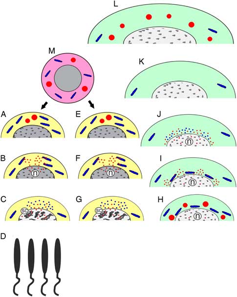
In this study, we wanted to figure out if GPRS transformation was similar or different between females and males during meiosis initiation. In the ovary of R. philippinarum, oocytes are arrested during first meiotic prophase (Lippai et al., Reference Lippai, Gobet, Tomkowiak, Durocher, Leclerc, Moreau and Guerrier1995). Thus, to verify if female and male early germ cells could be considered as equal background for GPRS comparison, we firstly investigated early oogenesis using TEM. We found that meiotic arrest does not occur up to zygotene–pachytene stage. It is more likely that it occurs during the following steps of prophase I. Therefore, in R. philippinarum, early meiotic cells are comparable in the two sexes and the GPRS comparison is meaningful. We found that both oogonia and spermatogonia have VASA-positive GG that are located in the vicinity of mitochondria (Fig. 9A, E). We did not find any sign of GG formation and suggest that these structures are inherited by premeiotic cells from PGC (Fig. 9M) following a preformation way of germ plasm inheritance, a mechanism previously proposed for bivalve molluscs (Milani et al., Reference Milani, Ghiselli, Maurizii and Passamonti2011, Reference Milani, Pecci, Ghiselli, Passamonti, Bettini, Franceschini and Maurizii2017 and references therein). GG were found in both compact and dispersed forms and, based on quantification, we concluded that during meiotic differentiation of oogonia and spermatogonia their compact VASA-positive GG undergo fragmentation and dispersal (Fig. 9B, F). As we did not find cytoplasmic VASA in oogonia and spermatogonia before GG dispersion, we suggest that GG are the unique source of VASA at this stage.
Schematic drawing of GPRS dynamics in the meiotic differentiation of males (A–D) and females (E–L) of the Manila clam Ruditapes philippinarum. Note that ‘yellow phase’ of meiosis is similar in males (A–C) and females (E–G) and is characterized by the presence of VASA-positive compact germ plasm granules (A, E, red), their fragmentation and dispersion of their VASA-positive substance (red) into mitochondria (blue) and into the nucleus (n) (B, F). The next step is characterized by the disassembly of some mitochondria, with the appearance of cytoplasmic substance that includes CYTB-positive mitochondrial material (blue) and VASA (red), together with intranuclear VASA (red) located in synaptonemal complexes (sc) of zygotene/pachytene germ cells (C, G). Note that the ‘yellow phase’ of meiotic differentiation is sufficient for male type of meiosis and spermatogenesis lacked any further GPRS up to the spermatozoon stage (D). Growing oocytes are characterized by the activation of the germ plasm granule formation complex (for details, see also Fig. 10) resulted in: (H) the production of new VASA-positive germ plasm granules (red) that aggregate together with some mitochondria (blue) in close proximity with the nucleus (n); (I) fragmentation of perinuclear germinal granules (red) and intramitochondrial localization of these fragments (red); and (J) disassembly of perinuclear mitochondria with the emission of CYTB-positive mitochondrial material (blue) and VASA entrance into the nucleus. (K) The following stage of oocyte growth lacks any GPRS. (L) Maturing oocytes are peculiar in resuming the germ plasm granule formation complex with the production of new germ plasm granules (red). (M) During embryo development the germ plasm granules (red) formed by mature oocytes are likely inherited by primordial germinal cells (pink) giving rise to meiotic lines of both sexes (arrows).

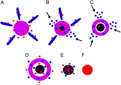
Schematic drawing of the progressive stages of the germ plasm granule formation complex of the Manila clam Ruditapes philippinarum. (A) A granular substance (purple) is surrounded by mitochondria (blue); note cytoplasmic VASA (red) that penetrates both into the granular substance and mitochondria. (B) Some mitochondria are disassembled (shown by arrows) and their CYTB-positive material (blue dots) penetrates into the granular substance; note a germ plasm granule (black) that appears at the centre of granular substance and contains both VASA and mitochondrial material. (C) Mitochondria that are penetrated by VASA undergo disassembly (arrows), then both the CYTB-positive mitochondrial material and VASA saturate the granular substance and growing germ plasm granule; note that a light cavity appears between the granular substance and the germ plasm granule. (D) Mitochondria are absent although some CYTB-positive mitochondrial material is seen around the granular substance together with cytoplasmic VASA; both VASA and CYTB-positive mitochondrial material penetrate into the granular substance, are present in the light cavity, and are included in the growing germ plasm granule. (E) A granular substance is absent; note a germ plasm granule that is filled with both CYTB-positive mitochondrial material and VASA that are also present around the germ plasm granule. (F) A mature germ plasm granule that is VASA-positive and presumably lacks any visible mitochondrial material.

VASA is an essential factor in meiotic progression and obviously is a universal key germ plasm component both of invertebrate as well as vertebrate animals and humans (Lasko and Ashburner, Reference Lasko and Ashburner1988; Raz, Reference Raz2000; Castrillon et al., Reference Castrillon, Quade, Wang, Quigley and Crum2000; Carré et al., Reference Carré, Djediat and Sardet2002; Gustafson and Wessel, Reference Gustafson and Wessel2010; Medrano et al., Reference Medrano, Reijo Pera and Simon2013; Nikolic et al., Reference Nikolic, Volarevic, Armstrong, Lako and Stojkovic2016). However, a role for this protein remained obscure in terms of germ plasm formation and functioning.
Our study of germ cells during their shift from mitosis to meiosis showed that VASA was found in contact with some mitochondria and inside mitochondria (Fig. 9B, F). Using anti-CYTB antibody we noticed that mitochondria adjacent to VASA-positive GG substance are disassembled (Fig. 9C, G). Actually, premeiotic disassembly of mitochondria induced by GG substance was previously described by plain electron microscopy (Reunov et al., Reference Reunov, Isaeva, Au and Wu2000). Being confirmed here with the use of specific mitochondrial marker, this fact may provide new clues on the mechanism of extramitochondrial localization of mitochondrial material as suggested in mice spermatogenesis (Villegas et al., Reference Villegas, Araya, Bustos-Obregon and Burzio2002). Amikura et al. (Reference Amikura, Kashikawa, Nakamura and Kobayashi2001, Reference Amikura, Sato and Kobayashi2005), as well as Gur and Breitbart (Reference Gur and Breitbart2006, Reference Gur and Breitbart2008), proposed that mitochondrial ribosomes could translate nuclear RNAs during germ line cells differentiation. Based on our study, mitochondria may release their ribosomes by demolishing their own structure and a role of VASA in mitochondrial disassembling could be supposed.
VASA labelling was also found close to nuclei and inside them (Fig. 9B, F). Interestingly, the presence of VASA both in the cytoplasm and nucleus was also reported for oocytes of Drosophila (Findley et al., Reference Findley, Tamanaha, Clegg and Ruohola-Baker2003). Pek and Kai (Reference Pek and Kai2011) reported the perichromosomal localization of VASA and its role in Drosophila mitotic chromosome condensation. In primary spermatocytes and primary oocytes of the Manila clam, VASA was observed in synaptonemal complexes (Fig. 9C, G). Based on these findings, we suggest that during meiosis onset VASA participates in pro-meiotic chromatin transformation.
Our comparative study showed that the meiotic onset is similar in both sexes (‘yellow phase’ in Fig. 9A–C, E–G). In Manila clam males, the dispersion of VASA-positive GG followed by intramitochondrial and intranuclear VASA entrance is a single event of GPRS action. Indeed, no other pattern of GPRS is present in late stages of spermatogenesis (Fig. 9D). So, we suggest sufficiency of prezygotene/pachytene GPRS for the completion of male meiotic differentiation in this clam.
During oogenesis the formation of GG is resumed twice
According to our observations, the VASA-positive GG appeared twice during oogenesis. Firstly, these structures form at stages 1 and 2 (go1 and go2) of the growing oocytes, being formed by the GG formation complex (see next paragraph). The GG initially appeared in the cytoplasm of go1 oocytes. In go2, these bodies were located perinuclearly together with some mitochondria (Fig. 9H). The perinuclear location of GG is known for the germline cells of various animal species (Carré et al., Reference Carré, Djediat and Sardet2002; Yajima and Wessel, Reference Yajima and Wessel2011; Yakovlev, Reference Yakovlev2016) although a role for this phenomenon has not been hypothesized. We found that this pattern of GG localization was followed by dispersion of GG and penetration of their VASA-positive substance into perinuclear mitochondria (Fig. 9I). Using an anti-CYTB antibody, we showed that, after VASA penetration in mitochondria, the organelles disassemble and the mitochondrial substance as well as VASA are dispersed in the perinuclear cytoplasm. Also, the VASA-positive labelling is present in the nucleus (Fig. 9J). We suggest that this process is shared by prezygotene/pachytene meiotic stages of both sexes (Fig. 9A–C, E–G). Perinuclear localization of GG and assigned mitochondria may be aimed to create tighter link of these GPRS with a nucleus in the oocytes that increases in size. Probably this link helps intranuclear penetration of GG originated VASA, although we still do not know if cytoplasmic VASA may also penetrate into the nucleus. Interestingly, in growing oocytes at stage 3 (go3), no GPRS were found (Fig. 9K). GPRS absence should mean that they are no more involved in meiosis promotion.
The second time, GG were formed by the GG formation complex (see next paragraph) in late oocytes (Fig. 9L). Considering that in formed oocytes the GG do not undergo any transformations, it seems that their accumulation can be related to germ plasm assignment during embryogenesis, following a preformation type of germline cell determination (Milani et al., Reference Milani, Ghiselli, Maurizii and Passamonti2011, Reference Milani, Pecci, Ghiselli, Passamonti, Bettini, Franceschini and Maurizii2017 and reference therein). According to this mechanism, an early segregation of VASA-positive GG occurs in late oocytes and, during embryogenesis, GG are sequestered into the cytoplasm of PGC (Fig. 9M).
Germ plasm granule formation complex is a novel type of germ plasm arising
The process of germ plasm formation has not been ultrastructurally investigated yet in any bivalve species. We took advantage of our study with R. philippinarum to characterize how germ plasm arises in this bivalve representative. Here, we showed that GG are formed by a complex mechanism that we called the GG formation complex (GGFC). Both the GG and elements of GGFC are present in the post-zygotene/pachytene oocytes at stages go1 and go2, as well as in MO.
Comparative study of MO based on the ultrastructure of their cortical layer allowed characterization of consecutive stages of GGFC performance. The initial GGFC morphologic pattern appeared as a granular substance (GS) that arose in the cytoplasm by a mechanism that is as yet unclear. A study of serial sections showed that GS typically has an irregular 3D shape and is surrounded by several mitochondria situated in close vicinity to the GS periphery. The iEM study showed that VASA could be found in the cytoplasm (GSM stage; Fig. 10A). So, it seems likely that VASA is translated in MO as well as in go1 and go2 that are also peculiar in GGFC functioning. VASA located close to these mitochondria and inside them (GSM stage; Fig. 10A), therefore we suggest that VASA contributes to the mechanism of mitochondrial disassembly and incorporation of mitochondrial material into the GS. Indeed, some mitochondria were seen to have deteriorated and CYTB-positive material was found around the GS. Both VASA and CYTB were also found inside the GS and inside the initial form of GG that appeared at the centre of the GS (GSMgg stage; Fig. 10B). We suggested that, as a result of mitochondrial disassembly and incorporation of mitochondrial material in the GS, the following stage of GGFC arose: it lacked adjacent mitochondria, is bigger and the forming GG is separated from the GS by a thin cavity (GSgg; Fig. 10C). The next stage was characterized by the development of a large cavity around the GG (GSggL stage; Fig. 10D) followed by the appearance of GG that lacked the GS envelope (Fig. 10E, F). We did not track down in details the steps of GG release from the GS envelope. However, GG were often found in close vicinity to these capsules, and late oocytes contain many more, free GG than enveloped ones.
The GGFC described the process of GG formation that appeared morphologically different in comparison with the Balbiani body (mitochondrial cloud) that is usually connected with formation of multiple GG (Kalt, Reference Kalt1973; Kloc et al., Reference Kloc, Dougherty, Bilinski, Chan, Brey, King, Patrick and Etkin2002; Saffman and Lasko, Reference Saffman and Lasko1999; Cox and Spradling, Reference Cox and Spradling2003; Chang et al., Reference Chang, Torres, Lewis, Mowry, Houliston and King2004; Wilk et al., Reference Wilk, Bilinski, Dougherty and Kloc2004). Indeed, in each GGFC only one GG is formed and multiple GG are formed by corresponding multiple GGFC. The novel pattern of germ plasm formation that we discovered may be specific for bivalve molluscs. This pattern could be added to the set of the known Balbiani body-like structures that may morphologically vary even among closely related species (Bilinski et al., Reference Bilinski, Kloc and Tworzydlo2017).
It should be stressed that the exact mechanism of germ plasm formation is not clear in relation to Balbiani body characteristic for Xenopus, Drosophila and the other species. As shown here for Manila clam, the mechanism of GGFC work obviously included the incorporation of mitochondrial material to germ plasm substance. Indeed, it is worth noting that the developing GG took up not only VASA but also CYTB (Fig. 10E). It was suggested that mitochondrial material contribution is a universal event of germ plasm formation across multicellular animals (Reunov, Reference Reunov2004). More detailed research is required to verify if mitochondria may be precursors of GG formed in the Balbiani body-like structures of various living organisms.
Conclusion
In the present study of Manila clam gametogenesis, we described germ plasm-related structures (GPRS) that appeared to accompany meiosis. Using ultrastructural analysis of GPRS typical for successive steps of oogenesis and spermatogenesis, as well as immunological analysis, we found that male and female meiotic differentiation was similar regarding the prezygotene/pachytene stage.
In both sexes, a shift from mitosis to meiosis is characterized by three steps: (1) the dispersion of VASA-positive GG occurs, (2) VASA-positive substance enters neighbouring mitochondria and induces disassembly of these organelles, and (3) intranuclear entrance of VASA-positive substance occurs. The rupture of mitochondria is in accordance with the cytoplasmic localization of mitochondrial ribosomes as previously described in germ cells of Drosophila, Xenopus and mice. VASA-positive substance entering the nucleus was suggested to promote meiotic remodelling of chromatin.
A novel pattern of germ plasm formation, the GG formation complex or GGFC, was described in the Manila clam. The process may be specific for bivalve molluscs, however some of its features, such as the structural role of mitochondria in the formation of germ plasm, might be a more general mechanism, at least in the light of the similarities observed with some other animals as reported above.
A better knowledge of premeiotic GPRS functioning inside the gonad niche can be useful in the development of technologies for artificial gamete production. For example, the presence of VASA-positive GG might be necessary for successful meiosis onset in stem cells triggered for artificial meiosis. Considering that female post-zygotene/pachytene meiosis is promoted by the occurrence of GGFC followed with formation of additional GG, artificial activation of the GGFC might theoretically be considered: the search for inductors enabling GGFC activation appears reasonable if stem cells used for artificial meiosis lack inherited GG.
Acknowledgements
We are deeply indebted to Mr D.F. Fomin (National Scientific Centre of Marine Biology, Far Eastern Branch of Russian Academy of Sciences, Vladivostok, Russia) for helpful assistance during our observations using the electron microscope. Many thanks to Irina Reunova – a student at St. Joseph Catholic High School (Ottawa, Canada) for accurate proofing of the English language and critical reading of this manuscript.
Financial support
This study was funded by the Russian program for fundamental research ‘The Far East’ (Project No. 15 I 6 007 о) funded to AR, and by the Italian Ministry of Education, University and Research MIUR-SIR Programme (Grant No. RBSI14G0P5) funded to LM.
Declaration of interest
We declare we have no competing interests.
Ethical standards
Not applicable.