Introduction
Unrestrained human immunodeficiency virus type 1 (HIV-1) replication in humans ultimately leads to the onset of acquired immunodeficiency syndrome (AIDS) by destroying cells critical for effective immune function. The administration of highly active antiretroviral therapy (HAART) has transformed the prognosis of an HIV-1 infection from a once deadly illness to a chronic, manageable disease by preventing progression to AIDS (Ref. Reference Sterne1). HAART is a combination therapy of small molecule inhibitors that primarily target the essential enzymes of HIV-1 replication, which include the viral protease (PR), reverse transcriptase (RT) and integrase (IN) (Ref. Reference Engelman and Cherepanov2). Despite the exceptional success of HAART, new HIV-1 infections continue to spread due to the emergence of drug-resistant viral strains (Ref. Reference Frentz, Boucher and van de Vijver3), thereby emphasising the need for novel classes of inhibitors that target currently unexploited aspects of the HIV-1 lifecycle.
The HIV-1 enzymes, which are encoded by the viral pol gene, are incorporated into nascent particles as the C-terminal part of the Gag-Pol protein precursor during virus assembly. Concomitant with virus budding or shortly after particle release, auto-processing of PR from Gag-Pol initiates virion maturation (Ref. Reference Debouck4). PR cleaves a total of 11 sites within the Gag and Gag-Pol polyproteins in step-wise fashion to release the functional enzymes and structural matrix, capsid (CA), and nucleocapsid (NC) proteins that constitute the HIV-1 virion. Differential cleavage rates by PR define an ordered process that is critical for the formation of mature, infectious virus (Refs Reference Pettit5, Reference Könnyű6). Approximately one-half of the complement of the CA protein condenses during maturation (Refs Reference Briggs7, Reference Lanman8) to form the cone-shaped HIV-1 core that houses the viral RNA-NC ribonucleoprotein (RNP) complex in association with the RT and IN enzymes (Refs Reference Pornillos, Ganser-Pornillos and Yeager9, Reference Zhao10, Reference Sundquist and Kräusslich11).
RT and IN provide critical functions during the early phase of retroviral replication. RT converts viral genomic RNA into a linear, double-stranded DNA molecule, while IN integrates the viral DNA (vDNA) into a cell chromosome (Ref. Reference Engelman and Cherepanov2). IN is a highly dynamic protein, which in solution yields a multitude of higher-order species (Ref. Reference McKee12). Four IN molecules will interact in the presence of the long terminal repeat (LTR) ends of vDNA to assemble the catalytically active stable synaptic complex (SSC) or intasome that mediates retroviral DNA integration (Refs Reference Bao13, Reference Faure14, Reference Li15, Reference Bera16, Reference Kotova17, Reference Hare18, Reference Maertens, Hare and Cherepanov19, Reference Hare, Maertens and Cherepanov20). IN catalyses two distinct chemical reactions, 3′ processing and DNA strand transfer. The initial reaction prepares 3′ hydroxyl groups at the vDNA ends by excising dinucleotides adjacent to conserved CA sequences (Refs Reference Katzman21, Reference Pauza22, Reference Sherman and Fyfe23, Reference Bushman and Craigie24, Reference Engelman, Mizuuchi and Craigie25). After nuclear import and association with host chromatin, IN facilitates the nucleophilic attack of the reactive CAOH-3′ ends to create a staggered cut, which concomitantly joins the vDNA ends to the 5′ phosphates of the target DNA (Refs Reference Engelman, Mizuuchi and Craigie25, Reference Bushman, Fujiwara and Craigie26). The unjoined 5′ vDNA ends are connected to the 3′ ends of target DNA by host cell DNA repair machinery, resulting in the proviral template necessary for productive HIV-1 replication (Fig. 1) (reviewed in Ref. Reference Krishnan and Engelman27). A small percentage of linear vDNA is cyclised by host cell DNA repair machineries within the nucleus (Refs Reference Li28, Reference Kilzer29), leading to the formation of one- and two-LTR containing circles that can serve as indirect markers of vDNA nuclear import (Ref. Reference Munir30).
Mechanism of human immunodeficiency virus type 1 (HIV-1) DNA integration. The stable synaptic complex (SSC) is formed when an IN tetramer (green and purple) assembles onto the vDNA ends (thick blue lines). The canonical CCD–CCD dimer interface between purple and green IN protomers is indicated by a white line; the inner green protomers mediate all DNA contacts in the prototype foamy virus (PFV) intasome co-crystal structures (Refs Reference Hare18, Reference Maertens, Hare and Cherepanov19). The removal of a dinucleotide from each DNA end (3′ processing) yields activated 3′ hydroxyl (OH) groups. The target capture complex (TCC) is formed after nuclear import and intasome association with host cell DNA (grey lines). The green IN molecules subsequently facilitate the nucleophilic attack of the activated 3′-OH ends onto the DNA target, which creates a staggered double-stranded DNA cut and concomitantly joins the processed 3′ viral ends to the 5′ phosphates of cellular DNA (strand transfer). The integration intermediate is then repaired via host cell machinery, effectively joining the 5′ viral ends to the 3′ ends of target DNA to form the integrated provirus flanked by a duplicated sequence of the staggered DNA cut.

IN strand transfer inhibitors (INSTIs) disarm the SSC after 3′ processing of the vDNA ends by displacing the resulting terminal deoxyadenylate residue from the IN active site (Ref. Reference Hare18). Raltegravir (RAL), which was the first INSTI approved by the US Food and Drug Administration (FDA) (Ref. 31), is an effective component of HAART cocktails (Ref. Reference Markowitz32). Yet as anticipated, RAL-resistant mutant viruses emerge within infected patients during therapy (Refs Reference Sichtig33, Reference Steigbigel34). The INSTI elvitegravir (EVG), a component of a once-a-day single tablet antiretroviral regime, was subsequently approved by the FDA (Ref. Reference Olin, Spooner and Klibanov35). In the course of evaluating novel antiretroviral drugs, it is critical to establish patterns of cross-resistance to the currently available inhibitors. Unfortunately, EVG selects for similar drug resistant mutations as RAL (Ref. Reference Metifiot36), thereby preventing the use of EVG for the majority of patients failing RAL-containing therapies. The third and most recent INSTI to be licensed, dolutegravir, is a second-generation drug that remains effective in the face of most RAL-resistance mutations, and accordingly may prove useful for patients that fail RAL/EVG-based therapies (Ref. Reference Eron37). Drugs that engage the SSC at sites that are distinct from the enzyme active site should lack cross-resistance to all INSTIs, and therefore should be a priority for prospective therapeutic strategies that target the IN protein.
Inhibition of IN activity through the targeting of protein–protein interfaces
There is great therapeutic potential in identifying interacting protein interfaces that are drug-targetable, but for several reasons the discovery of small molecules that can disrupt protein–protein interactions is an enormous challenge. The relatively large size of contact surfaces of a typical protein–protein interface (~1500–3000 Å) compared with the interface formed between a protein and a small molecule (~300–1000 Å), the lack of contiguity of interacting amino acids and the interface landscape, which can often be devoid of divots or pockets for small molecules to occupy, are a few examples of such concerns (Ref. Reference Wells and McClendon38). Therefore, a full characterisation of the interaction interface is crucial to assess the true potential for drugability.
HIV-1 IN harbours three functional domains, the N-terminal domain (NTD), catalytic core domain (CCD) and the C-terminal domain (CTD) (reviewed in Ref. Reference Li39). The CCD, which contains the Asp and Glu residues that comprise the enzyme active site (configured as the D,D-35-E amino acid motif), has been studied extensively using structural biology approaches. The CCD adopts an RNase H-fold and invariably crystallises as a dimer with a large protein–protein interface between monomers (Refs Reference Li39, Reference Dyda40) (Fig. 2). The positioning of the CCD–CCD interface within the active IN tetramer remained unknown until the structure of the active intasome from the prototype foamy virus was solved by X-ray crystallography (Ref. Reference Hare18). The intasomal IN tetramer is comprised of a dimer of IN dimers. The ‘inner’ subunit in each dimer contacts both vDNA and target DNA, whereas the ‘outer’ subunit does not interact with DNA but engages the ‘inner’ subunit through the canonical CCD–CCD interface (Fig. 1). Furthermore, the two ‘inner’ subunits bring the two dimers together through non-canonical protein–protein and protein–DNA interactions (Refs Reference Hare18, Reference Maertens, Hare and Cherepanov19). Small molecules that engage the CCD–CCD interface could in theory impale the architecture of the intasome by perturbing the key interactions that link the ‘outer’ IN molecules to the ‘inner’ workhorse IN protomers.
HIV-1 IN CCD structure and binding sites of small molecule inhibitors at the CCD–CCD dimer interface. (a) Surface representation of the IN CCD dimer from protein database code 2B4J (Ref. Reference Cherepanov68). IN amino acids Glu-170 and Ala-128, which when mutated confer resistance to the antiviral effects of an overexpressed LEDGF/p75 IN-binding domain (IBD) containing-fragment (Ref. Reference Hombrouck73), are coloured in hot pink and indicate the location of the host factor-binding pocket (encircled). (b–d) Orange residues indicate amino acids that when mutated confer resistance to coumarin-containing compounds (Cys-130 and Trp-132) (Ref. Reference Al-Mawsawi75) (b), tetra-acetylated chicoric acid (Lys-173) and 1-pyrrolidineacetamide (Lys-103, Lys-173, Thr-174) (Refs Reference Shkriabai43, Reference Du45) (c) and allosteric IN inhibitors (ALLINIs) (tert-butoxy-[4-phenyl-quinolin-3-yl]-acetic acids and LEDGIN 6) (Tyr-99, Ala-128, Ala-129 and Thr-174) (Refs Reference Christ79, Reference Tsiang83, Reference Feng112) (d). The enzyme active site residues of the D,D-35-E amino acid motif are coloured red. Purple in panel d highlights Ala-128, which when mutated confers resistance to a dominant interfering LEDGF/p75 IBD-containing fusion protein (Ref. Reference Hombrouck73) and ALLINIs (Refs Reference Christ79, Reference Tsiang83, Reference Feng112).
Small molecules that target the HIV-1 IN CCD–CCD interface
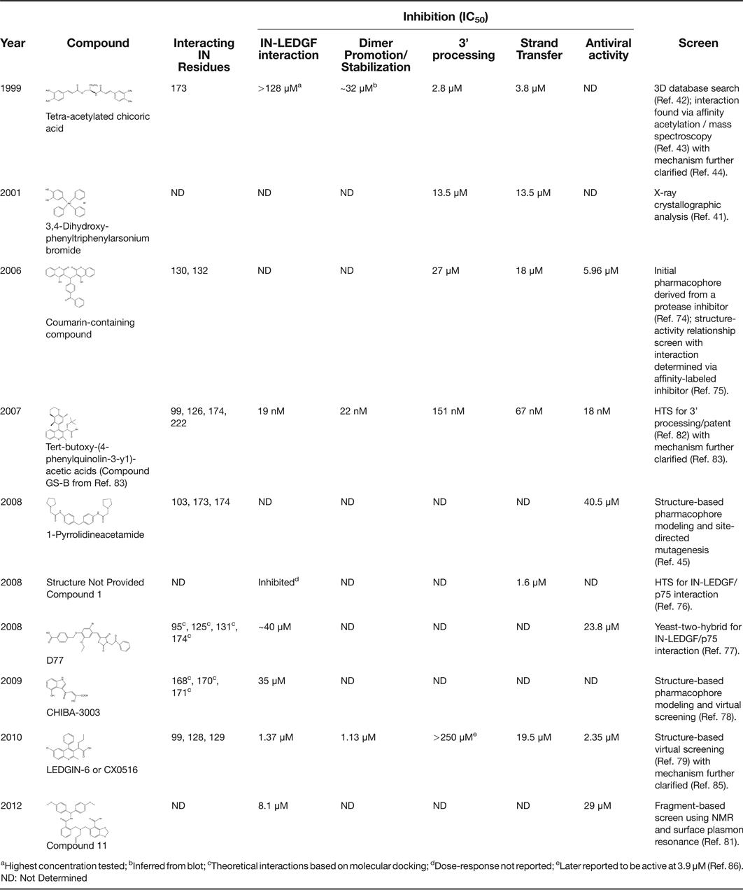
The IN CCD–CCD interface was initially acknowledged as a putative drug-binding site through X-ray crystallography, which was used to screen for novel binding compounds of soaked CCD apocrystals (Ref. Reference Molteni41). One identified compound, 3,4-dihydroxy-phenyltriphenylarsonium bromide, inhibited IN 3′ processing and DNA strand transfer activities in vitro at low micromolar concentrations (Ref. Reference Molteni41) (Table 1). The use of affinity acetylation and mass spectroscopy helped to subsequently clarify that a tetra-acetylated-chicoric acid derivative that had previously been identified as an IN inhibitor (Ref. Reference Lin42) also engaged the IN dimer interface. The compound selectively modified IN residue Lys-173, and was predicted through molecular docking to additionally interact with Lys-103 (Ref. Reference Shkriabai43) (Fig. 2c and Table 1). Interestingly, in addition to inhibiting both 3′ processing and DNA strand transfer activities, the compound modulated the dynamic exchange between IN subunits that occurs in solution (Refs Reference McKee12, Reference Kessl44). A separate group subsequently identified 1-pyrrolidineacetamide through its ability to inhibit the binding of IN to vDNA substrate in vitro. Results of molecular modelling, and the testing of recombinant IN mutant proteins that harboured changes at predicted sites of compound contact, indicated that 1-pyrrolidineacetamide engaged the IN dimer interface at residues Lys-103, Lys-173 and Thr-174) (Ref. Reference Du45) (Fig. 2c). Since the IN active site lies apposed from the CCD–CCD dimer interface (Fig. 2), these studies established precedence for allosteric inhibition of IN catalytic activity through the binding of small molecules to the protein–protein interaction site.
Chronological depiction of the discovery of small molecules inhibitors targeting the IN dimer interface.
aHighest concentration tested; bInferred from blot; cTheoretical interactions based on molecular docking; dDose-response not reported; eLater reported to be active at 3.9 μM (Ref. Reference Kessl86).
ND: Not Determined
The LEDGF/p75–IN interaction as a novel target for antiretroviral therapy
Although IN has been shown to interact with several host cell proteins, few have been confirmed to play an important role in the context of HIV-1 infection (reviewed in Refs Reference Turlure46–Reference Engelman49). Lens epithelium-derived growth factor (LEDGF)/p75, which is a chromatin-associated (Ref. Reference Nishizawa50) transcriptional co-activator (Ref. Reference Ge, Si and Roeder51), by contrast plays a critical role in directing HIV-1 integration into active transcription units (reviewed in Refs Reference Engelman and Cherepanov52, Reference Poeschla53). LEDGF/p75 was independently determined to interact with HIV-1 IN through co-immunoprecipitation analysis of cell extracts that harboured ectopically expressed viral protein (Refs Reference Turlure46, Reference Cherepanov54) and through a yeast two-hybrid screen (Ref. Reference Emiliani55). The protein interaction is mediated by an evolutionary conserved IN-binding domain (IBD) within the C-terminal region of LEDGF/p75 that spans amino acids 347–429 of the 530-residue human protein (Ref. Reference Cherepanov56). LEDGF/p75 belongs to the hepatoma-derived growth factor related protein (HRP) family, which is defined by the amino acid sequence conservation of an N-terminal Pro-Trp-Trp-Pro (PWWP) domain (Refs Reference Cherepanov56, Reference Izumoto57). Among the six members of the family, HRP2 is the only protein that in addition to LEDGF/p75 harbours a downstream IBD (Refs Reference Cherepanov56, Reference Vanegas58).
When expressed in animal cells in the absence of other viral proteins, IN accumulates in the nucleus and associates with chromatin throughout mitosis (Ref. Reference Cherepanov59). Upon endogenous LEDGF/p75 knockdown by RNA interference (RNAi), IN loses chromosomal association and relocates to the cytoplasm (Refs Reference Emiliani55, Reference Maertens60). These results suggested that LEDGF/p75 could play a functional role in tethering HIV-1 IN to chromatin. Although purified LEDGF/p75 protein also potently stimulated IN activity in vitro (Refs Reference Cherepanov54, Reference Cherepanov56), the importance of the host factor for HIV-1 replication remained a debated topic for several years because little-to-no HIV-1 infectivity defects were observed despite achieving efficient reductions in LEDGF/p75 protein by RNAi (Refs Reference Llano61, Reference Vandegraaff62, Reference Vandekerckhove63). Residual chromatin-associated LEDGF/p75 protein was soon implicated as the responsible culprit, as short-hairpin RNA-mediated ‘deep’ knockdown correlated the extent of residual LEDGF/p75 protein in the cell with virus infectivity (Ref. Reference Llano64). In agreement, the ablation of LEDGF/p75 protein from mouse cells through the use of genetic knockouts revealed significant (~10-fold) reductions in HIV-1 infectivity (Refs Reference Shun65, Reference Marshall66). Such observations were central to solidifying the crucial role of LEDGF/p75 in HIV-1 integration and virus replication, which by extension highlighted the IN–LEDGF/p75 protein interaction as a potential drug target.
An NMR structure of the IBD led to the identification of three hotspot contact residues on LEDGF/p75 (Ile-365, Asp-366 and Phe-406) as crucial for the interaction with HIV-1 IN (Ref. Reference Cherepanov67). The IBD forms a compact right-handed bundle of five alpha helices, with the contact residues mapping to the interhelical loops on one side of the globular structure (Ref. Reference Cherepanov67). A subsequent X-ray co-crystal structure of the IN CCD dimer in complex with the IBD afforded an in-depth look at the protein–protein interaction (Ref. Reference Cherepanov68). The CCD–IBD interface buries ~1280 Å of protein surface within a hydrophobic pocket that is formed by the dimerisation of two IN CCD chains (A and B) and accommodates IBD contact resides Ile-365 and Asp-366 (Fig. 3a and b). LEDGF/p75 residue Asp-366 forms hydrogen (H) bonds with the backbone amides of IN residues Glu-170 and His-171 in the A-chain of IN, while residue Ile-365 on LEDGF/p75 makes critical contacts with both IN chains: Leu-102, Ala-128, Ala-129 and Trp-132 within the B-chain and Met-178 from chain A (Fig. 3b). Substitution of Asn for LEDGF/p75 hotspot residue Asp-366, which in theory would disrupt only one of the two H bond contacts with IN chain A, abolished the IN–LEDGF/p75 interaction in vitro and in cells engineered to express the viral protein (Ref. Reference Cherepanov67). LEDGF/p75 mutants carrying substitutions of critical hotspot residue Asp-366 moreover failed to back complement the infection defects instilled through deep LEDGF/p75 knockdown or genetic knockout (Refs Reference Llano64, Reference Shun65). Results of additional structure-based mutagenesis experiments highlight that amino acid side chains crucial for the interaction are chiefly donated by the host factor, since most single amino acid substitutions within IN reduce the apparent affinity of the protein–protein interaction without grossly affecting IN function (Refs Reference Rahman69, Reference Busschots70). The IN side chains therefore primarily make up the structural integrity of the LEDGF/p75 binding pocket, as compared with mediating direct contacts with the LEDGF/p75 protein.
Comparative binding of the IN-binding domain (IBD) and ALLINI compound BI-D within the LEDGF/p75 binding pocket. (a) Cartoon representation of the IBD (raspberry) bound at the CCD interface of two IN molecules (green and purple) from protein database code 2B4J (Ref. Reference Cherepanov68). The H bonds between LEDGF/p75 IBD residue Asp-366 and the backbone amides of IN residues Glu-170 and His-171 (shown as sticks, with oxygen and nitrogen atoms in red and blue, respectively) are depicted by dashed lines. The region within the box is expanded in panel b. (b) The details of the IBD–CCD interaction. LEDGF/p75 contact residues Ile-365 and Asp-366 are labelled in italics; IN amino acid residues that participate in IBD binding pocket formation are also indicated. (c) A representative ALLINI, BI-D (raspberry backbone), bound to the CCD dimer interface, from protein database code 4ID1 (Ref. Reference Jurado89). IN residues that directly interact with or lie nearby the compound are indicated. H bond interactions between BI-D and IN residues Glu-170, His-171 and Thr-174 are shown by dashed lines.
The isolated IBD fragment or a short oligopeptide containing the critical IBD hotspot residues competitively counteracted the stimulatory effect of full-length LEDGF/p75 on IN activity in vitro (Refs Reference Cherepanov56, Reference Al-Mawsawi71). Additionally, expression of IBD-containing fusion proteins in cells potently inhibited HIV-1 replication, with no effect observed for proteins carrying the IBD interaction-deficient D366A mutation (Refs Reference Llano64, Reference De Rijck72). Quantitative analysis of the DNA replication intermediates formed during the early phase of HIV-1 infection pinpointed the replication defect to integration (Refs Reference Llano64, Reference De Rijck72). When HIV-1 was serial passaged in cells overexpressing an IBD-containing region of the LEDGF/p75 CTD, two resistance mutations within the IN reading frame were selected: A128T in the B chain and E170G in chain A (Ref. Reference Hombrouck73) (Fig. 2a). Both residues mapped to the IN–LEDGF/p75 interface (Fig. 3b) and thus provided ex vivo data consistent with the IBD–CCD crystallographic structure (Refs Reference Cherepanov68, Reference Hombrouck73). These observations indicated that premature engagement of the IBD fragment by the virus prevents critical downstream events, including the interaction of IN with endogenous LEDGF/p75 protein.
Because the interface formed through the dimerisation of two IN molecules creates a hydrophobic cavity at the LEDGF/p75 binding site that is capable of hydrogen bonding (Fig. 3), it is an ideal region for small molecule design. As IBD-containing fusion proteins in addition potently inhibited HIV-1 integration (Refs Reference Llano64, Reference De Rijck72), it became evident that the host–virus interaction was an attractive target for antiretroviral drug development.
Targeting the LEDGF/p75 binding pocket with small molecule inhibitors
Through the screening of novel HIV-1 PR inhibitors (PIs), Mazumder and colleagues (Ref. Reference Mazumder74) identified a coumarin-containing compound that also inhibited HIV-1 IN activity in vitro. The binding site of the compound, which was determined through the use of an affinity-label, mapped to IN amino acid residues 128–136, and recombinant mutant proteins containing changes at IN residues Cys-130 and Trp-132 concordantly resisted drug action in vitro (Ref. Reference Al-Mawsawi75) (Fig. 2b). Although Trp-132 also helps to comprise the LEDGF/p75 binding pocket (Fig. 3), the complete mechanism of inhibition, or potential role of LEDGF/p75 in determining drug potency, was not assessed.
The discovery of small molecules that disrupt the HIV-1 IN–LEDGF/p75 interaction has been reported by numerous groups, each of which used a unique experimental approach. These include: (i) a fluorescence-based high-throughput screen (HTS) for the IN–LEDGF/p75 interaction (Ref. Reference Hou76), (ii) a yeast two-hybrid screen for the interaction (Ref. Reference Du77), (iii) virtual library screening of structure-based pharmacophores based on the IBD–IN CCD co-crystal structure (Refs Reference De Luca78, Reference Christ79), (iv) an HTS for IN 3′ processing activity (Ref. Reference Fenwick80) and (v) a fragment-based screen for compounds that bind the IN CCD using NMR and surface plasmon resonance (Ref. Reference Peat81). These efforts led to the discovery of lead compounds that inhibited the LEDGF/p75–IN interaction at low micromolar concentrations (Refs Reference Hou76, Reference Du77, Reference De Luca78, Reference Christ79, Reference Fenwick80, Reference Peat81) (Table 1). Hou et al. (Ref. Reference Hou76) characterised one compound that additionally inhibited IN activity in vitro, while the D77 compound identified from the yeast screen influenced the intracellular distribution of IN and possessed antiviral activity. Relatively robust cellular cytotoxicity, however, yielded a selectivity index (ratio of cytotoxicity to effective concentration 50% [EC50]) of only ~3 for D77 (Ref. Reference Du77). The fragment-based screen also returned a lead compound with a selectivity index of ~3 (Ref. Reference Peat81).
To date, the most promising class of small molecule inhibitors that target the LEDGF/p75–IN binding interface are quinoline-based acetic acid derivatives, which were independently discovered using two of the aforementioned strategies (Refs Reference Christ79, Reference Tsantrizos, Boes, Brochu, Fenwick, Malenfant, Mason and Pesant82). Scientists at Boehringer Ingelheim Pharmaceuticals, Inc., utilised a fluorescence-based HTS for inhibition of IN 3′ processing activity to discover and patent a series of tert-butoxy-(4-phenyl-quinolin-3-yl)-acetic acids (tBPQAs), which were subsequently licensed by Gilead Sciences, Inc. (Refs Reference Tsantrizos, Boes, Brochu, Fenwick, Malenfant, Mason and Pesant82, Reference Tsiang83). Inhibition of IN 3′ processing versus DNA strand transfer activity has distinct predicted sequence signatures at the 2-LTR circle junction (Ref. Reference Svarovskaia84), and the sequencing of 2-LTR circle junction DNAs confirmed that the 3′ processing activity of IN is most likely inhibited by tBPQAs during the early phase of HIV-1 infection (Ref. Reference Tsiang83). The second approach utilised structure-based virtual screening to identify a compound with modest antiviral activity, LEDGIN-3 (LEDGIN for ‘LEDGF/p75-IN inhibitor’), which ultimately drove the synthesis of sub-micromolar inhibitors CX05045 (LEDGIN-7) and CX14442 (Refs Reference Christ79, Reference Christ85).
Despite similar pharmacophores, initial reports suggested distinct mechanisms of action for LEDGINs and tBPQAs, as LEDGIN-6 selectively impaired the LEDGF/p75–IN interaction (inhibitory concentration 50% [IC50] of 1.37 µM) in vitro while not affecting IN 3′ processing activity (IC50 > 250 µM) (Ref. Reference Christ79). Representative inhibitors from both groups (LEDGIN-6 and BI-1001) were subsequently compared in a series assays, which revealed that the compounds inhibited both LEDGF/p75–IN binding and LEDGF/p75-independent 3′ processing and DNA strand transfer activities in vitro at similar concentrations (4–10 µM for LEDGIN-6 and 1–2 µM for BI-1001), supporting the notion that these two independently discovered small molecules are likely to inhibit HIV-1 replication with identical mechanisms of action (Ref. Reference Kessl86). X-ray crystal structures of compound-IN CCD complexes revealed the binding of these inhibitors to the LEDGF/p75 binding pocket, a region distinct from that of the active site (Fig. 2a), yet they still inhibit IN catalytic activity in a LEDGF/p75-independent manner. The field somewhat unfortunately has yet to converge on a unified name for these compounds. Given that they inhibit LEDGF/p75–IN binding in vitro, Christ et al. adopted the ‘LEDGIN’ term (Ref. Reference Christ79), while scientists from Gilead Sciences, Inc., have utilised both NCINI (for ‘non-catalytic IN inhibitor’) and tBPQA (Refs Reference Tsiang83, Reference Balakrishnan87). We and our co-workers by contrast refer to this now-witnessed single class of small molecule IN inhibitors as ALLINIs, for ‘allosteric IN inhibitors’, to emphasise the mechanistic basis through which they inhibit IN activity (Refs Reference Kessl86, Reference Wang88, Reference Jurado89).
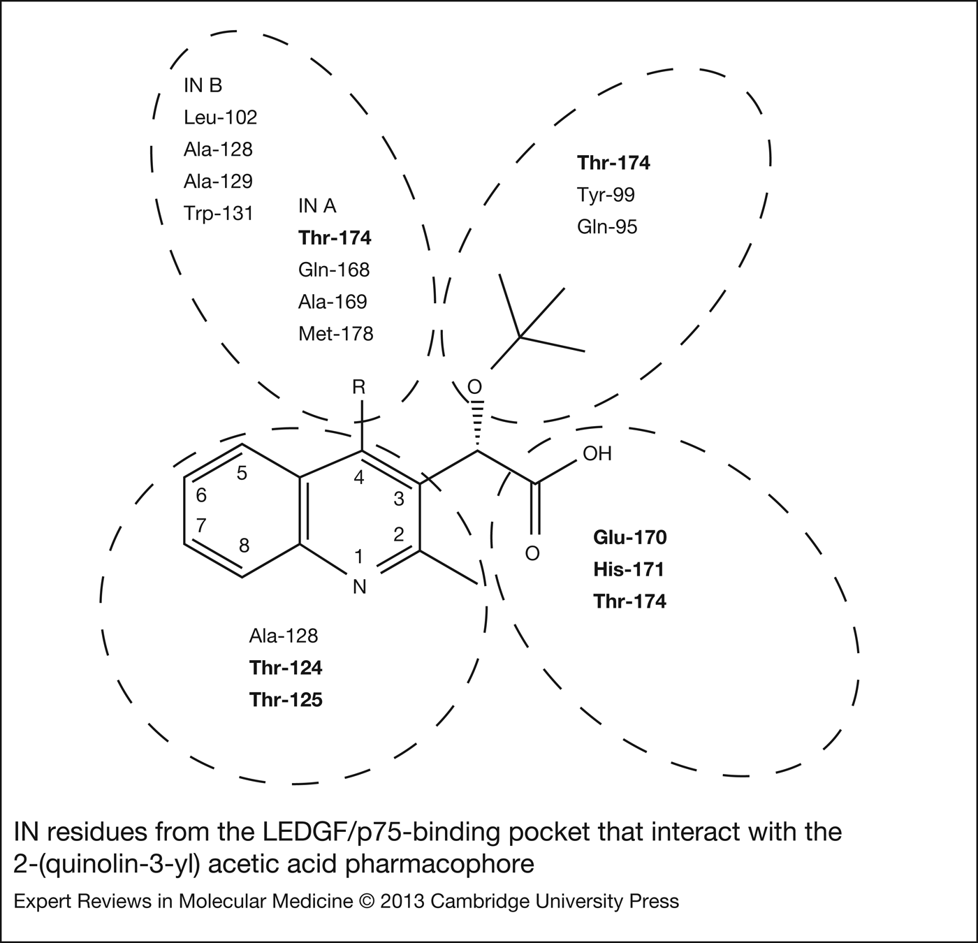
The ALLINI carboxylic acid moiety that extends from position 3 of the fused-ring quinoline (Fig. 4) forms H bonds with the IN backbone nitrogen atoms of residues Glu-170 and His-171, which is the same interaction mediated by LEDGF/p75 hotspot residue Asp-366 (Refs Reference Christ79, Reference Tsiang83, Reference Kessl86, Reference Jurado89) (Fig. 3b and c). The acid group in the context of the small molecules additionally mediates an H bond with the side-chain oxygen of Thr-174 (Refs Reference Tsiang83, Reference Kessl86) (Fig. 3c). The methoxy or tert-butoxy moiety that lies between the quinolone ring and the carboxylic acid positions in the vicinity of IN residues Tyr-99, Gln-95 and Thr-174 (Refs Reference Tsiang83, Reference Kessl86) (Table 1, Figs 3c and 4). The quinoline core itself situates near IN residues Ala-128, Thr-124 and Thr-125, which make up one face of the host factor-binding pocket (Fig. 3c). The functional group attached at position 4 of the quinoline is situated within the dimer interface cleft made up of IN chain B residues Leu-102, Ala-128, Ala-129 and Trp-131, and chain A residues Thr-174, Gln-168, Ala-169 and Met-178, and thereby establishes contacts with both CCD molecules (Figs 3c and 4). The majority of mutations that confer resistance to ALLINIs, selected through ex vivo viral challenge, map to several of these residues (Y99H, L102F, A128T, H171T and T174I), confirming the interaction site analyses of the ALLINI-IN CCD co-crystal structures (Refs Reference Christ79, Reference Tsiang83, Reference Kessl86, Reference Jurado89). Importantly, with resistance mutations mapping to the LEDGF/p75-binding pocket of IN and not to the enzyme active site, ALLINIs retained potency against viral strains resistant to INSTIs (Refs Reference Christ79, Reference Tsiang83). Furthermore, a computer-simulated program along with impact of drug combinations on HIV-1 replication predicted that the inhibitory effects of ALLINIs and INSTIs are unlikely to be antagonistic and likely to be additive, indicating combination therapy should be feasible (Refs Reference Christ79, Reference Christ85). ALLINIs remain active across a broad range of HIV-1 subtypes, but are not effective against HIV-2 (Refs Reference Christ79, Reference Christ85). The most potent compounds described to date yield antiviral EC50 values in the range of 10–90 nM with selectivity indices of ~1400–8800 (Refs Reference Tsiang83, Reference Christ85, Reference Jurado89).
IN residues from the LEDGF/p75-binding pocket that interact with the 2-(quinolin-3-yl) acetic acid pharmacophore. Dash-lined circles indicate sets of IN amino acid residues that either directly interact or are within a close vicinity of the indicated ALLINI functional group. The R moiety, which among potent allosteric IN inhibitors (ALLINIs) harbours minimally one 6-carbon ring (Table 1), varies among the various 2-(quinolin-3-yl) acetic acid derivatives (Ref. Reference Engelman, Kessl and Kvaratskhelia119). R is capable of interacting with both IN chains (A and B) within the CCD–CCD complex. The bolded amino acids represent IN residues that participate in H bonding with the ALLINI carboxylic acid functional group.
The affect of ALLINIs on the dynamic exchange of IN subunits was investigated (Ref. Reference Kessl86) based on the precedence that the previously discussed chicoric acid derivative deregulated IN–IN interactions important for the formation of the SSC (Ref. Reference Kessl44). Impressively, ALLINIs promote IN multimerisation in the absence of vDNA, and in doing so inhibit SSC formation in vitro (Refs Reference Tsiang83, Reference Christ85, Reference Kessl86). Preformed SSCs moreover are recalcitrant to ALLINI challenge (Ref. Reference Kessl86). These data are fully consistent with the inhibition of 3′ processing activity during the early phase of HIV-1 infection (Ref. Reference Tsiang83). To test if the LEDGF/p75–IN interaction contributes to ALLINI potency during the early phase of HIV-1 infection, compound EC50 values were assessed in cells knocked down (Ref. Reference Jurado89) or knocked out (Refs Reference Wang88, Reference Schrijvers90) for LEDGF/p75 expression. These conditions interestingly yielded significant enhancements in drug potency. ALLINIs and LEDGF/p75 therefore compete for binding to the IN CCD interface during the early phase of HIV-1 infection when IN is known to engage LEDGF/p75 to facilitate integration (Refs Reference Wang88, Reference Jurado89). Yet, the host–virus interaction does not underlie antiviral activity. If it did, inhibitor potency would weaken in the absence of the drug target, which was not observed. HRP2, the other IBD-containing member of the HRP protein family, can stimulate HIV-1 IN activity in vitro (Ref. Reference Cherepanov56) and act as an integration co-factor during HIV-1 infection, though this has only been observed in the backdrop of a LEDGF/p75 knockout (Refs Reference Llano64, Reference Wang88, Reference Schrijvers90, Reference Schrijvers91). Though HRP2 was suggested to contribute to ALLINI potency during HIV-1 infection (Ref. Reference Schrijvers90), subsequent work discounted an important role for the HRP2–IN interaction (Ref. Reference Wang88). It was nevertheless posited in 2012 that two integration-related functions, SSC formation and the association of LEDGF/p75 with the SSC, determined the antiviral activity of ALLINIs (Refs Reference Tsiang83, Reference Christ85, Reference Kessl86). Subsequent discoveries that treatment of virus-infected cells yields non-infectious progeny virions have since revamped this model (Refs Reference Balakrishnan87, Reference Jurado89, Reference Desimmie92).
ALLINI potency is determined through the inhibition of late-stage HIV-1 replication events
Single amino acid substitutions within IN that can abrogate LEDGF/p75 binding, such as Q168A, were initially proposed as evidence for the importance of the LEDGF/p75–IN interaction during HIV-1 infection (Ref. Reference Emiliani55). Upon further study, the replication blocks associated with this mutation mapped to preintegrative steps. Although IN catalytic activity remained near the levels of the wild-type enzyme (Refs Reference Emiliani55, Reference Rahman69), the ability for the mutant virus to synthesise vDNA was impaired (Ref. Reference Rahman69). Various deletion and missense mutations within the IN region of the HIV-1 pol gene can cause defects within the virus lifecycle at steps other than integration. Whereas reverse transcription is invariably affected, particle assembly and/or release from virus producer cells can also be impaired. The behaviours of different HIV-1 IN mutant viruses led to the following classification system: mutants that are specifically blocked at the integration step are defined as class I, whereas mutants that harbour replication defects at steps other than integration are referred to as class II (reviewed in Refs Reference Engelman93, Reference Engelman and Neamati94). The underlying aspects of IN biology that extend beyond its catalytic activity and yield replication defects at steps other than integration are incompletely understood. As the majority of HIV-1 IN mutations, including those in and around the LEDGF/p75 binding pocket, elicit the class II mutant viral phenotype (Ref. Reference Engelman and Neamati94), it was predicted a number of years ago that small molecule inhibitors that target this pocket might induce the class II mutant phenotype, resulting in pleiotropic perturbations in the HIV-1 lifecycle (Refs Reference Cherepanov68, Reference Rahman69).
Drug treatment of target cells infected with virus constructs that are restricted to a single round of replication, and are thereby acute indicators of the level of HIV-1 DNA integration, surprisingly revealed a significant loss of potency with the ALLINI compound BI-D as compared with the strength observed during multiple rounds of HIV-1 replication (EC50 values of 1.1 and 0.09 µM, respectively). This observation prompted the limitation of ALLINI exposure to virus producing cells, where, unexpectedly, full drug potency was recovered (EC50: 0.09 µM) (Ref. Reference Jurado89). Thus, the main antiviral effect of ALLINIs occurs during a late-stage event of HIV-1 replication (Ref. Reference Jurado89). This finding was further demonstrated with two NCINIs (GS-A and GS-B) (Ref. Reference Balakrishnan87), and time-of-addition experiments also corroborated the interpretation that ALLINIs act predominantly during virus production (Ref. Reference Desimmie92). With class II mutants providing precedence for both preintegrative and postintegrative defects, the effects of ALLINIs were compared with the behaviour of class II HIV-1 IN mutant viruses.
The most common pleiotropic defect associated with class II IN mutations occurs at reverse transcription (Refs Reference Shin95, Reference Masuda96, Reference Engelman97, Reference Wiskerchen and Muesing98, Reference Leavitt99, Reference Wu100; reviewed in Refs Reference Engelman93 and Reference Engelman and Neamati94). Consistent with prior reports (Refs Reference Christ79, Reference Tsiang83), exposing target cells to ALLINIs during the early phase of HIV-1 infection did not influence reverse transcription (Ref. Reference Jurado89). In contrast, when virus made in the presence of ALLINIs was used to infect drug-free target cells, reverse transcription was severely impaired (Refs Reference Jurado89, Reference Desimmie92). Interestingly, endogenous reverse transcription, an artificial induction of RT activity within viral particles, was not affected by ALLINI treatment (Ref. Reference Balakrishnan87), indicating that the compounds do not inhibit RT enzyme activity or its association with viral RNA within the virion. HIV-1 requires IN expression for proper reverse transcription, and IN provided in trans during virus production in the form of a Vpr-IN fusion protein can complement the reverse transcription defect of a class II IN deletion mutant virus (Ref. Reference Wu100). Interestingly, the trans-complemented IN does not need to be enzymatically active, as a Vpr-IN fusion protein harbouring the D116A mutation in the IN active site efficiently complemented the DNA synthesis defect (Ref. Reference Wu100). This observation suggests that it is the structure of IN protein as compared with its enzymatic function that in some way contributes to the overall process of reverse transcription. The CTD of IN directly interacts with RT (Refs Reference Wu100, Reference Zhu, Dobard and Chow101, Reference Hehl102, Reference Wilkinson103), and IN can augment RT initiation, elongation and processivity activities in vitro (Ref. Reference Dobard, Briones and Chow104), but the significance of the RT–IN interaction within the context of HIV-1 infection has eluded direct demonstration. Because ALLINIs only affect reverse transcription when present during virus production and the RT within drug-treated viral particles is inherently active, the influence on vDNA synthesis seems likely to occur prior to or immediately upon virus entry into target cells. Altered protein–protein and/or protein–RNA interactions during viral assembly or after Gag-Pol processing by the viral PR to allow for proper RT and IN positioning within the maturing virion could account for the subsequent reverse transcription defect. A clearer mechanistic understanding of the influence of IN on reverse transcription awaits further dissection.
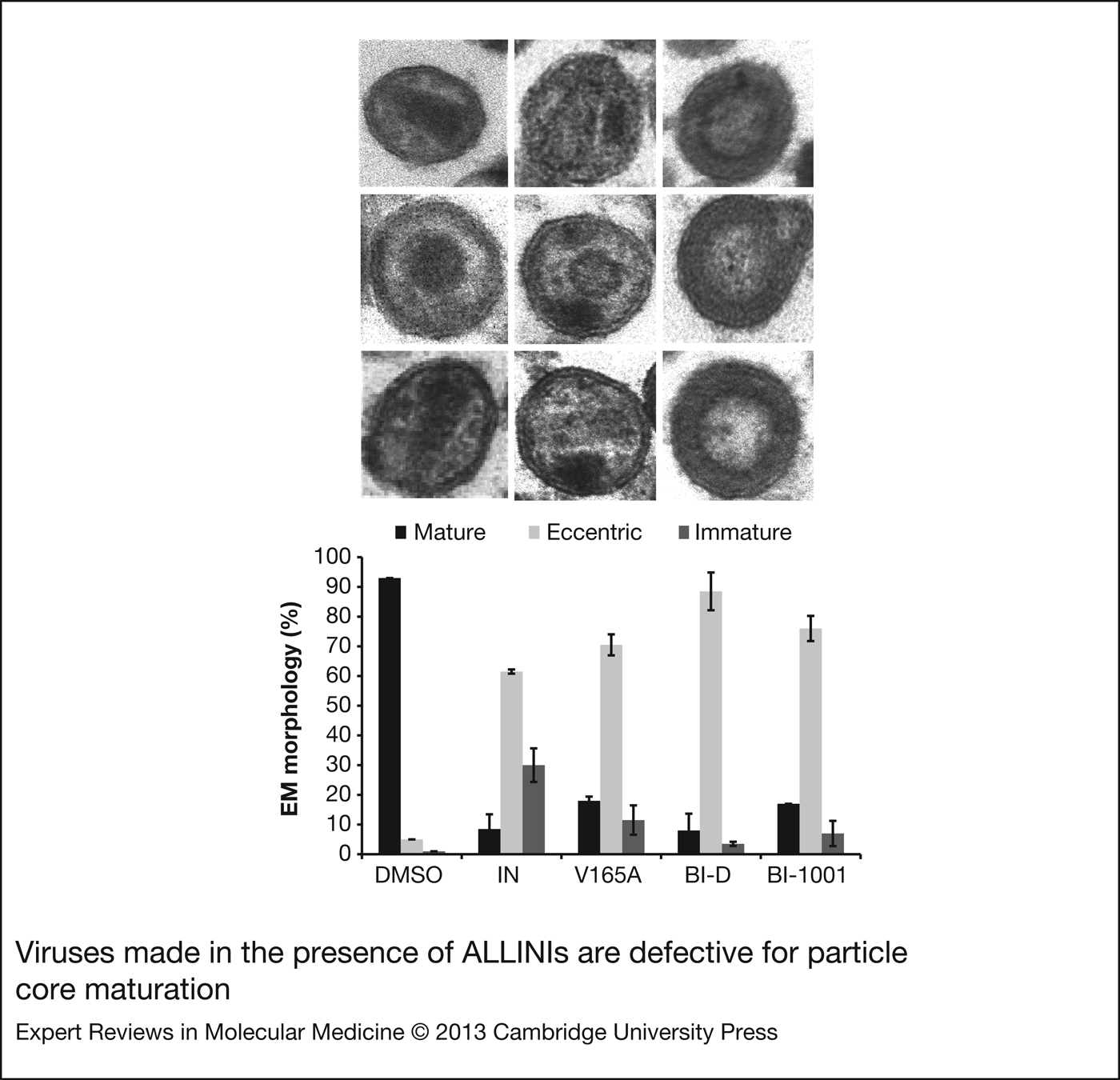
Although ALLINI potency mapped to late-stage replication events, the compounds did not affect the release of virus from cells, viral protein processing or viral protein or genomic RNA incorporation into virions (Refs Reference Balakrishnan87, Reference Jurado89, Reference Desimmie92). With class II mutant viruses providing precedence for defects in virion morphology (Refs Reference Engelman97, Reference Shehu-Xhilaga105), viral particles were evaluated by transmission electron microscopy. Viruses made in the presence of ALLINIs were defective for particle core maturation, with ~75–90% of virions having an eccentric phenotype as defined by the positioning of the electron dense material, which is usually encaged within the conical core, between the virus membrane and an electron-lucent or empty core (Fig. 5) (Ref. Reference Jurado89). Similar results have been reported for the related compounds CX05045 and GS-B (Refs Reference Balakrishnan87, Reference Desimmie92). Localisation of electron-dense material to a region outside of the electron-lucent core suggests misplacement of the viral RNP complex. IN can bind random-pooled RNA with different affinities in vitro (Ref. Reference Allen, Worland and Gold106), and IN has been implicated in proper genomic RNA dimerisation within the maturing virion (Refs Reference Shehu-Xhilaga105, Reference Buxton, Tachedjian and Mak107). This may not require proper IN oligomerisation, as a multimerisation-deficient IN mutant, V260E, formed the wild-type RNA dimer profile (Ref. Reference Buxton, Tachedjian and Mak107). In addition to class II mutant viruses (Refs Reference Engelman97, Reference Shehu-Xhilaga105), the eccentric virion core morphology has been seen under conditions of suboptimal doses of PR inhibitors (Refs Reference Moore108, Reference Kaplan109) and improper Gag polyprotein processing (Ref. Reference Ohishi110).
Viruses made in the presence of allosteric IN inhibitors (ALLINIs) are defective for particle core maturation.Upper, representative images of three discernable virion morphologies: mature, eccentric and immature. The eccentric phenotype is characterised by electron dense material situated between the virus membrane and electron-lucent core; the electron-dense ribonucleoprotein complex usually associates with the conical core (mature). Lower, quantitation of virion phenotypic frequencies (average ± SD for 100 virions; n = 2 experiments) in the presence of ALLINI BI-D (10 µM), BI-1001 (50 µM) or DMSO solvent control. V165A and ΔIN are class II IN mutant virus controls produced in the absence of drug. Figure from Jurado et al. (Ref. Reference Jurado89).
The presence of ALLINIs during virus production induced a phenotype that is surprisingly similar to class II IN mutant viruses. In an effort to uncover the molecular basis behind this effect, the influence of ALLINIs on IN oligomerisation within nascent virions was investigated. Consistent with in vitro data utilising purified, recombinant IN protein (Refs Reference Tsiang83, Reference Christ85, Reference Kessl86), ALLINIs were indeed discovered to enhance IN multimerisation within virions (Refs Reference Balakrishnan87, Reference Jurado89, Reference Desimmie92). Importantly, the use of resistance mutations that negate drug action also conferred resistance to compound-induced IN multimerisation (Refs Reference Balakrishnan87, Reference Jurado89, Reference Desimmie92). The estimated 100 molecules of IN per virion translates to an approximate protein concentration of 20 mg/ml, which invariably favours IN multimerisation (Ref. Reference Cherepanov111). Although the exact oligomeric state of IN within virions is unknown, ALLINI-induced stabilisation of IN oligomers appears to be detrimental for proper particle maturation, likely through the formation of a higher order IN oligomer or aggregate, or through the disruption of critical IN–protein or IN–RNA interactions.
The LEDGF/p75–IN interaction is not thought to play a role in HIV-1 replication after the integration step. Over-expression of an IBD-containing fusion protein in virus producer cells, for example, does not influence virus release or the infectivity of the progeny virions, but does potently inhibit infection at the integration step during viral ingress (Ref. Reference De Rijck72). To determine whether LEDGF/p75 might influence the potency of ALLINIs, drug EC50 values were determined during the late stage of HIV-1 replication using control versus LEDGF/p75 knockdown cells (Refs Reference Balakrishnan87, Reference Jurado89) or with cells engineered to overexpress the IN co-factor (Ref. Reference Balakrishnan87). Drug strength under these conditions was LEDGF/p75-independent, indicating that the absence of competing LEDGF/p75 protein during virion production determines ALLINI potency (Refs Reference Balakrishnan87, Reference Jurado89). Consistent with this interpretation, a detailed study of the ALLINI BI-1001 resistance mutation A128T in IN indicated that stimulation of IN multimerisation is the primary mode of drug action. The A128T mutation conferred significant resistance to the multimerisation-inducing effects of the compounds, with little-to-no influence on the inhibition of the LEDGF/p75–IN interaction (Ref. Reference Feng112).
It has recently become clear that the main antiviral effect of ALLINIs occurs during the late phase of HIV-1 replication, and is accounted for by compound induced-multimerisation of IN during viral egress in the absence of competing LEDGF/p75 co-factor, ultimately resulting in defective particle maturation and impaired reverse transcription and integration in subsequently infected target cells (Refs Reference Balakrishnan87, Reference Jurado89, Reference Desimmie92). It is noteworthy that ALLINIs do not break apart a critical protein interaction. By contrast, it is their ability to enhance the interaction between two CCD molecules that underlies their unique pharmacology.
Pharmacodynamics of HIV-1 inhibitors and ALLINIs
In vitro and ex vivo drug potency is most widely measured as the concentration at which 50% (IC50 and EC50) of inhibition is achieved. Yet, drug concentrations in patients are much higher than the amount needed to inhibit 50% of infection, and virus replication is an exponential process. Therefore, plots of antiviral drug activity are underappreciated using linear scales, and should instead be evaluated on a logarithmic scale. Analogous to the Hill coefficient, the shape of the dose–response curve is influenced through cooperative interactions, and is mathematically defined as the slope (Ref. Reference Shen, Rabi and Siliciano113). Inclusion of the slope in quantitating antiviral activity has proven to be an important variable to accurately predict clinical outcomes (Ref. Reference Rosenbloom114). Two drugs may have the same IC50, but the one with the steeper slope will be capable of achieving a much greater degree of inhibition at clinically relevant drug concentrations (Ref. Reference Shen115).
When the slope of the dose–response curve is steep, small changes in drug concentration will have a large effect on inhibitory potential (Ref. Reference Shen115). For antiretroviral drugs, slopes have been shown to vary in a class-dependent manner, with PIs and non-nucleoside RT inhibitors (NNRTIs) characterszed by steep slopes (≥1.7), while nucleoside RT inhibitors (NRTIs) and INSTIs have slopes of one (Ref. Reference Shen115). PIs have notably steep dose–response curves, with some drugs reaching slopes of 4.5 (Ref. Reference Shen115), and consistent with the importance of this parameter in evaluating antiretroviral activity in the clinic, PIs are the only class of HIV-1 drugs that are successful in monotherapy (Ref. Reference Katlama116). The class-associated variation suggests that drugs which act through different mechanisms show distinct slopes (Ref. Reference Shen115). Considered alongside the fact that clinical INSTIs that target the enzyme active site posses slopes of one, it is intriguing that ALLINIs display slopes of significantly greater than one (Refs Reference Kessl86, Reference Jurado89).
Since viral enzymes targeted by HAART are univalent with respect to their inhibitor, the slope parameter was largely ignored within the antiretroviral field until the pioneering work of Robert Siliciano established its relevance (Ref. Reference Shen115). The critical subset model provides a conceptual basis for the observation of steep slopes with univalent drug targets (Ref. Reference Shen117). Infectivity may require participation of multiple copies of a drug target at a relevant stage of inhibition within the viral lifecycle, and it is this critical subset of drug targets that must remain drug-free for infection to occur. The model suggests that when there are multiple drug targets involved in a particular step of replication, intermolecular cooperativity occurs independent of the classical alteration in binding properties of individual sites observed under conditions of intramolecular cooperativity (Ref. Reference Shen117). The model suggests that intermolecular cooperativity underlies multivalent drug targets and accounts for steep slopes. The theoretical equation of the mathematical representation of the critical subset model was confirmed experimentally through modulating the number of functional drug targets and observing changes in EC50 with drugs that have steep slopes, with no changes observed for drugs that display slopes of one (Ref. Reference Shen117). If this model holds true, the steep slope of ALLINIs suggests that there must be a critical subset of IN molecules required for proper particle maturation during viral egress.
Although ALLINIs have two binding sites per drug target (two binding-pockets per IN CCD dimer; Fig. 3), their steep slopes are not attributed to intramolecular cooperativity: ALLINIs importantly display steep slopes only during the late stage of HIV-1 replication; when assessed in target cells, where they act as weak integration inhibitors, the slope was ~1 (Ref. Reference Jurado89). If the steepness of the slope were due to intramolecular cooperativity, the slope of the dose–response curve would be expected to be independent of the time point of drug action. Another explanation for the steep slopes could be attributed to the inhibition of multiple steps along the viral lifecycle, which for ALLINIs include particle maturation, reverse transcription and integration (Ref. Reference Jurado89). The ability for PIs to inhibit multiple replication steps appears to contribute to the steep slopes observed for this drug class (Ref. Reference Rabi118).
The slope parameter has further been implicated in determining the evolution patterns of resistance mutations (Ref. Reference Rosenbloom114). Since the inhibitory potential of drugs with steep dose–response curves are particularly sensitive to changes in drug concentration, the amount of time a virus will exist within a suboptimal drug concentration that pressures the outgrowth of resistance mutants is narrowed (Ref. Reference Rosenbloom114). Therefore, the relative risk of mutant outgrowth is curtailed, while the relative risk of wild-type virus growth is enhanced. This prioritises the essentiality of adherence to drug regimes of inhibitors with steep slopes, but in theory decreases the frequency of selection of drug resistant mutant viruses (Ref. Reference Rosenbloom114). The evolution of resistance to current ALLINI compounds occurs relatively quickly in vitro (over ~5 to 6 serial passages) under conditions where drug strength is maintained at concentrations ≥EC50 (Refs Reference Christ79, Reference Tsiang83). If ALLINIs are to be used within the clinic, compounds that are less prone to the generation of drug resistance will need to be developed.
Steep slopes assist in identifying appealing drug targets for clinical suppression. Detection of an IN inhibitor with a steep slope thus further highlights ALLINIs as promising candidates for future antiretroviral development.
Research in progress and outstanding questions
The observation that IN inhibitors can induce the class II IN mutant virus phenotype revives several interesting questions regarding the potential role for IN during the late stage of HIV-1 replication. It is tempting to interpret the ALLINI mechanism of action to suggest that IN plays an active role in the formation of the electron-dense HIV-1 core. Alternatively, particle maturation is particularly sensitive to deregulated IN multimerisation. This sensitivity could stem from a protein–protein or protein–RNA interaction during viral assembly that is necessary for proper positioning of the RNP within the viral core. The structural rearrangements of IN and other viral constituents subsequent to Gag and Gag-Pol proteolysis require further elucidation.
The current ALLINIs inhibit two integration-related functions in vitro, SSC assembly and the SSC–LEDGF/p75 interaction, yet their ability to drive IN multimerisation at a point in the virus lifecycle when endogenous LEDGF/p75 protein is apparently unable to compete for IN binding determines their antiretroviral activity (Refs Reference Balakrishnan87, Reference Jurado89). It will be instructive to ascertain the mechanisms of action of future compounds that separate the two in vitro IN-related inhibitory activities of ALLINIs. As LEDGF/p75-independent, IN multimerisation-inducing inhibitors have been described (Ref. Reference Kessl44), it will be informative to see if they influence the late events of HIV-1 replication. The description of compounds that specifically disrupt LEDGF/p75–IN binding without concomitant perturbation of IN multimerisation will allow investigators to assess the potential clinical relevance of blocking the virus–host interaction.
The finding of steep ALLINI dose–response curve slopes is consistent with the inhibition of a multivalent target (Refs Reference Kessl86, Reference Jurado89). NRTIs and INSTIs both target a single molecular complex of an enzyme and nucleic acid and have slopes of one, whereas PIs, NNRTIs and ALLINIs target the enzyme alone and have slopes greater than one (Refs Reference Kessl86, Reference Jurado89, Reference Shen115). The events inhibited by NRTIs and INSTIs solely depend on the targeted univalent enzyme–nucleic acid complex, with the extraneous substrate-free enzyme molecules being irrelevant. The targets of PIs, NNRTIs and ALLINIs involve multiple copies of the enzyme target that are participating in the same event of virus replication. Understanding whether steep ALLINI slopes are mechanistically determined through the inhibition of multiple steps within the viral lifecycle, or are indicative of a critical subset of molecules required to carry out a particular event, necessitates further attention.
Financial support
The authors acknowledge funding support from the US National Institutes of Health grants AI039394 and GM103368. K.A.J. is a recipient of a National Academies' Ford Foundation Predoctoral Fellowship.
Conflicts of interest
None