Alzheimer disease (AD) is a progressive and irreversible neurodegenerative disorder that is characterised by the presence of senile plaques (SPs), neurofibrillary tangles, persistent neuronal loss and the deterioration of cognitive functions (Ref. Reference Querfurth and LaFerla1). AD represents the most common form of dementia and affects around 27 million people worldwide (Ref. Reference Ferri2). It is one of the most severe socioeconomic and medical burdens, with the annual cost of care currently estimated to be over $100 billion in the USA alone (Ref. Reference Smith3). Moreover, over 5 million new cases of AD are reported each year, with incidence increasing from 1% between the ages of 60 and 70 to 6–8% by 85 years (Refs Reference Ferri2, Reference Kukull4). Over the past two decades, considerable effort has been aimed at gaining a better understanding of the cause of AD, with the ultimate hope of developing safe and effective pharmacological treatments. However, irrespective of the form of therapy, current approaches for the treatment of AD provide only temporary symptomatic relief and do not inhibit or reverse the underlying disease mechanisms. Therefore, the treatment of AD still remains a great challenge, and the development of new strategies is an active area of research.
The amyloid-β cascade, oxidative stress and neuroinflammation in AD
The amyloid-β peptide (Aβ) and tau are both hallmark proteins in AD, with the fibrillar Aβ aggregates representing the main components of SPs. Aβ is produced as a result of enzymatic proteolysis of the mammalian transmembrane amyloid precursor protein (APP). APP undergoes proteolytic processing through either the nonamyloidogenic or the amyloidogenic pathway (Ref. Reference Nunan and Small5). In the nonamyloidogenic pathway, the membrane-bound enzyme α-secretase cleaves APP within its Aβ domain, resulting in the extracellular secretion of soluble APPα (sAPPα) and the production of a short membrane-bound C-terminal fragment (CTF), α-CTF or C83 (Ref. Reference Panegyres6). Subsequent γ-secretase cleavage of C83 results in the secretion of a 3 kDa peptide termed p3 (Ref. Reference Haass7) and release of the APP intracellular domain (AICD) into the cytoplasm (Ref. Reference Black8). The amyloidogenic pathway is initiated when β-secretase cleaves APP at the N-terminal part of the Aβ domain. This cleavage leads to the extracellular release of the soluble fragment sAPPβ, whereas the β-CTF or C99 fragment remains membrane bound. Subsequent cleavage by the γ-secretase enzyme of the C99 fragment, at the C-terminal of Aβ, allows shedding of the AICD and secretion of Aβ species of variable length into the lumen or extracellular space (Ref. Reference Seubert9).
Following their characterisation, the APP secretases became attractive targets to reduce the pathogenesis of Aβ by stimulating α-secretase cleavage in order to direct APP processing towards the nonamyloidogenic pathway or by suppressing β- or δ-secretase cleavage to reduce the amount of Aβ produced. Therefore, numerous γ- and β-secretase inhibitors or modulators have also been designed (see Ref. Reference De Strooper, Vassar and Golde10 for a review). However, most of these agents are not specific for the secretase cleavage of APP and thus might prevent the cleavage and processing of additional substrates, which could result in various adverse effects (Refs Reference Evin, Sernee and Masters11, Reference Tschape and Hartmann12).
Aβ proteins and oxidative stress are believed to have central roles in the development of AD preceding Aβ pathology and neurofibrillary tangles. Interestingly, results have indicated that lipid peroxidation occurs in the brain upon myocardial infarction, indicating that oxidative damage could have an early role in the pathogenesis of AD. For instance, F-4 isoprostanes, derived from free radical oxidation of docosahexaenoic acid, are increased in the brain cortex regions (Ref. Reference Nourooz-Zadeh13). Moreover, the level of lipid peroxidation in the hippocampus and frontal cortex in AD is dependent on the apolipoprotein E (ApoE) genotype and the levels of this protein in these brain regions (Refs Reference Ramassamy14, Reference Ramassamy15). Protein carbonyls are present in neurons both with and without tangles, and hippocampal neurons demonstrate intense cytoplasmic staining with 8-hydro-2-deoxyguanosine (Ref. Reference Smith16). F-2 isoprostanes, prostaglandin-like compounds derived from free-radical-catalysed peroxidation of arachidonic acid, are also elevated in the plasma, urine and cerebrospinal fluid (CSF) of patients suffering from AD (Ref. Reference Pratico17). In brains from patients with AD, acrolein, which is a byproduct of lipid peroxidation, has been found to be increased in the hippocampus and temporal cortex, where oxidative stress is high (see Ref. Reference Nam18 for a review).
Oxidative stress is considered as both a cause and a consequence of neuroinflammation. Indeed, reactive oxygen species (ROS) and reactive nitrogen species (RNS) act as signals to activate proinflammatory genes such as induced nitric oxide synthase (iNOS), interleukin-1β (IL-1β), tumour necrosis factor-α (TNF-α) and nuclear factor-κB (NF-κB), and thereby establish a chronic inflammation state. In turn, chronic inflammation leads to the generation of ROS and RNS, and therefore amplifies the damage in target cells (Ref. Reference Sarkar and Fisher19). Despite early epidemiological evidence that chronic use of nonsteroidal anti-inflammatory drugs diminished the risk for AD, clinical intervention trials have thus far failed to slow down disease progression or provide convincing evidence of symptomatic improvements in patients with early or moderate AD (Ref. Reference Shehzad, Wahid and Lee20). Several in vitro and in vivo studies have repeatedly reported that Aβ production, oxidative markers and neuroinflammation are attenuated by the administration of different polyphenolic compounds with pleiotropic properties (e.g. curcumin) (see Ref. Reference Singh21 for a review). In this review, we will summarise the challenges associated with curcumin therapy in AD.
Curcumin: an Eastern traditional medicine for modern drug application
The Curcuma genus belongs to the Zingiberaceae, the same family as Zingiber (ginger). Curcuma longa (C. longa) is a short-stemmed (~100 cm in height), perennial plant that grows naturally throughout the Indian subcontinent and in tropical Asia, particularly in South East Asia. Turmeric, the dried ground rhizome of C. longa, is a spice present in curry and is widely used for its flavouring in many food preparations and recipes. The synonyms for turmeric are ‘haldi’ in Hindi, ‘chiang huang’ in Chinese, ‘ukon’ in Japanese, ‘haridra’ or ‘gauri’ in Sanskrit and ‘kurkum’ in Arabic. In addition to its applications as a spice, dye, food additive and preservative in traditional Indian cooking, turmeric has been used for thousands of years in Ayurvedic and Chinese medicine. In the 12th to 13th century, it was introduced, along with other spices, to Western countries by Arab traders and Marco Polo, after they visited India. Currently, turmeric continues to be widely used as an alternative medicinal agent in many parts of southern and eastern Asia. It has been hypothesised that its extensive use might account for the significantly lower prevalence of AD in the Asian-Indian population (Ref. Reference Ganguli22) and that curcumin treatment might be beneficial for AD patients. Over the past decade, the number of cell culture and animal model studies with curcumin has increased significantly, and these have led to the identification of several pharmacological targets; some clinical trials have also been conducted.
Pharmacological aspects of curcumin
Structure–activity relationship and antioxidant activity
The major curcuminoids present in turmeric are demethoxycurcumin (curcumin II), bisdemethoxycurcumin (curcumin III) and cyclocurcumin (Ref. Reference Kiuchi23). Commercial curcumin, that is curcumin I (77%), curcumin II (17%) and curcumin III (3%) (Ref. Reference Goel, Kunnumakkara and Aggarwal24), is commonly referred to as ‘the curcumin complex’ or ‘the curcumins’ (Fig. 1). Several in vitro studies have shown that the curcumins are powerful antioxidants. For instance, they are potent scavengers of superoxide and hydroxyl radicals, conserving glutathione (GSH) levels and decreasing the concentration of circulating free-radical end products (Ref. Reference Soni and Kuttan25). They also protect against lipid peroxidation, GSH peroxidase inactivation and DNA oxidative attack (Refs Reference Strimpakos and Sharma26, Reference Venkatesan, Punithavathi and Arumugam27). In several animal models, the curcumins protect the brain, liver, lungs, kidneys and heart from oxidative agents. For example, curcumins inhibit ROS generation in activated rat peritoneal macrophages (Ref. Reference Joe and Lokesh28) and lipid peroxidation in rat brain homogenates and rat liver microsomes (Ref. Reference Sreejayan and Rao29); they also protect against acetaminophen-induced nephrotoxicity in male Wistar albino rats through reduction of the malondialdehyde level, increasing GSH, GSH peroxidase, catalase and superoxide dismutase activities in renal tissue (Ref. Reference Cekmen30). The curcumins can protect the liver, testis, brain, kidneys and lungs of male rats against sodium-arsenite-induced oxidative damage by enhancing GSH S-transferase, superoxide dismutase and catalase activities (Ref. Reference El-Demerdash, Yousef and Radwan31), reduced oxidative DNA and protein damage in diabetic rat heart (Ref. Reference Farhangkhoee32). By contrast, in the presence of high concentrations of free iron or copper, the curcumins behave like pro-oxidants; they contribute to ROS generation and hence ROS-induced apoptosis. This property might be relevant to their beneficial effects on cancer cells (Refs Reference Bhaumik33, Reference Aggarwal, Kumar and Bharti34). Electron withdrawal from the keto-enol group to the phenolic hydroxyl moieties (Fig. 1) has been strongly associated with curcumin antioxidant activity in both free cells (Refs Reference Cai35, Reference Jayaprakasha36, Reference Chen37) and biological systems (Ref. Reference Somparn38). Indeed, scavenging effects are connected with the formation of phenoxyl radicals formed by deprotonation of the phenolic hydroxyl groups (Ref. Reference Gorman39), and metal chelation is highly affected by enol proton mobility or acidity (Ref. Reference Schaich, Ficher and King40). The differences in antioxidant activities between curcumin and its analogues suggest that the phenolic methoxy substituents have an important role. Therefore, curcumin is more efficient than demethoxycurcumin, and bisdemethoxycurcumin is almost inactive. A comparative study was carried out between the antioxidant activity of curcumin and those of its hydrogenated derivatives. Surprisingly, tetrahydrocurcumin, hexahydrocurcumin and octahydrocurcumin were significantly more efficient than curcumin, suggesting that hydrogenation at conjugated double bonds of the central seven carbon chain and the β-diketone of curcumin lead to the highest antioxidant activity (Ref. Reference Somparn38). Curcuminoid antioxidant activities are also described in in vivo models. In male Wistar rats, curcumin and demethoxycurcumin, but not bisdemethoxycurcumin, prevent the decrease of GSH level and protein oxidation (Ref. Reference Dairam41). By contrast, bisdemethoxycurcumin was shown to be more efficient than curcumin regarding ROS generation in different human cancer cell lines (Ref. Reference Ravindran42). In the same study, bisdemethoxycurcumin was found to be more potent than curcumin regarding its ability to suppress TNF-induced NF-κB activation; however, others found that curcumin was more efficient (Ref. Reference Sandur43). The mechanisms of action underlying the antioxidant activities by radical scavenging are still not fully understood and remain controversial. However, a recent study suggested that both phenolic OH and the β-diketone moiety of curcumin are responsible for the antioxidant effects, depending on the nature of the free radical. Indeed, phenolic OH is responsible for the reaction with DPPH and CCl3O2• radicals, whereas the β-diketone moiety acts with O2•− and 1O2 (Ref. Reference Singh44).
Chemical structure of curcuminoids and their reactive groups. Curcumins include curcumin (curcumin I, 77%), demethoxycurcumin (curcumin II, 17%) and bisdemethoxycurcumin (curcumin III, 3%). Depending on their structures, curcumins can possess up to three reactive groups with a role in oxidative stress through radical scavenging or metal chelation, including the keto-enol group and the phenolic hydroxyl and phenolic methoxy groups.

Curcumin as an anti-inflammatory drug
Neuroinflammation has a key role in the origins and development of AD. Several studies report that NF-κB has a critical role in signal transduction pathways involved in inflammation. In numerous studies, curcumin was shown to possess strong anti-inflammatory activity. Aggarwal's group was the first to demonstrate that curcumin can inhibit the transcription factor NF-κB. Indeed, this ability is demonstrated using different inducers of NF-κB, including hydrogen peroxide on myelomonoblastic leukaemia cell line (ML-1a), probably through its free radical scavenging ability (Ref. Reference Singh and Aggarwal45). In addition to its action in ROS-related inflammation, curcumin can also prevent the activation of NF-κB induced by lipopolysaccharide (LPS) (Ref. Reference Jin46) or TNF-α (Ref. Reference Singh and Aggarwal45). The decrease in inflammatory mediators after curcumin treatment has been widely reported. For instance, in LPS-stimulated BV2 microglia, curcumin reduces, in a dose-dependent manner, the expression of IL-1β, IL-6 and TNF-α (Refs Reference Xu47, Reference Cho, Lee and Kim48). Furthermore, the effects of curcumin on proinflammatory enzymes, as well as their products, have been investigated. For instance, in addition to its effect on the suppression of prostaglandin E2 (PGE2) and nitric oxide release, curcumin also decreases the expression of iNOS and cyclooxygenase-2 (COX-2) mRNA and their corresponding proteins (Ref. Reference Jin46). Similarly, curcumin affects arachidonic acid metabolism through lipoxygenase (LOX) activity. In fact, curcumin is able to block the phosphorylation of cytosolic phospholipase (cPLA2) and inhibit the catalytic activity of 5-LOX in murine macrophages (RAW264.7 cells) and HT-29 human colon cancer cells (Ref. Reference Hong49). The mechanisms underlying the reduction of cPLA2 by curcumin have been investigated in vitro and involve reducing phosphorylation at the mitogen-activated protein kinase (MAPK)-sensitive site. Curcumin is also a PPARγ agonist. The enhancement of PPARγ activity by curcumin has been demonstrated in Aβ25−35-treated astrocytes, resulting in the inhibition of proinflammatory pathways (Ref. Reference Wang50). The in vivo anti-inflammatory effect of curcumin and tetrahydrocurcumin was demonstrated in an acute inflammatory model and in AD models. In the APPsw mouse model Tg2576, it was reported that curcumin reduced IL-1β levels (Refs Reference Lim51, Reference Chainani-Wu52). In the same transgenic mice model receiving the equivalent of 1.25 mg/day of curcumin for 4 months during the accelerated plaque formation period (12–16 months of age), a marked reduction in plaques and Aβ levels was described, whereas tetrahydrocurcumin did not have any effect (Ref. Reference Begum53).
Effect of curcumin on the Aβ cascade
One of the strategies developed to treat AD is the inhibition of amyloid plaque burden, Aβ aggregation and fibril formation because for many years it was believed that the toxic effects of Aβ were a result of mature Aβ fibrils. Recent studies suggest that low-molecular-weight, soluble, oligomeric forms of Aβ1–42 rather than Aβ1–40 (Ref. Reference Lambert54) are more neurotoxic than mature Aβ fibrils (Ref. Reference Hartley55). Therefore, it is necessary to consider the inhibition of Aβ oligomer formation and Aβ fibrils for the treatment of AD (Ref. Reference Necula56). Several in vitro studies have shown that curcumin can bind to Aβ and inhibit Aβ aggregation. Of 214 antioxidant compounds tested, curcumin had the strongest inhibitor effect on the formation of Aβ fibrils (Ref. Reference Kim57). Curcumins have a dose-dependent effect on the inhibition of Aβ1−40/1−42 fibrils and destabilise preformed fibrils with an EC50 of 0.09–0.63 µM (Ref. Reference Ono58). The effect of curcumin on Aβ oligomers and Aβ-derived diffusible ligand has been recently reviewed (Ref. Reference Yanagisawa59). Moreover, in mouse primary cortical neurons, curcumin decreased both Aβ levels and APP maturation (Ref. Reference Zhang60). This mechanism describes a possible cellular property underlying in vivo observations showing the decrease of Aβ deposition and plaque burden in an AD transgenic mice model after curcumin treatment.
Despite its poor bioavailability, some studies reported that curcumin can cross the blood–brain barrier (BBB) and label SPs (Ref. Reference Garcia-Alloza61). Indeed, natural products such as curcumin can cross the BBB using the substrate-binding site of the efflux transporter P-glycoprotein (P-gp) (Refs Reference Demeule62, Reference Romiti63). Aβ binding of curcumin was demonstrated in vivo using a multiphoton imaging system with a 30% reduction of Aβ plaque size after 1 week of injection (Ref. Reference Garcia-Alloza61). A low (160 ppm) and a high (5000 ppm) dose of curcumin administered orally for 6 months significantly reduced the Aβ plaque burden and lowered the levels of oxidised protein and proinflammatory cytokine IL-1β. Interestingly, only the low dose of curcumin decreased the level of insoluble Aβ, soluble Aβ and plaque burden in mice cortex and hippocampus. In the same transgenic mice, a daily single dose (500 ppm) of curcumin administered orally for 5 months significantly reduced the level of Aβ, insoluble Aβ and Aβ plaques (Refs Reference Yang64, Reference Begum65). In old female Sprague–Dawley rats receiving an intracerebroventricular infusion of Aβ, treatment with a low dose of curcumin (500 ppm) administered orally for 2 months before infusion of Aβ reduced Aβ deposition and increased both pre- and postsynaptic protein levels (Ref. Reference Frautschy66).
Bioavailability, pharmacokinetics and metabolism of curcumin
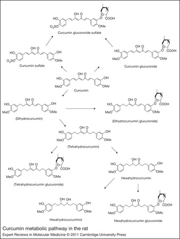
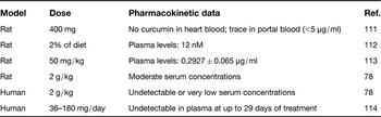
Curcumin has a molecular mass of 368.38 Da and a melting point of 183°C. In both solid and in soluble form, curcumin undergoes photodegradation when exposed to light (Ref. Reference Ansari67). It is soluble in acetone, ethanol and glacial acetic acid, and particularly insoluble in water. At acid pH (between 1 and 6) the degradation of curcumin is extremely low; however, at neutral and basic pH curcumin is unstable and is degraded to ferulic acid and feruloylmethane. Therefore, it would be stable in the stomach and small intestine (Ref. Reference Wang68). Curcumin is safe and well tolerated even at very high doses. Indeed, in a phase I clinical trial, a single daily oral dose ranging from 500 to 8000 mg/day for 3 months showed no toxicity (Ref. Reference Cheng69). The acceptable daily intake of curcumin as an additive has been defined by the World Health Organization as 0–1 mg/kg body weight (Ref. Reference Cheng69). Several factors, including its insolubility in water, lead to the poor systemic bioavailability of curcumin and make it a challenging molecule for oral delivery (see Table 1). Indeed, particularly low serum levels are observed after oral administration. It has been reported that 500 mg/kg of curcumin administered orally in rats gave a maximum serum level of 0.06 ± 0.01 µg/ml, whereas a 50-fold lower curcumin concentration given intravenously results in 0.36 ± 0.05 µg/ml. Moreover, when given orally, 40% of the curcumin is excreted unchanged in the faeces. In addition, in aqueous solution and at neutral and alkaline pH, curcumin undergoes degradation. That might contribute to its short half-life, which is reported to be 28.1 ± 5.6 and 44 ± 7.5 h in the rat for an intravenous dose of 10 mg/kg and an oral dose of 500 mg/kg, respectively (Ref. Reference Shehzad, Wahid and Lee20). Recently, the permeability of curcumin in Caco-2 cell monolayers showed its poor permeability across these cells and excludes the possible role of P-gp efflux in its permeability. Degradation in Hank's balanced salts solutions buffer, metabolism and accumulation within the cells are the main factors restraining its transport across Caco-2 cells. Moreover, curcumin tends to accumulate in cells, which can contribute to its slight absorption through the gastrointestinal tract (Ref. Reference Wahlang, Pawar and Bansal70). Curcumin also undergoes extensive enterohepatic recirculation, resulting in its rapid elimination in bile and urine. The metabolism of curcumin involves, on the one hand, successive reductions, which transform curcumin to hexahydrocurcuminol and hexahydrocurcumin (probably through the intermediates dihydrocurcumin and tetrahydrocurcumin), and, on the other hand, rapid molecular modification by conjugation, mostly in the liver, to glucuronide, sulfate and glucuronide–sulfate forms (Fig. 2) (Ref. Reference Ireson71). However, the main curcumin metabolites remain controversial. Some authors report that dihydrocurcumin and tetrahydrocurcumin are the main metabolites (Ref. Reference Pan, Huang and Lin72), whereas others propose that curcumin glucuronide and curcumin sulfate are predominant (Ref. Reference Vareed73).
Curcumin metabolic pathway in the rat. Curcumin undergoes successive reductions to hexahydrocurcuminol and hexahydrocurcumin, probably through intermediates dihydrocurcumin and tetrahydrocurcumin. Curcumin also undergoes rapid molecular modification by conjugation, mostly in the liver, to the glucuronide, sulfate and glucuronide–sulfate forms. Data from Ireson et al. (Ref. Reference Ireson71).

Systemic bioavailability of curcumin after oral administration

The tissue distribution of curcumin is important for its pharmacological activities. In mice, peak levels were, respectively, 73 ± 20, 200 ± 23, 9.1 ± 1.1, 16 ± 3, 8.4 ± 6.0, 78 ± 03 and 2.9 ± 0.4 nmol/g in the liver, intestinal mucosa, heart, lungs, muscle, kidneys and brain after a single intraperitoneal (i.p.) injection of 100 mg/kg (Ref. Reference Perkins74), and 117.04, 26.06, 26.90, 7.51 and 0.41 µg/g in the intestines, spleen, liver, kidneys and brain 1 h after an i.p. administration of 100 mg/kg of curcumin (Ref. Reference Pan, Huang and Lin72). The level of curcumin declined rapidly and was unquantifiable within 3–6 h after intake (Ref. Reference Perkins74). Unfortunately, there are few studies regarding curcumin metabolites on oxidative stress activity. As shown above, the scavenging activities of tetrahydrocurcumin, hexahydrocurcumin and octahydrocurcumin are higher than that of curcumin in a free-cell system using the 2,2-dephenyl-1-picryldrazyl radical, and hexahydrocurcumin was more potent than curcumin in inhibiting 2,2′-azobis(2-amidinopropane)dihydrochloride (AAPH)-induced linoleic acid oxidation (Ref. Reference Somparn38). Moreover, in streptozotocin–nicotinamide-induced diabetic rats, tetrahydrocurcumin leads to a significant reduction in lipid peroxidation indexed by thiobarbituric acid reactive substances (TBARS) in serum and tissues (Ref. Reference Murugan and Pari75), and showed higher efficacy compared with curcumin in enzyme activation (superoxide dismutase, catalase, GSH peroxidase and glutathione-S-transferase) and TBARS formation in the liver and kidney of these mice (Ref. Reference Murugan and Pari76). However, compared with curcumin, hexahydrocurcumin and curcumin sulfate showed lower PGE2 and COX-2 inhibitory activity and hexahydrocurcuminol was completely inactive (Ref. Reference Ireson71). Furthermore, unlike curcumin, tetrahydrocurcumin did not induce haem oxygenase-1 (HO-1) expression and Nrf2 nuclear translocation in RAW264.7 macrophages (Ref. Reference Jeong77). The HO-1 protein resides within the endoplasmic reticulum where it functions, in association with NADPH cytochrome P450 reductase, to oxidise haem to free ferrous iron, carbon monoxide and biliverdin. It is generally accepted that the elevated HO-1 levels represent an attempt to restore redox homeostasis and downmodulate inflammation.
Taking this into consideration, enhancement of curcumin solubility and stability following its oral bioavailability represents a pharmacological challenge for its therapeutic applications. To this end, several methods are proposed, including the use of adjuvants such as piperine to block metabolic pathways (Ref. Reference Shoba78); solid dispersion with polyvinylpyrrolidone (Ref. Reference Manju and Sreenivasan79); phospholipid complex formation (Ref. Reference Gupta and Dixit80); self-microemulsifying drug delivery systems (SMEDDSs) (Ref. Reference Setthacheewakul81); loading curcumin into liposomes (Ref. Reference Takahashi82); curcumin nanodisk formulation (Ref. Reference Ghosh83); synthesis of structural analogues of curcumin (Ref. Reference Anand84); and nanoparticle encapsulation (Ref. Reference Shaikh85).
Nanotechnology to enhance the bioavailability and activity of curcumin
To improve the bioavailability of curcumin, many approaches have been investigated. Recently, nanotechnologies have been shown to increase the solubility and stability of different chemical compounds such as curcumin. In fact, curcumin is so poorly soluble in water at acid and neutral pH (Ref. Reference Tonnesen, Masson and Loftsson86) that this reduces its gastrointestinal absorption. The short half-life of curcumin and systemic elimination from the body are also important factors for its pharmacological activity (Ref. Reference Shehzad, Wahid and Lee20). Various methods have raised the solubility of curcumin, such as encapsulation in polymeric micelles, liposomes, polymeric nanoparticles, lipid-based nanoparticles and hydrogels. Curcumin-loaded nanoparticles were completely dispersed or dissolved in aqueous solution compared with free curcumin (Refs Reference Yallapu87, Reference Mukerjee and Vishwanatha88, Reference Mohanty and Sahoo89, Reference Bisht90). The capacity of curcumin solubility has also been increased to 21 mg/g using a SMEDDS containing 57.5% surfactant, 30.0% cosurfactant (PEG 400) and 12.5% ethyl oleate oil (Ref. Reference Cui91). More than 95% of curcumin from SMEDDSs was dissolved after 20 min and gives a clear solution (Ref. Reference Cui91). Also, gastrointestinal absorption of curcumin-loaded SMEDDS was increased fourfold at 24 h after an oral administration in mice. These data show that the encapsulation of curcumin increases its absorption despite its poor solubility in water. In addition, sustained release of curcumin in nanoparticles has been shown over 48 h, whereas free curcumin in suspension or in suspension with piperine, an inhibitor of hepatic and intestinal glucoronidation, was not detectable beyond 6 h (Ref. Reference Shaikh85). Furthermore, with piperine, which is used to enhance the oral bioavailability of curcumin, curcumin levels increased to three times that of the native form of curcumin, whereas nanoparticles raised the level to nine times that of the native form. The kinetics of curcumin release show a biphasic release, with a rapid response in 24 h of about 40–60%, followed by a slower and constant curcumin release after 20 days (Refs Reference Shaikh85, Reference Yallapu87, Reference Mohanty and Sahoo89, Reference Bisht90, Reference Mukerjee and Vishwanatha92). The first burst is attributed to the drug bound to the surface of the nanoparticles and the second to release of the drug from within the nanoparticles.
Curcumin-loaded micelles can be internalised by HeLa cells (Ref. Reference Sahu93), pancreatic cancer cells (BxPC3 cells) (Ref. Reference Bisht90), prostate cancer cell lines (Ref. Reference Mukerjee and Vishwanatha88) or human chronic leukaemia cells (Ref. Reference Anand94). For example, after 5 min of exposure, the fluorescence corresponding to curcumin-loaded micelles is observed within cells, whereas for free curcumin, the uptake occurs only after 30 min (Ref. Reference Anand95). Thus, curcumin-loaded nanoparticles enhance the anticancer effect of the drug. Indeed, they are more efficient in blocking cell growth than free curcumin, particularly in proliferating cells in comparison to normal cells (Ref. Reference Mukerjee and Vishwanatha88). In this regard, several studies report that curcumin-loaded nanoparticles induce the cellular mechanism to manage apoptosis by blocking nuclear factor NF-κB, which regulates the gene expression of Iκb and Akt, blocks the expression of cyclin D1, downregulates proinflammatory cytokines (IL-6, IL8, TNFα) and inhibits tumour angiogenesis factors (Cox2, VEGF) (Refs Reference Mohanty and Sahoo89, Reference Bisht90, Reference Duan96). However, the anticancer effect of curcumin-loaded nanoparticles remains controversial. Some studies showed no difference between curcumin and curcumin-loaded nanoparticles regarding their efficacy (Ref. Reference Bisht90). A dose-dependent decrease in the cell viability of HeLa cells, human hepatocarcinoma, and ovarian and metastatic breast cancer cells by curcumin-loaded nanoparticles was comparable to free curcumin (Refs Reference Yallapu87, Reference Sahu93, Reference Duan96, Reference Sahu, Kasoju and Bora97).
Nanoparticles of curcumin have been widely investigated for specific drug delivery and show promising results for cancer treatment. However, studies targeting brain delivery and neuronal cells for neurodegenerative disease therapy such as AD remain limited. Owing to the specific properties of the BBB, little curcumin is transported to the brain. Biocompatible and degradable curcumin-loaded PLGA (poly(d,l-lactide-co-glycolide)–PEG–PLGA micelles have been developed and compared with free curcumin. Intravenous administration in mice showed that these micelles delivered curcumin to the brain 4 h after administration. The drug concentrations for these nanocarriers reached a peak value at 161.9 ng/g, which was 6.28 times less than that for curcumin in solution (25.8 ng/g), demonstrating that PLGA–PEG–PLGA micelles could distinctly enhance drug distribution into the brain owing to the capacity of the BBB to allow particles with a size of about 50 nm through (Ref. Reference Song98). Curcumin-loaded polybutylcyanoacrylate nanoparticles (PBCNs) coated with polysorbate 80 (CUR-PBCN) have been developed as a delivery system to carry curcumin across the BBB. These nanoparticles release the drug in vitro from the nanoparticles with two-phase kinetics. After intravenous injection of CUR-PBCN at 5 mg/kg, the levels of curcumin in the brain were 2.53-fold higher compared with free curcumin solution at 10 mg/kg (Ref. Reference Sun99). The mechanism of drug delivery transport through the BBB is unclear. However, ApoE isoforms can enter the CSF by crossing the choroidal epithelium and can ultimately be taken up by the brain parenchymal cells through LDL-receptor-mediated endocytosis (Ref. Reference Martel100). Therefore, the binding of ApoE isoforms could enhance the cellular uptake in brain (Ref. Reference Mulik101). Other studies focus on increasing the neuronal uptake of curcumin by using ApoE3-mediated poly(butyl)cyanocrylate nanoparticles containing curcumin (ApoE3-C-PBCA) (Refs Reference Khumsupan102, Reference Mulik103). Results on SH-SY5Y neuroblastoma cells indicated that ApoE3-C-PBCA has a higher efficacy against Aβ cytotoxicity compared with free curcumin by reducing ROS levels. In differentiated SK-N-SH human neuroblastoma cells, NanoCurc polymeric nanoparticles, which encapsulate curcumin, protected the cells against H2O2-mediated insult by increasing cell viability in a dose-dependent manner and by reducing ROS generation. In addition, an increase in GSH concentration and a decrease in the activities of caspase-3 and caspase-7 were observed in brains from athymic mice treated with NanoCurc (Ref. Reference Ray104). Taken together, these results suggest that curcumin entrapped in nanoparticles represents an optimised drug delivery system for assessing the therapeutic value of curcumin in AD.
Over the past decade, nanoparticles have been widely investigated. However, the in vivo safety and the toxicity aspects are still not well documented, and it is important to seriously consider these aspects. Some polymers such as polylactides (PLAs) and PLGA have been investigated for drug delivery because of their biocompatibility, biodegradability and restorability through the natural pathway. These polymers have been tested for toxicity and safety in different animal models, and are presently used in humans for resorbable sutures, bone implants and screws, and contraceptive implants (Ref. Reference Panyam and Labhasetwar105). However, some nanoparticles can be neurotoxic after brain penetration (see Ref Reference Hu and Gao106 for a review). For instance, titanium dioxide nanoparticles can promote Aβ fibril formation in vitro (Ref. Reference Wu107), and mice exposed to these nanoparticles display an impairment of spatial recognition memory (Ref. Reference Hu108). More effort is required to enable us to understand the risks associated with exposure to nanoparticles and how they are cleared from the brain.
Clinical trials with curcumin in AD
Two clinical trials with curcumin in AD are complete. A pilot study with combined curcumin (1 g/day) and a standardised extract of Ginkgo biloba (120 mg/day) was conducted in Hong Kong (China) for 6 months in 34 patients with probable or possible AD; these patients showed no better score in the Mini-Mental State Examination and unchanged plasma Aβ levels compared with the control group (Ref. Reference Baum109). The second study, a phase II double-blind study on mild to moderate AD, was conducted in California (USA), where patients receiving 2–10 mg curcumin for 6 months also showed no significant improvement in cognitive function or changes in the levels of Aβ, total tau and phosphorylated tau in plasma and CSF (Ref. Reference Ringman110). Two other studies are active: one phase II study in India using 2 g/day of the curcumin formulation Longvidal, and the second, an early intervention conducted in the USA with a combination of 5.4 g of curcumin and bioperine. It is therefore premature to conclude any effect of curcumin in AD. Additional studies with larger numbers of patients and longer durations of treatment are required.
Conclusion
Curcumin has been shown to display complex and multifaceted activities as an antioxidant and an anti-inflammatory agent and in the inhibition of β-secretase and Aβ aggregation. It therefore represents one of the most promising compounds for the development of AD therapies. Several pharmacological targets of curcumin have been identified. However, its poor absorption and low bioavailability make the development of curcumin-related drugs very challenging. Different strategies have been investigated to increase its solubility and bioavailability. Some analogues have been synthesised and others are under investigation. Using nanotechnology, some studies demonstrate higher efficacy and bioavailability with sustained release. However, further studies are necessary to validate the long-term application and safety of nanomaterials for delivery of curcumin to the brain. Curcumin is quite safe in humans, but more studies are required for its application as a therapeutic agent for AD.
Acknowledgements
We thank the anonymous reviewers for their constructive comments and suggestions. The financial support from NSERC (C.R., L.D.) and the fellowship from the Foundation Armand Frappier are gratefully acknowledged (S.D.).