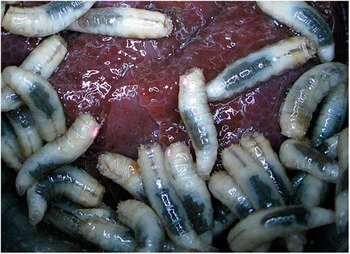
The ability to distinguish individual animals in a population has helped to progress many areas of zoology, and where natural appearance does not facilitate this, artificial marking or tagging can be a great help. Henderson (Reference Henderson2003) defines “tagging” in relation to identifying animals individually, whereas “marking” is an umbrella term which also includes batch marking of cohorts.
A variety of marking methods has been used in population studies of insects (Southwood and Henderson Reference Southwood and Henderson2000; Hagler and Jackson Reference Hagler and Jackson2001), particularly to distinguish adult insects. Such techniques may involve the ingestion of batch-marking media as larvae, including protein markers (Hagler Reference Hagler1997), radio isotopes (Service Reference Service1993), and rubidium (Berry et al. Reference Berry, Stimman and Wolf1972) among others. Many of these have the advantage that the mark persists within the insect across moulting episodes. However, they are not designed to distinguish between individuals of a cohort and detection often requires specialist equipment and sophisticated techniques.
Tagging of insect larvae for its own sake, however, has received less attention and presents additional challenges. Many endopterygote larvae have a thin, translucent integument, which flexes continually and is shed repeatedly as the insect develops. While external marks can be applied to such larvae (e.g., White and Singer Reference White and Singer1987), or prolegs (where present) can be clipped (Weseloh Reference Weseloh1985), these tags will not survive moulting.
Currently, there appears to be no method of tagging insects that can survive moulting and allow discernment of individuals within a cohort. It was decided to attempt to address this using an injected visible implant in an endopterygote insect larva.
Visible Implant Elastomer (VIE), produced by Northwest Marine Technology (NMT) Inc. (Washington, United States of America), has been used for some time to tag marine organisms including fish (e.g., Frederick Reference Frederick1997), shrimp (e.g., Godin et al. Reference Godin, Carr, Hagino, Segura, Sweeney and Blankenship1996), lobster (e.g., Uglem et al. Reference Uglem, Noess, Farestveit and Jorstad1996), and crab (e.g., Davis et al. Reference Davis, Young-Williams, Hines and Zmora2004). More recently, it has been used with squid (e.g., Replinger and Wood Reference Replinger and Wood2007), frogs (e.g., Nauwelaerts et al. Reference Nauwelaerts, Coeck and Aerts2000), salamanders (e.g., Kinkead et al. Reference Kinkead, Lanham and Montanucci2006), snakes (Hutchens et al. Reference Hutchens, Deperno, Matthews, Pollock and Woodward2008), and turtles (Davy et al. Reference Davy, Coombes, Whitear and MacKenzie2010). It comprises a brightly coloured, medical-grade liquid polymer, and curing agent, which when mixed produce a harmless, gelatinous substance after around 24 hours (longer at lower temperatures). The elastomer can be injected hypodermically before curing, and remains visible through integument that is sufficiently translucent. A range of colours of VIE are available, some of which are fluorescent, facilitating observation in different light conditions with the use of an ultraviolet (black) lamp.
Butt and Lowe (Reference Butt and Lowe2007) used VIE for marking earthworms and found that it had only a limited effect on survival, and tags were visible after many months. The method has been successfully extended to a group of terrestrial arthropods – scorpions (Chapin Reference Chapin2011). Based upon these successes, trials on an endopterygote insect were attempted to assess the potential use of the method with insects in general. The results of those trials are presented here.
While a “typical” endopterygote insect does not exist, blow flies (Diptera: Calliphoridae) were chosen based upon certain characteristics. Like all Cyclorrhapha, blow flies lack appendages and have three distinct and discrete larval developmental phases (instars) separated by moulting episodes (Ferrar Reference Ferrar1987). Many calliphorid larvae feed in large intermingling aggregations – “maggot masses” – until reaching a point of satiation during the third instar phase, after which they move away from their food source (Erzinclioglu Reference Erzinclioglu1996), and undergo complete metamorphosis. Such an insect seemed to present a good test of tagging, while having the advantage of being relatively easy to culture. The tagging method specifically was required to: (i) allow a marked individual to be distinguishable in real time from otherwise ostensibly identical individuals in the maggot mass and (ii) allow the tag to be preserved across life stages, so having been retained as the insect moults.
Calliphora vicina Robineau-Desvoidy (a bluebottle fly), was used for all trials reported here, as it is common and relatively large (reaching a maximum larval size of about 18 mm (Greenberg and Kunich Reference Greenberg and Kunich2002)). Adult cultures were established from wild-caught individuals into which porcine liver was introduced as an oviposition medium. The liver was removed and kept at ∼22 °C at 55% relative humidity as the larvae hatched and began to develop.
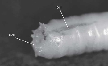
After preliminary trials it was established that first and second instars were too small to inject successfully with VIE as the tip of the syringe needle was too large relative to the larvae. The gauge of the needle (a 29-gauge needle is supplied with the VIE) is limited by the viscosity of the elastomer. Young third instars (∼8 mm long), however, showed more promise particularly at two injection sites (Fig. 1). The first, referred to here as the posterior ventral process (PVP), is the swollen area below the posterior spiracles on the terminal (12th) segment (abdominal segment 8). The second site was in the 11th segment, dorsally in the midline between two tissue masses (D11). These locations can only be approximate as it is difficult to maintain precise control over the tip of the syringe needle at a finer scale so more precise anatomical descriptions are superfluous.
Rear of blow fly larva showing Visible Implant Elastomer injection locations. PVP, posterior ventral process; D11, dorsum of segment 11.

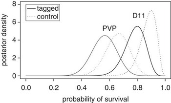
A comparison of mortality of insects injected at the PVP and D11 sites was made using 120 feeding third instars. The larvae were randomly divided into six groups of 20. Ten of each group were injected with VIE at either PVP or D11, with each injection site represented by three of the six groups. Each group was then reared separately on 30 g of fresh porcine liver. The numbers that perished before the adult stage were significantly different for the two locations (generalised linear mixed-effects model: z = 2.82, P = 0.005); as Figure 2 shows, lower mortality resulted from injection into the D11 site. Tagging made no statistically significant difference to mortality (z = 1.28, P = 0.20), though Figure 2 suggests it may have had some effect (odds ratio = 1.7). The mortality of nontagged individuals appears to have been influenced by the mortality rate of the tagged individuals of the same cohort.
Posterior β density plots of the probability of survival for larvae tagged in one of two locations and their untagged cohabitants.

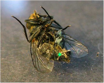
The tags were not readily visible through the cuticle of living adults, necessitating killing and dissection to establish if the tags were retained. Tags were visible in every case in the adult fly's abdomen (Fig. 3) in both male and female insects as a single, intact “blob” in almost all cases. However, in a few cases the tag had become a diffuse constellation of minute globules referred to as “scatter tags” by Butt et al. (Reference Butt, Briones and Lowe2009). These were more difficult to see than an intact tag but were readily detectable under close scrutiny, again facilitated by using fluorescent tags and ultraviolet lighting.
Green Visible Implant Elastomer tag clearly visible in a dissected adult blow fly.

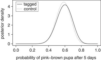
Whether VIE injection affected rate of development was investigated using a further 60 larvae. The larvae were divided randomly into 12 groups of five, and larvae in six of the groups given an injection of VIE at D11. Each group was kept separately on a 30 g piece of fresh porcine liver and allowed to develop. Once a reasonable number of insects had reached an arbitrary pink–brown colour of puparium (mid-way through pupariation), this was used to simply classify insects into two developmental stages. Ignoring dead larvae (five tagged and one untagged; Fisher's exact test, P = 0.19) there was no difference in how these developmental groups were distributed in the tagged and untagged cohorts (z = 0.21, P = 0.83) (see Fig. 4).
Posterior β density plot showing rate of development – measured by numbers reaching an arbitrary stage of pupation – was not affected by tagging.

Injecting the VIE was not straightforward. The small size of the larvae, and their constant movement made locating the syringe needle a challenge. Holding the larva between finger and thumb of one hand against a bench, with its posterior exposed was the most successful method. As the syringe needle pierces the integument, there is a release of fluid in every case. When first observed, this led to speculation that subsequent death was very likely, and it was at first surprising to discover this was not the case.
Individual tagged larvae were discernible within an untagged group, though tags are small, and it takes close inspection rather than a quick glance to pick them out (Fig. 5). However, use of a more contrasting and fluorescent coloured VIE (pink) under ultraviolet light made this somewhat easier.
Visible Implant Elastomer tagged larvae are discernible among untagged individuals. Here, three larvae have been tagged with fluorescent pink elastomer and photographed under an ultraviolet lamp.

Visible Implant Elastomer clearly has potential as a tag for use with insect larvae. Once injected under the integument it is visible and does not appear to affect subsequent development. It does affect mortality but not markedly, which is also what Butt and Lowe (Reference Butt and Lowe2007) found when tagging earthworms, and like those authors it is believed that as the researcher becomes more experienced in the technique rates of mortality in the subjects would decrease. Thus, VIE injection into blow fly larvae satisfies at least partially, those requirements stipulated by Southwood and Henderson (Reference Southwood and Henderson2000). However, there is a size limitation for the subjects tagged by this method, with sizes of much less than about 8 mm likely to prove too difficult.
Acknowledgements
The author thanks Kevin Butt for introducing him to Visible Implant Elastomer and to Michael Wilson who collected some of the preliminary data.