Non-technical Summary
During the Middle Devonian period, around 390 million years ago, the seas of southwestern Gondwana (today’s parts of South America, Antarctica, and South Africa) hosted a unique marine ecosystem, known as the Malvinoxhosan Realm. Many groups of organisms that were common elsewhere, like tiny conodonts, colonial graptolites, or reef-building stromatoporoids, were extremely rare or absent in this region, which makes it difficult for scientists to compare rock layers across these basins. Among the rare groups were ammonoids, ancient relatives of squids and octopuses with coiled shells, previously found in only a few locations in Argentina and Bolivia. Here, we report a new discovery of ammonoids from the northernmost Precordillera Basin in Argentina. Based on 15 preserved fossil specimens, we identified three distinct species: one previously known species from Argentina, Epitornoceras baldisi, and two new species, Trimaenioceras ivanae and Trimaenioceras alegria. Based on evidence from Morocco, these ammonoid genera (Epitornoceras and Trimaenioceras) are only found together worldwide in a specific time interval of the middle Givetian stage. This allows us to date the Argentine fossils with high precision, providing one of the clearest correlations for this period in southwestern Gondwana. This interval corresponds to the upper part of the Taghanic Crisis, a time of environmental changes in the late Middle Devonian. Our findings suggest that ammonoids expanded into the cold-water basins of the Malvinoxhosan Realm during this time interval, showing how rare fossils can provide key insights into the history of ancient oceans.
Introduction
During the Early and Middle Devonian, Argentina was located in the southwestern part of Gondwana (Fig. 1; Cocks and Torsvik, Reference Cocks, Torsvik, Gee and Stephenson2006; Torsvik et al., Reference Torsvik, Cocks, Domeier, Marcilly and Dowding2025), in circumpolar cold water marine settings, recording a high proportion of faunal endemism along with the near-absence of a number of typical Paleozoic groups (mainly conodonts, graptolites and stromatoporoids) (Penn-Clarke and Harper Reference Penn-Clarke and Harper2021, Reference Penn-Clarke and Harper2023). These features characterized the Malvinoxhosan Bioregion (= Malvinokaffric Realm), a major austral paleobiogeographic biochorema (Fig. 1) recognized in South America (except Colombia and Venezuela), Antarctica, and South Africa (Dowding and Ebach, Reference Dowding and Ebach2019; Penn Clarke and Harper, Reference Penn-Clarke and Harper2021, Reference Penn-Clarke and Harper2023; Dowding et al., Reference Dowding, Ebach and Mavrodiev2022).
Simplified Early–Middle Devonian paleogeographic map showing the Malvinoxhosan Realm (red circle), modified from Cocks and Torsvik (Reference Cocks, Torsvik, Gee and Stephenson2006); the white star indicates the location of the Argentine Precordillera. Old Red Sandstone continents in Laurussia and Gondwana are indicated in light yellow; terrane names are labeled (RH: Rheno-Hercynian Terrane).

Figure 1. Long description
A circular global map centered on the Southern Hemisphere.
* Northern Hemisphere: Siberia, Kazakh, Tarim, North China, and Annamia are shown as isolated landmasses in the North. The Equator is marked by a curved line across the upper third of the globe.
* Western Region: The continent of Laurussia is positioned vertically on the left, with Canada labeled in the Northwest. A large light yellow area indicates Old Red Sandstone.
* Central Region: The Rheic Ocean separates Laurussia from Gondwana. Small terranes including R H, Perunica, Armorica, Adria, Pontides, Hellenic, and Moesia are clustered between the major continents.
* Southern Hemisphere: The massive continent of Gondwana dominates the South and East. A large red circle highlights the Malvinoxhosan Realm, which encompasses the South Pole. Within this circle, a white star marks the Precordillera on the Western edge of Gondwana, near Morocco and South Africa.
* Eastern Region: A string of smaller terranes lines the Eastern edge of Gondwana, including South China, Taurides, Apulia, Sanand, Alborz, Lut, Afghan, and Himalayan Terranes.
* Geological Features: Light yellow shading indicates Old Red Sandstone continents. Red dashed lines and black jagged lines represent tectonic boundaries and ridges.
The rarity of biostratigraphic marker groups has historically limited stratigraphic correlations in Malvinoxhosan basins. Ammonoids represent one of these very rare clades, since only a few occurrences have been reported from Argentina (Leanza, Reference Leanza1968; Allaire et al., Reference Allaire, Rustán, Korn, Vaccari, Ezpeleta and Balseiro2024) and Bolivia (Hünicken et al., Reference Hünicken, Kullmann and Suárez-Riglos1980; Babin et al., Reference Babin, Racheboeuf, Le Hérissé and Riglos1991; Troth et al., Reference Troth, Marshall, Racey and Becker2011), in deposits ranging from the early Eifelian to (probably) the early Frasnian (see Allaire et al., Reference Allaire, Rustán, Korn, Vaccari, Ezpeleta and Balseiro2024, for a review). Their scarcity has been interpreted as evidence of low water temperatures, and their occurrences have therefore been regarded as indicators of warm water input and of the breakdown of the biogeographic barriers that sustained Malvinoxhosan endemism, particularly during the Givetian (Babin et al., Reference Babin, Racheboeuf, Le Hérissé and Riglos1991; Troth et al., Reference Troth, Marshall, Racey and Becker2011; Penn-Clarke and Harper, Reference Penn-Clarke and Harper2023). However, the presence of ammonoids in South America is documented as early as the earliest Eifelian, based on the recognition of the genus Mimotornoceras Klug, Reference Klug2002, in the lower part of the Huamampampa Formation in Bolivia (Hünicken et al., Reference Hünicken, Kullmann and Suárez-Riglos1980; Troth et al., Reference Troth, Marshall, Racey and Becker2011).
Recently, a revision led to the recognition of the genus Epitornoceras Frech, Reference Frech1902, in the Middle Devonian of the Chigua Formation in the Argentine Precordillera Basin from San Juan Province (Allaire et al., Reference Allaire, Rustán, Korn, Vaccari, Ezpeleta and Balseiro2024). Epitornoceras is a typical late Givetian ammonoid that is already present in the uppermost middle Givetian of Moroccan sections (Aboussalam and Becker, Reference Aboussalam and Becker2011). Until now, the endemic species Epitornoceras baldisi (Leanza, Reference Leanza1968) constituted the only Devonian ammonoid known in Argentina (Leanza, Reference Leanza1968; Allaire et al., Reference Allaire, Rustán, Korn, Vaccari, Ezpeleta and Balseiro2024).
Here we report an additional ammonoid record coming from sedimentary successions recently recognized in the northernmost Precordillera, La Rioja Province. We present a preliminary mapping and stratigraphic description of these Middle Devonian outcrops in Argentina for the first time. Ammonoid taxa recorded from these successions include Epitornoceras baldisi (Leanza, Reference Leanza1968) and two new species of the genus Trimaenioceras Bockwinkel and Korn, Reference Bockwinkel and Korn2024. The new species are described, and the description of E. baldisi is expanded and refined.
Based on biostratigraphic correlations with well-known Moroccan sections, we strengthen the biostratigraphic significance of the Epitornoceras–Trimaenioceras assemblage, refining the age of the fossil-bearing interval and its implications for recognizing the middle upper Givetian Taghanic Crisis interval. Aside from discussing regional correlations with other stratigraphically equivalent units, we provide some biogeographic remarks. In particular, we critically evaluate the hypothesis that these ammonoids are indicators of warm-water conditions and constitute evidence for the demise of the Malvinoxhosan Realm during the Middle Devonian in southwestern Gondwana.
Locality and geological setting
The study area is located in the Sierra de Las Minitas, La Rioja Province, northwestern Argentina (Fig. 2), which corresponds to the northernmost extent of the Precordillera Basin (Caminos et al., Reference Caminos, Fauque, Cingolani, Varela and Morel1993). The middle to upper Paleozoic marine strata there exhibit intense folding and faulting and are intruded by Upper Devonian to Mississippian igneous dikes (Ar–Ar ages ca. 346–364 Ma; Coughlin, Reference Coughlin2000). Evidence of Paleozoic deformation events is also seen in the stratigraphic truncations and angular unconformities separating the successions (Coughlin, Reference Coughlin2000; Astini et al., Reference Astini, Dávila, Lopez-Gamundi, Gomez, Collo, Ezpeleta, Martina, Ortiz, Chebli, Cortiñas and Spalletti2005; Ezpeleta, Reference Ezpeleta2009).
Locality information. (1) Geological map showing the studied area (red square) in the Sierra de las Minitas, Precordillera of La Rioja Province, Argentina. (2) Satellite image showing the location of the ammonoid sites. (3) Regional map showing the location of the studied area (red square) within La Rioja Province. (4) Map of South America showing the location of the studied locality (red square) within Argentina.

Figure 2. Long description
Panel 1 is a geological map of Sierra de las Minitas. A red square in the center-left highlights the study area containing blue star icons for ammonoid sites. The map includes geological formations such as Mulas Muertas Fm (Permian), Rio del Peñon Fm (Pennsylvanian), and various Mississippian and Devonian units. Structural features like faults and folds are marked. A scale bar indicates 0 to 5 kilometers. Panel 2 is a satellite image of the rugged terrain within the red square, pinpointing specific sites labeled S M 23-6 B, S M 23-16, S M 23-18, S M 23-25, S M 23-36, S M 23-37, S M 23-38, and S M 23-49. Panel 3 is a regional map of western Argentina showing La Rioja, San Juan, and Mendoza provinces, with a red square in northern La Rioja. Panel 4 is a map of South America with a red square indicating the study location within western Argentina.
Extensive deformation of the Devonian and Carboniferous successions in the Sierra de Las Minitas, along with the scarcity and scattered nature of paleontological findings, has historically led to the use of multiple lithostratigraphic schemes (e.g., Fauqué and Limarino, Reference Fauqué and Limarino1991; Caminos et al., Reference Caminos, Fauque, Cingolani, Varela and Morel1993; Azcuy and Carrizo, Reference Azcuy and Carrizo1995; Carrizo and Azcuy, Reference Carrizo and Azcuy1995; Coughlin, Reference Coughlin2000; Limarino et al., Reference Limarino, Tripaldi, Marenssi and Fauqué2006). Figure 2 summarizes the various lithostratigraphic schemes previously applied at both local and regional scales.
Devonian units recognized in the Sierra de Las Minitas include the Lower Devonian Talacasto Formation and the Middle Devonian Chigua Formation (Rustán et al., Reference Rustán, Vaccari and Astini2011; Carrizo and Azcuy, Reference Carrizo and Azcuy2015), which typically crop out farther to the south, in the San Juan Province. The Talacasto Formation has been recognized mainly based on Pragian trilobites, but in a restricted small area with no stratigraphic continuity with the overlying Chigua Formation due to faulting (Rustán et al., Reference Rustán, Vaccari and Astini2011; Holloway and Rustán, Reference Holloway and Rustán2012; Rustán and Peralta, Reference Rustán and Peralta2024).
The sedimentary succession of the Chigua Formation studied herein exceeds 300 meters in thickness. The base of the unit is difficult to recognize because of faulting and folding. The upper boundary is marked by a sharp angular unconformity, which is overlaid by the conglomeratic base of the Mississippian Agua de Lucho Formation. In the homonymous type locality of the Chigua Formation (San Juan Province, to the South), this same stratigraphic contact has been recognized and interpreted to be linked to the Chanic Orogeny (see Astini et al., Reference Astini, Ramos, Benedetto, Vaccari and Cañas1996, and Ramos, Reference Ramos2004, for a collisional model or Dahlquist et al., Reference Dahlquist, Morales Cámera, Alasino, Pankhurst, Basei, Rapela, Moreno, Baldo and Galindo2021, for a subduction-related tectonic setting).
In the studied locality, the upper part of the Chigua Formation consists of a dominantly dark greenish fine-grained succession characterized by interbedded, variably bioturbated, and fossiliferous nodular mudstones (containing most of the ammonoid specimens), black carbonaceous shales, ripple cross-laminated siltstones, and localized lenses of polymictic conglomerates (Fig. 3). The depositional environment is interpreted as an outer-shelf setting dominated by suspension settling and intermittent turbidity currents, with localized scour features and the accumulation of coarse-grained sediments within wide, shallow channels. These occasional conglomeratic lenses suggest episodes of high-energy conditions related to unidirectional tractive currents. Some soft-sediment deformation structures are interpreted as the result of overlying mass-transport processes.
Stratigraphic column showing the positions of the fossil sites and the distribution of ammonoid species.

Figure 3. Long description
A vertical stratigraphic column with a Y-axis measuring height in meters from 0 to 350.
From 0 to 100 meters, the column consists of alternating green or gray mudstones and conglomerate layers with channels and intraclasts. Fossil site S M 23-6 at the base contains Punillaspis argentina and Epitornoceras baldisi. Site S M 23-25 at 95 meters contains Trimaenioceras ivanae n. sp.
From 100 to 130 meters, a thick conglomerate layer is present.
From 130 to 330 meters, the Chigua Formation (Givetian) is dominated by black mudstones with frequent parallel lamination, ripples, and bioturbation. Fossil sites S M 23-16 and S M 23-18 at approximately 140 meters contain Punillaspis argentina, Epitornoceras baldisi, and Trimaenioceras ivanae n. sp. Near the top of this section, sites S M 23-36, S M 23-37/38, and S M 23-49 show the presence of Epitornoceras baldisi and Trimaenioceras alegria n. sp.
At 330 meters, an angular unconformity greater than 60 degrees separates the Chigua Formation from the overlying Agua de Lucho Formation (Tournaisian), which begins with a conglomerate layer.
A References box on the right defines symbols:
- Lithology: Green/gray mudstones, Black mudstones, S m plus S l, and C g l plus D m.
- Structures: Bioturbation, Ripples, Convolute lamination, Intraclasts, Lense lamination, Parallel lamination, and Channels.
- Species: Blue spiral for Epitornoceras baldisi, green spiral for Trimaenioceras ivanae n. sp., red spiral for Trimaenioceras alegria n. sp., and a trilobite icon for Punillaspis argentina.
Material and methods
The material described here consists of 15 ammonoid specimens coming from eight different stratigraphic sampling levels from the Chigua Formation at the Sierra de Las Minitas (see Table 1 for GPS coordinates, repository information, and taxonomic determination). All specimens, numbered PULR-I-161 to PULR-I-174, are housed in the collection of the Museo de Antropología y Ciencias Naturales of the Universidad Nacional de La Rioja (Argentina). Fossil remains are preserved in slightly carbonaceous, dark-gray to black nodules within dark greenish fine-grained intervals. Associated biota include trilobites, conulariids, gastropods, bivalves, orthoconic cephalopods, corals, hyoliths, fishes, and lycopsids.
Material inventory, taxonomic identification, and GPS coordinates of the fossil localities

Table 1. Long description
The table contains four columns: Specimen number, Identification, Locality, and G P S coordinates.
* P U L R dash I dash 161: Epitornoceras baldisi, Locality S M 23 dash 6 B, G P S 28 degrees 44 minutes 49.36 seconds S, 68 degrees 42 minutes 32.47 seconds W.
* P U L R dash I dash 162: Epitornoceras baldisi?, Locality S M 23 dash 6 B, G P S 28 degrees 44 minutes 49.36 seconds S, 68 degrees 42 minutes 32.47 seconds W.
* P U L R dash I dash 163: Trimaenioceras ivanae n. sp., Locality S M 23 dash 16, G P S 28 degrees 45 minutes 3.8 seconds S, 68 degrees 42 minutes 38.3 seconds W.
* P U L R dash I dash 164: Epitornoceras baldisi, Locality S M 23 dash 18, G P S 28 degrees 45 minutes 5 seconds S, 68 degrees 42 minutes 40.8 seconds W.
* P U L R dash I dash 165: Trimaenioceras ivanae n. sp., Locality S M 23 dash 25, G P S 28 degrees 45 minutes 19.44 seconds S, 68 degrees 42 minutes 21.6 seconds W.
* P U L R dash I dash 166: Trimaenioceras?, Locality S M 23 dash 25, G P S 28 degrees 45 minutes 19.44 seconds S, 68 degrees 42 minutes 21.6 seconds W.
* P U L R dash I dash 167: Ammonoid indet., Locality S M 23 dash 25, G P S 28 degrees 45 minutes 19.44 seconds S, 68 degrees 42 minutes 21.6 seconds W.
* P U L R dash I dash 168: Epitornoceras baldisi, Locality S M 23 dash 36, G P S 28 degrees 45 minutes 11.9 seconds S, 68 degrees 43 minutes 2.57 seconds W.
* P U L R dash I dash 169: Trimaenioceras alegria n. sp., Locality S M 23 dash 36, G P S 28 degrees 45 minutes 11.9 seconds S, 68 degrees 43 minutes 2.57 seconds W.
* P U L R dash I dash 170: Ammonoid indet., Locality S M 23 dash 36, G P S 28 degrees 45 minutes 11.9 seconds S, 68 degrees 43 minutes 2.57 seconds W.
* P U L R dash I dash 171 dash 1: Epitornoceras baldisi, Locality S M 23 dash 37, G P S 28 degrees 45 minutes 14.50548 seconds S, 68 degrees 43 minutes 0.47424 seconds W.
* P U L R dash I dash 171 dash 2: Epitornoceras baldisi, Locality S M 23 dash 37, G P S 28 degrees 45 minutes 14.50548 seconds S, 68 degrees 43 minutes 0.47424 seconds W.
* P U L R dash I dash 172: Epitornoceras baldisi, Locality S M 23 dash 38, G P S 28 degrees 45 minutes 13.30164 seconds S, 68 degrees 43 minutes 3.88452 seconds W.
* P U L R dash I dash 173: Epitornoceras baldisi, Locality S M 23 dash 49, G P S 28 degrees 46 minutes 3.86076 seconds S, 68 degrees 43 minutes 7.6296 seconds W.
* P U L R dash I dash 174: Epitornoceras baldisi?, Locality S M 23 dash 49, G P S 28 degrees 46 minutes 3.86076 seconds S, 68 degrees 43 minutes 7.6296 seconds W.
Fossils were prepared using pneumatic jacks and needles. Latex casts of external molds and natural internal molds were inked and whitened with ammonium chloride (NH4Cl) sublimate for taking pictures under raking light using a Canon Power Shot S50 digital camera mounted on a Leica MZ75 binocular microscope. The suture lines were drawn under a stereomicroscope with a camera lucida.
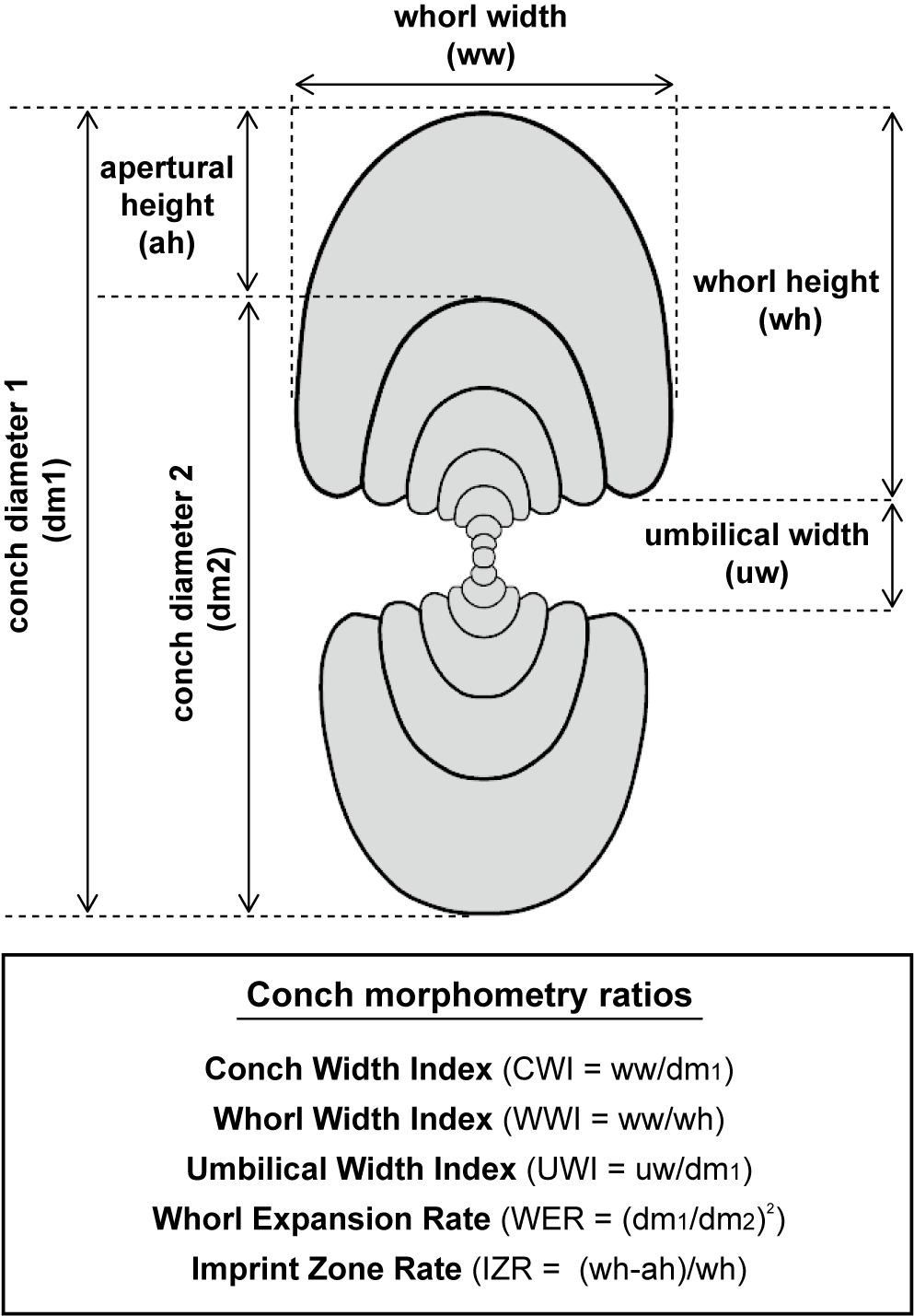
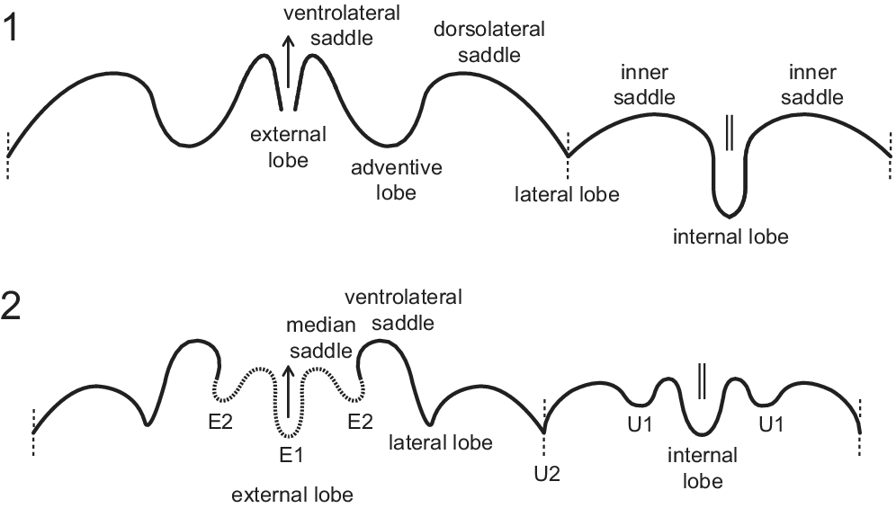
Description of the material follows the scheme for Paleozoic ammonoids outlined by Korn (Reference Korn2010) and Klug et al. (Reference Klug, Korn, Landman, Tanabe, De Baets, Naglik, Klug, Korn, De Baets, Kruta and Mapes2015) (see Fig. 4 for conch measurements and morphometry ratios, and Fig. 5 for suture line terminology). Unfortunately, due to the limited ontogenetic data available, fully detailed descriptions remain incomplete.
Measurements and conch morphometry ratios.

Figure 4. Long description
The diagram shows a cross-section of a coiled shell with concentric whorls. Dimensional arrows define the following parameters. At the top, whorl width w w is the horizontal span of the outermost whorl. On the left side, conch diameter 1 d m 1 spans the total vertical height, while conch diameter 2 d m 2 spans from the bottom to the top of the penultimate whorl. Apertural height a h is the distance from the top of the penultimate whorl to the top of the outermost whorl. On the right side, whorl height w h is the vertical span of the outermost whorl, and umbilical width u w is the vertical gap in the center between the whorl sections.
A box at the bottom titled Conch morphometry ratios lists five formulas.
* Conch Width Index C W I equals w w forward slash d m 1.
* Whorl Width Index W W I equals w w forward slash w h.
* Umbilical Width Index U W I equals u w forward slash d m 1.
* Whorl Expansion Rate W E R equals open parenthesis d m 1 forward slash d m 2 close parenthesis squared.
* Imprint Zone Rate I Z R equals open parenthesis w h minus a h close parenthesis forward slash w h.
Suture line terminology. (1) Tornoceratid ammonoid (Epitornoceras), suture formula: E A L I. (2) Maenioceratid ammonoid (Trimaenioceras), suture formula: (E2 E1 E2) L U2 U1 I.

Figure 5. Long description
The diagram consists of two panels labeled 1 and 2.
Panel 1 shows the suture line of a Tornoceratid ammonoid (Epitornoceras). From left to right, the undulating line features a broad curve leading to a sharp downward external lobe with an upward arrow. This is flanked by a ventrolateral saddle. Moving right, there is a deep adventive lobe, followed by a dorsolateral saddle. A dashed vertical line separates this from the lateral lobe. To the right of the lateral lobe is an inner saddle, followed by a deep, rounded internal lobe marked with a double vertical line, and another inner saddle.
Panel 2 shows the suture line of a Maenioceratid ammonoid (Trimaenioceras). The line is more complex. From left to right, it begins with a curve leading to a tripartite external lobe. This section includes a central E 1 lobe with an upward arrow and a median saddle, flanked by two E 2 lobes shown with dotted lines. To the right is a ventrolateral saddle, followed by a lateral lobe. A dashed vertical line marks the transition to the umbilical lobes, labeled U 2 and U 1. At the far right is the internal lobe, marked with a double vertical line, flanked by U 1 lobes on either side.
Abbreviations
dm = conch diameter; ww = whorl width; wh = whorl height; uw = umbilical width; ah = apertural height; CWI = conch width index (ww/dm); WWI = whorl width index (ww/wh); UWI = umbilical width index (uw/dm); WER = whorl expansion rate (dm1/dm2)2; IZR = imprint zone rate (wh-ah/wh); E lobe = external lobe; A lobe = adventive lobe; L lobe = lateral lobe; U lobe = umbilical lobe; I lobe = internal lobe.
Repository and institutional abbreviation
All types and other specimens examined in this study are deposited in the Museo de Antropología y Ciencias Naturales of the Universidad Nacional de La Rioja (Argentina), under the catalogue numbers PULR-I-161 to PULR-I-174.
Systematic paleontology
Class Cephalopoda Cuvier, Reference Cuvier1795
Subclass Coleoidea Bather, Reference Bather1888
Superorder Ammonoida Haeckel, Reference Haeckel1866
Order Goniatitida Hyatt, Reference Hyatt1884
Suborder Tornoceratina Wedekind, Reference Wedekind1914
Superfamily Tornoceratoidea von Arthaber, Reference von Arthaber1911
Family Tornoceratidae Hyatt, Reference Hyatt1884
Subfamily Tornoceratinae von Arthaber, Reference von Arthaber1911
Genus Epitornoceras Frech, Reference Frech1902
Type species
Tornoceras (Tornoceras) mithracoides Frech, Reference Frech1888, by original designation. Rhenish Mountains, upper Givetian (Pseudoprobeloceras pernai Zone). For illustrations and synonymy, see Bockwinkel et al. (Reference Bockwinkel, Becker and Ebbighausen2013), Bockwinkel and Korn (Reference Bockwinkel and Korn2015), and Korn and Bockwinkel (Reference Korn and Bockwinkel2022).
Included species
Epitornoceras mithracoides (Frech, Reference Frech1888), Germany; Epitornoceras peracutum (Hall, Reference Hall1876; holotype described by Miller, Reference Miller1938), North America; Epitornoceras baldisi (Leanza, Reference Leanza1968), Argentina; Epitornoceras dennisoni Kirchgasser, Baird, and Brett, Reference Kirchgasser, Baird, Brett, Avary, Hasson and Diecchio2020, North America; Epitornoceras transmediterraneum Korn and Bockwinkel, Reference Korn and Bockwinkel2022, Morocco. For a comparison of the morphological characters and ontogenetic trajectories of the different species of the genus Epitornoceras, see Appendix Figure A1 (and table 1 in Allaire et al., Reference Allaire, Rustán, Korn, Vaccari, Ezpeleta and Balseiro2024).
Diagnosis (emended by Korn and Bockwinkel, Reference Korn and Bockwinkel2022, p. 40)
Genus of the subfamily Tornoceratinae with compressed conch, umbilicus closed since the pre-adult stage but slightly opened in the juvenile stage. Usually without ventrolateral grooves. Ornament with biconvex growth lines, without ribs. Sutural formula E A L I; adventive lobe broadly rounded, asymmetric with steep dorsal flank; ventrolateral saddle narrow, subacute.
Occurrence
Epitornoceras has been reported in Germany, Morocco, North America (New York), and Argentina, in deposits ranging from the latest middle Givetian to the early Frasnian (House, Reference House1965, Reference House1978; Becker and House, Reference Becker and House2000; Bockwinkel et al., Reference Bockwinkel, Becker and Ebbighausen2009, Reference Bockwinkel, Becker and Ebbighausen2013, Reference Bockwinkel, Becker and Ebbighausen2015, Reference Bockwinkel, Becker and Aboussalam2017; Aboussalam and Becker, Reference Aboussalam and Becker2011; Becker et al., Reference Becker, Aboussalam, Hartenfels, El Hassani and Fischer2013; Aboussalam et al., Reference Aboussalam, Becker, Mayer, Gibb, Hüneke, Hartenfels, Becker, El Hassani and Lüddecke2018; Kirchgasser et al., Reference Kirchgasser, Baird, Brett, Avary, Hasson and Diecchio2020; Korn and Bockwinkel, Reference Korn and Bockwinkel2022; Allaire et al., Reference Allaire, Rustán, Korn, Vaccari, Ezpeleta and Balseiro2024).
Epitornoceras baldisi (Leanza, Reference Leanza1968)
Epitornoceras baldisi (Leanza, Reference Leanza1968). (1) Specimen PULR-I-173. (2, 3) Specimen PULR-I-168. (4, 5) Specimen PULR-I-172. (6–8) Specimen PULR-I-161. (9, 10) Suture lines of PULR-I-168, at wh = 11.0 mm (9), and at wh = 10.4 mm (10). (11) Suture line of PULR-I-173 at wh = 10.2 mm. (12, 13) Suture line of PULR-I-172, external part at wh = 7.9 mm (12), internal part at wh = 7.9 mm plus one whorl (13). (14) Suture line of PULR-I-171-2 at wh = 5.0 mm, showing the external and internal parts. (15–17) Specimen PULR-I-164. (18, 19) Latex cast of the inner whorls of PULR-I-164. (20) Specimen PULR-I-171-2. Scale bar units = 1 mm.

Figure 6. Long description
The plate consists of 20 numbered panels.
Panels 1 through 8, 15 through 17, and 20 are black and white photographs of Epitornoceras baldisi fossil specimens.
- Panel 1 shows a large lateral view of specimen P U L R I 173.
- Panels 2 and 3 show lateral and ventral views of specimen P U L R I 168, highlighting the curved suture lines on the shell surface.
- Panels 4 and 5 show specimen P U L R I 172 in frontal and oblique views.
- Panels 6, 7, and 8 show specimen P U L R I 161 in lateral, ventral, and apertural views.
- Panels 15, 16, and 17 show specimen P U L R I 164 in lateral, ventral, and apertural views.
- Panel 20 shows specimen P U L R I 171 2.
Panels 9 through 14 are line drawings of suture lines, which are the complex wavy lines where the internal septa meet the outer shell.
- Diagrams 9 and 10 represent specimen P U L R I 168 at whorl heights of 11.0 mm and 10.4 mm respectively.
- Diagram 11 represents specimen P U L R I 173 at whorl height 10.2 mm.
- Diagrams 12 and 13 show the external and internal suture parts of specimen P U L R I 172 at whorl height 7.9 mm.
- Diagram 14 shows the full suture of specimen P U L R I 171 2 at whorl height 5.0 mm.
- Each diagram includes a vertical arrow indicating the direction of the aperture and a small black and white scale bar.
Panels 18 and 19 show a latex cast of the inner whorls of specimen P U L R I 164 in lateral and ventral views.
All fossil photographs include a vertical black and white segmented scale bar for size reference.
Conch measurements (in mm) and ratios of Epitornoceras baldisi (Leanza, Reference Leanza1968); *values marked with an asterisk are estimated or based on estimates.

Table 2. Long description
The table is divided into two main sections based on specimen origin.
Section 1: Epitornoceras baldisi from the Sierra de Las Minitas (La Rioja Province).
Columns include Specimen, d m 1, d m 2, u w, w w, w h, a h, C W I, U W I, W E R, W W I, and I Z R.
- P U L R dash I dash 161: d m 1 24.0, d m 2 16.8, u w 0, w w 9.2, w h 14.0, a h 7.2, C W I 0.38, U W I 0.00, W E R 2.04, W W I 0.66, I Z R 0.49.
- P U L R dash I dash 173: d m 1 19.7, u w 0, w w 6.6 asterisk, w h 11.5, C W I 0.34 asterisk, U W I 0.00, W W I 0.57 asterisk.
- P U L R dash I dash 168: d m 1 18.5, u w 0, w w 6.1 asterisk, w h 11.2, C W I 0.33 asterisk, U W I 0.00, W W I 0.54 asterisk.
- P U L R dash I dash 164 (three growth stages): d m 1 values of 18.4, 12.5, and 7.5. At the largest stage (18.4), w w is 8.3, w h is 11.4, and C W I is 0.45.
Section 2: Epitornoceras baldisi from the Chigua Formation (San Juan Province).
- I N G E O dash P I dash 2004: d m 1 50.0, u w 0, w w 12.0 asterisk, w h 35.0, C W I 0.24 asterisk, U W I 0.00, W W I 0.34 asterisk.
- I N G E O dash P I dash 2011: d m 1 30.0, u w 0, w w 7.5 asterisk, w h 19.0 asterisk, C W I 0.25 asterisk, U W I 0.00, W W I 0.39 asterisk.
- I N G E O dash P I dash 2003 (three growth stages): d m 1 values of 20.0, 13.0 asterisk, and 9.0 asterisk. At the 20.0 stage, w w is 6.0 asterisk, w h is 12.0, and C W I is 0.30 asterisk.
Asterisks denote estimated values.
Reference Leanza1968 Tornoceras baldisi Leanza, figs. 1–4.
non Reference House1978 Tornoceras (Tornoceras) cf. baldisi; House, pl. 6, figs. 9, 10.
Reference Allaire, Rustán, Korn, Vaccari, Ezpeleta and Balseiro2024 Epitornoceras baldisi; Allaire et al., figs. 6, 7.
Holotype
Specimen CPUBA 8088 designated by Leanza (Reference Leanza1968, figs. 1–4), stored at the University of Buenos Aires, Argentina (re-illustrated in Allaire et al., Reference Allaire, Rustán, Korn, Vaccari, Ezpeleta and Balseiro2024, fig. 6). Type locality and horizon: uppermost part of the Chavela Member, uppermost part of the Chigua Formation, Chinguillos Group, north of Angualasto town, San Juan Province, Argentina (see Allaire et al., Reference Allaire, Rustán, Korn, Vaccari, Ezpeleta and Balseiro2024, for further details).
Material
Nine specimens are housed in the collection of the Museo de Antropología y Ciencias Naturales (Universidad Nacional de La Rioja): PULR-I-161, PULR-I-162, PULR-I-164, PULR-I-168, PULR-I-171-1, PULR-I-171-2, PULR-I-172, PULR-I-173, and PULR-I-174. They all come from the Sierra de Las Minitas, La Rioja Province, Argentina (Figs. 2, 3) (see Table 1 for GPS coordinates of the fossil localities).
Diagnosis (emended from Allaire et al., Reference Allaire, Rustán, Korn, Vaccari, Ezpeleta and Balseiro2024, p. 334)
Species of Epitornoceras reaching at least 50 mm in conch diameter. Extremely discoidal and involute adult stage, with strongly compressed whorl profile (at 30 mm dm, CWI ~0.25, WWI ~0.40, and UWI = 0.00). Thinly discoidal and involute pre-adult stage, with weakly compressed whorl profile (at around 18 mm dm, CWI ~0.35–0.45, WWI = 0.55–0.70, umbilicus very narrow or already closed). Flanks weakly convex, convergent, umbilical margin continuously rounded. Without any ventrolateral grooves. Suture line with broadly rounded, nearly symmetric adventive lobe, wider than deep, with convergent flanks.
Occurrence
Epitornoceras baldisi is known only from the Argentine Precordillera, where it occurs in Givetian successions (Middle Devonian), in its type locality (San Juan Province), and in the Sierra de Las Minitas (La Rioja Province, this study).
Description
Specimens PULR-I-173 (19.7 mm dm; Fig. 6.1), PULR-I-168 (18.5 mm dm; Fig. 6.2, 6.3), PULR-I-161 (24.0 mm dm; Fig. 6.6–6.8), and PULR-I-164 (18.4 mm dm; Fig. 6.15–6.17) can be considered pre-adults, as E. baldisi can reach up 50 mm in diameter (Allaire et al., Reference Allaire, Rustán, Korn, Vaccari, Ezpeleta and Balseiro2024); they all show a thinly discoidal and involute conch with a completely closed umbilicus (CWI = 0.35–0.45; UWI = 0.00), and a weakly compressed whorl profile (WWI = 0.55–0.70). Between the two specimens for which these parameters could be measured (i.e., PULR-I-161 and PULR-I-164), the whorl expansion rate is high (WER = 2.05–2.20) and the degree of whorl overlap is very strongly embracing (IZR = 0.50). Specimens PULR-I-168 and PULR-I-173 exhibit well-preserved suture lines, displaying the typical shape of E. baldisi (Fig. 6.9–6.11), consisting of a narrow and deep external lobe, a narrow and subacute E/A saddle, a broadly rounded, wide and deep nearly symmetric adventive lobe with convergent flanks, followed by a similarly broadly rounded and deep but wider saddle.
Specimen PULR-I-172 is a conch fragment with an estimated diameter of about 15 mm (Fig. 6.4, 6.5). Several septa are preserved, and both the external and internal parts of the suture line are visible. The external part (i.e., ventral side of the whorl) exhibits the characteristic shape of the species (Fig. 6.12). The internal part (i.e., dorsal side of the whorl), described here for the first time in the genus, shows a very deep and relatively narrow internal lobe, slightly pointed with subparallel flanks, followed by a very wide, shallow, and broadly rounded saddle that reaches the umbilicus (Fig. 6.13).
The morphology of the inner whorls of specimen PULR-I-164 (Fig. 6.15–6.17) was studied using a latex cast, which revealed a thickly discoidal (almost pachyconic) and involute conch of 7.5 mm dm (CWI = 0.57), with a weakly compressed whorl profile (WWI = 0.88); the umbilicus appears completely closed (Fig. 6.18, 6.19).
Specimen PULR-I-171-2 is a fragment of a small conch with an estimated diameter of about 7.5 mm (Fig. 6.20). Although CWI cannot be measured, the conch appears slightly stouter than the latex cast of the inner whorls of specimen PULR-I-164, which has a comparable diameter, suggesting it is most likely thinly pachyconic. The conch is involute; the umbilicus was probably extremely narrow or even entirely closed. Both outer and inner parts of the suture lines are visible; the external lobe cannot be observed, but the other sutural elements form the distinctive sutural morphology of Epitornoceras (Fig. 6.14). At this early stage, the internal lobe is considerably shallower than in specimen PULR-I-172 (compare Fig.6.13, 6.14).
Remarks
The newly collected specimens of Epitornoceras baldisi reveal intraspecific variation in conch compression, with some specimens exhibiting a stouter conch at equivalent growth stages (e.g., at around 18.5 mm dm, CWI = 0.45 in PULR-I-164, while CWI ~0.35 in PULR-I-168). Additionally, specimens PULR-I-164 and PULR-I-171-2 reveal a thickly discoidal, even thinly pachyconic, juvenile morphology (around 7.5–12.5 mm dm, CWI ~0.55–0.60), whereas the only other specimen of comparable size (from the species type locality, Precordillera of San Juan Province) shows a more thinly discoidal conch at a similar growth stage (around 10–15 mm dm, CWI ~0.35–0.40; Allaire et al., Reference Allaire, Rustán, Korn, Vaccari, Ezpeleta and Balseiro2024), see Table 2 and Reference Baldis and LongobuccoAppendix Figure A1 to visualize the intraspecific variation observed in E. baldisi.
The new material described here from the Sierra de Las Minitas (Precordillera of La Rioja) consists exclusively of pre-adult or juvenile specimens, with the largest individual measuring 24 mm in diameter (the lithology may have played a role in the size distributions of the specimens between the two localities; see De Baets et al., Reference De Baets, Jarochowska, Buchwald, Klug and Korn2022, for an analysis of the factors controlling the size distributions in ammonoids). This prevents assessment of whether such variation in compression also exists at the adult stage, or if the mature morphology consistently tends to be extremely discoidal. This kind of variation in conch compression corresponds to intraspecific (i.e., phenotypic) variability, which has also been observed within some other taxa (De Baets et al., Reference De Baets, Bert, Hoffmann, Monnet, Yacobucci, Klug, Klug, Korn, De Baets, Kruta and Mapes2015); for example, see how variable the CWI is in E. transmediterraneum (Reference Baldis and LongobuccoAppendix Figure A1).
Based on the new material described herein, the species diagnosis was updated to better reflect morphological changes throughout ontogeny. It now includes the morphological characteristics of the conch at its intermediate growth stages.
Order Agoniatitida Ruzhencev, Reference Ruzhencev1957
Suborder Pharciceratina Korn, Reference Korn1998
Superfamily Pharciceratoidea Hyatt, Reference Hyatt, Von Zittel and Eastman1900
Family Maenioceratidae Bogoslovsky, Reference Bogoslovsky1958
Genus Trimaenioceras Bockwinkel and Korn, Reference Bockwinkel and Korn2024
Type species
Trimaenioceras klugi Bockwinkel and Korn, Reference Bockwinkel and Korn2024. Anti-Atlas of Morocco, middle Givetian (Afromaenioceras sulcatostriatum to Pharciceras aff. P. amplexum zones).
Included species
Trimaenioceras klugi Bockwinkel and Korn, Reference Bockwinkel and Korn2024, Morocco; Trimaenioceras eculeus Bockwinkel and Korn, Reference Bockwinkel and Korn2024, Morocco; Trimaenioceras fuscina Bockwinkel and Korn, Reference Bockwinkel and Korn2024, Morocco; Trimaenioceras paucum Bockwinkel and Korn, Reference Bockwinkel and Korn2024, Morocco; Trimaenioceras alegria n. sp. and Trimaenioceras ivanae n. sp., Argentina (this study). For a comparison of the morphological characters and ontogenetic trajectories of the different species of the genus Trimaenioceras, see Reference Baldis and SarudianskyAppendix Figure A2.
Diagnosis (emended from Bockwinkel and Korn, Reference Bockwinkel and Korn2024, p. 25)
Genus of the family Maenioceratidae with or without ventrolateral grooves, shell without constrictions or internal thickenings. Sutural formula (E2 E1 E2) L U2 U1 I; external lobe wide to extremely wide; E2 lobe deep, cuneiform; ventrolateral saddle symmetrically rounded; lateral lobe V-shaped, angular.
Occurrence
Trimaenioceras occurs in the Anti-Atlas of Morocco (Becker et al., Reference Becker, Aboussalam, Bockwinkel, Ebbighausen, El Hassani and Nübel2004; Aboussalam and Becker, Reference Aboussalam and Becker2011; Bockwinkel and Korn, Reference Bockwinkel and Korn2024) and in the Argentine Precordillera (this study), in middle Givetian successions.
Remarks
Trimaenioceras mainly occurs in the Afromaenioceras zones (Bockwinkel and Korn, Reference Bockwinkel and Korn2024), however, the species T. klugi (= Maenioceras sp. II in Becker et al., Reference Becker, Aboussalam, Bockwinkel, Ebbighausen, El Hassani and Nübel2004, and Aboussalam and Becker, Reference Aboussalam and Becker2011) appears to reach into the subsequent and youngest Pharciceras aff. P. amplexum Zone (uppermost middle Givetian Moroccan ammonoid zone; see table 2 in Becker et al., Reference Becker, Aboussalam, Bockwinkel, Ebbighausen, El Hassani and Nübel2004; Pharciceras aff. P. amplexum zone = Unit MD III-A, sensu Becker et al., Reference Becker, Aboussalam, Bockwinkel, Ebbighausen, El Hassani and Nübel2004).
The diagnosis here is slightly modified from the original version provided by Bockwinkel and Korn (Reference Bockwinkel and Korn2024): “External lobe very wide or extremely wide” is replaced by “External lobe wide to extremely wide”, to better match the descriptions of the different species assigned to the genus (see Bockwinkel and Korn, Reference Bockwinkel and Korn2024, and this study). Trimaenioceras eculeus, T. fuscina, and T. paucum possess a wide E lobe, representing 19–20% of the total width of the mature suture (see Bockwinkel and Korn, Reference Bockwinkel and Korn2024, figs. 14D, E, 15C, 16C). Trimaenioceras klugi (the type species) and T. alegria n. sp. exhibit a very wide E lobe, representing 23–27% of the total width of the mature suture (see Bockwinkel and Korn, Reference Bockwinkel and Korn2024, fig. 13E, F; Fig. 7.12, this study). Finally, T. ivanae n. sp. is characterized by an extremely wide E lobe, representing around 40% of the total width of the mature suture (see Fig. 8.12, this study).
Trimaenioceras alegria new species, holotype PULR-I-169. (1–3) Part and (7, 8) counterpart of the reconstructed specimen. (4–6, 9, 10, 16) Details of the specimen before its reconstruction, some inner whorls were still visible. (11, 15) Magnified images showing the trident-shaped external lobe characteristic of the genus Trimaenioceras, adult stage (11), juvenile stage (15). (12–14) Suture line at wh = 16.2 mm (12), at wh = 8.0 mm (13), and at wh = 6.5 mm (14). Scale bar units = 1 mm.

Figure 7. Long description
The plate consists of sixteen panels.
* Panels 1 through 3: Photos of the reconstructed holotype specimen P U L R dash I dash 1 6 9 from different angles, showing a coiled shell with complex internal structures.
* Panels 4 through 6, 9, 10, and 16: Detailed views of the specimen before reconstruction, highlighting inner whorls and the jagged, tooth-like edges of the septa.
* Panels 7 and 8: Counterpart views of the reconstructed specimen showing the negative impression of the shell.
* Panels 11 and 15: High-magnification photographs of the trident-shaped external lobe. Panel 11 shows the adult stage with sharp, pointed lobes, while panel 15 shows the juvenile stage with a similar but smaller trident structure.
* Panels 12, 13, and 14: Black line drawings of suture lines. Each drawing features a central deep, trident-shaped trough with an upward-pointing arrow at the midline. Panel 12 represents the suture at a whorl height of 16.2 mm, panel 13 at 8.0 mm, and panel 14 at 6.5 mm. The complexity of the undulating line decreases from panel 12 to 14.
* Scale bars: Each photographic panel includes a vertical black-and-white segmented scale bar, and each line drawing includes a horizontal scale bar.
Trimaenioceras ivanae new species. (1–7) Holotype PULR-I-165; after reconstruction, the holotype PULR-I-165 consists of three parts, respectively corresponding to pictures (1–3), (4) and (6, 7) (they form the full specimen once put all together, corresponding to an internal mold of conch preserved within a nodule). (5, 11) Latex cast of the holotype revealing the shape of the umbilicus. (8–10, 15, 16) Details of some elements of the holotype before reconstruction. (12–14) Suture line of the holotype, at wh = 8.5 mm (12), at wh = 5.0 mm (13), and at wh = 4.5 mm (14). (17, 18) Additional specimen PULR-I-163, composed of two parts; part (17) and latex cast of the counterpart (18). Scale bar units = 1 mm.

Figure 8. Long description
The figure contains 18 panels.
Panels 1 through 4 show the holotype P U L R-I-165, an internal mold of a conch. Panel 1 is a lateral view, 2 is a ventral view showing the suture patterns, 3 is an apertural view, and 4 shows a cross-section of the nodule.
Panels 5 and 11 are latex casts of the holotype. Panel 5 shows the spiral umbilicus, and panel 11 is a high-magnification view of the innermost whorls of that umbilicus.
Panels 6 and 7 show the holotype after reconstruction, revealing the internal spiral structure and the relationship between the mold and the surrounding matrix.
Panels 8, 9, 10, 15, and 16 show fragmented details of the holotype before assembly, highlighting the ribbed texture and the jagged edges of the septa.
Panels 12, 13, and 14 are line drawings of the suture lines at different whorl heights (w h). Panel 12 is at w h equals 8.5 mm, 13 is at w h equals 5.0 mm, and 14 is at w h equals 4.5 mm. Each drawing shows a series of undulating lobes and saddles, with a central upward-pointing arrow indicating the ventral direction. To the right of panel 12 is a wider, dashed suture line diagram.
Panels 17 and 18 show specimen P U L R-I-163. Panel 17 is a physical part of the fossil, and 18 is a latex cast of its counterpart, showing an elongated, textured internal mold.
Scale bars consisting of black and white segments are placed next to each fossil specimen and diagram to indicate physical dimensions.
Trimaenioceras alegria Allaire, Korn, and Rustán, new species
Conch measurements (in mm) and ratios of Trimaenioceras alegria new species; *values marked with an asterisk are estimated or based on estimates.

Table 3. Long description
The table consists of 12 columns: Specimen, d m 1, d m 2, u w, w w, w h, a h, C W I, U W I, W E R, W W I, and I Z R.
Row 1: Holotype P U L R dash I dash 169. d m 1 is 32.0 asterisk. d m 2 is blank. u w is 0.8. w w is 12.8. w h is 16.2. a h is 6.6. C W I is 0.40 asterisk. U W I is 0.03 asterisk. W E R is blank. W W I is 0.79. I Z R is 0.59.
Row 2: Holotype P U L R dash I dash 169. d m 1 is 28.0 asterisk. d m 2 is blank. u w is 0.8 asterisk. w w is 12.2 asterisk. w h is 14.9. a h is 5.9. C W I is 0.44 asterisk. U W I is 0.03 asterisk. W E R is blank. W W I is 0.82. I Z R is 0.60.
Row 3: Holotype P U L R dash I dash 169. d m 1 is 15.0 asterisk. d m 2 is 11.5 asterisk. u w is 1.1. w w is 8.6. w h is 8.0. a h is 3.5 asterisk. C W I is 0.57 asterisk. U W I is 0.07 asterisk. W E R is 1.70 asterisk. W W I is 1.08. I Z R is 0.56 asterisk.
Asterisks indicate values corresponding to estimations or based on estimations.
Holotype
Specimen PULR-I-169, collection of the Museo de Antropología y Ciencias Naturales (Universidad Nacional de La Rioja). Type locality and horizon: Sierra de Las Minitas, Precordillera, La Rioja Province, Argentina; most probably uppermost middle Givetian.
Material
Only the holotype (PULR-I-169), housed in the collection of the Museo de Antropología y Ciencias Naturales (Universidad Nacional de La Rioja), which comes from the Sierra de Las Minitas (La Rioja Province, Argentina; Figs. 2, 3) (see Table 1 for GPS coordinates of the locality).
Diagnosis
Species of Trimaenioceras with thinly discoidal and involute conch at ~30 mm dm (CWI ~0.40–45, UWI ~0.03), with weakly compressed whorl profile (WWI ~0.80) and very strongly embracing whorl overlap (IZR ~0.60). At ~15 mm dm, the conch is involute and has an intermediate morphology between thickly discoidal and thinly pachyconic (CWI ~0.60, UWI ~0.07). Crescent-shaped whorl profile with convex and weakly convergent flanks; rounded ventrolateral shoulder and rounded venter. Ventrolateral shoulder without spiral groove. At ~30 mm dm, suture line with a very wide external lobe, showing a narrow, very deep, and pointed E1 lobe with gently convergent flanks, and a deep, slightly asymmetric, acute, and V-shaped E2 lobe; slightly asymmetric, acute, and V-shaped L lobe. E1 lobe about twice as deep as the E2 and L lobes.
Occurrence
Only known from the type locality.
Description
Holotype PULR-I-169 is an internal mold of a conch fragment with an estimated total diameter of about 32 mm (Fig. 7; Table 3); several septa are visible and morphological changes throughout ontogeny can be investigated. The suture line is visible at various growth stages; it is clearly characteristic of the Maenioceratidae (i.e., sutural formula: (E2 E1 E2) L U2 U1 I), and shows the typical shape of the genus Trimaenioceras, notably the trident configuration of the external lobe (Bockwinkel and Korn, Reference Bockwinkel and Korn2024). Considering the shape of the suture line corresponding to the last preserved septa, which can be regarded as ‘mature’ (Fig. 7.12), this specimen can be considered a pre-adult.
Around 32 mm dm, the conch is thinly discoidal and involute, with a very narrow umbilicus (CWI ~0.40, UWI ~0.03); the whorl profile is weakly compressed (WWI ~0.80), crescent-shaped, and shows a very strongly embracing degree of whorl overlap (IZR ~0.60). The flanks are convex and weakly convergent; the ventrolateral shoulder and the venter are continuously rounded. Around 28 mm in diameter, the proportions are very similar, but the conch is slightly stouter (CWI ~0.45, UWI ~0.03, WWI ~0.80, IZR ~0.60). Around 15 mm dm, the conch exhibits a morphology intermediate between thickly discoidal and thinly pachyconic (CWI ~0.60), the umbilicus is still very narrow but slightly more open (UWI ~0.07), the whorl profile is weakly depressed (WWI ~1.10), and the degree of whorl overlap very strongly embracing (IZR ~0.60). The whorl expansion rate is low (WER ~1.70).
The mature suture line shows a very wide, trident-shaped external lobe subdivided into three prongs (Fig. 7.12). The E1 lobe is very deep and narrow, with convergent flanks, while the E2 lobe is V-shaped and shallower; both lobes are acute. They are separated by a broadly rounded, symmetric, V-shaped median saddle. The lateral lobe, also V-shaped, is acute, wider and slightly shallower than the E2 lobe. The ventrolateral saddle is broadly rounded, V-shaped, and somewhat deeper and wider than the median saddle. The lateral lobe is followed by a wide and convex saddle that progressively joins the umbilicus.
At earlier growth stages, although the E1 and E2 lobes are much less developed, the overall sutural pattern remains similar (Fig. 7.13, 7.14). At wh ~8 mm (estimated conch diameter of 15 mm), the E1 and E2 lobes are rounded and U-shaped. The ventrolateral saddle is inverted, U-shaped, and symmetric, whereas the lateral lobe remains V-shaped and acute, but appears asymmetric with a nearly straight ventral flank and a concave dorsal flank. The inner part of the suture line consists of a relatively wide and moderately deep internal lobe, followed by a small, shallow saddle, and a similarly small and slightly wider U1 lobe; all these sutural elements are broadly rounded (Fig. 7.13). The U2 lobe, marking the umbilical margin, is V-shaped and very acute (Fig. 7.13). The U1 and U2 lobes are separated by a relatively wide and convex saddle (Fig. 7.13). At wh ~6.5 mm, the trident shape of the external lobe is already recognizable, although the E2 appears much less developed, being relatively small and shallow.
Etymology
From the Spanish word alegría, meaning “joy” or “happiness” in reference to the joy shared during fieldwork when finding fossils.
Remarks
At comparable growth stages (i.e., diameters), T. alegria n. sp. has a stouter conch with a much narrower umbilicus than T. ivanae n. sp., the other Trimaenioceras species from Argentina described herein (at ~15 mm dm, CWI ~0.60 and UWI ~0.07 in T. alegria n. sp., while CWI ~0.50 and UWI ~0.35 in T. ivanae n. sp.).
Among the species from the Anti-Atlas, T. alegria n. sp. resembles T. klugi in conch proportions but differs by its stouter conch at comparable diameter (at ~15 mm dm, CWI ~0.50 in T. klugi; Bockwinkel and Korn, Reference Bockwinkel and Korn2024). Trimaenioceras alegria n. sp. also shows similarities with T. eculeus, but can be distinguished by its narrower umbilicus and more compressed whorl profile (at ~15 mm dm, UWI ~0.07 and WWI ~1.10 in T. alegria n. sp., while UWI ~0.25 and WWI ~1.35 in T. eculeus; Bockwinkel and Korn, Reference Bockwinkel and Korn2024). Trimaenioceras alegria n. sp. further differs from T. fuscina by its stouter conch and its narrower umbilicus, at 20 mm dm, this latter exhibiting a thinly discoidal and more widely umbilicate conch (CWI ~0.40 and UWI ~0.15; Bockwinkel and Korn, Reference Bockwinkel and Korn2024). Finally, T. alegria n. sp. mainly differs from T. paucum by the absence of spiral ventral groove; moreover, T. paucum possesses a wider umbilicus and a more depressed whorl profile at comparable diameter (i.e., at ~15 mm dm, UWI ~0.15 and WWI ~1.25; Bockwinkel and Korn, Reference Bockwinkel and Korn2024).
Regarding the suture line, T. alegria n. sp. differs from all other species mainly by its much shallower lateral lobe and by an E1 lobe that is about twice as deep as the E2 and lateral lobes.
Trimaenioceras ivanae Allaire, Korn, and Rustán, new species
Conch measurements (in mm) and ratios of Trimaenioceras ivanae new species; *values marked with an asterisk are estimated or based on estimates.

Table 4. Long description
The table contains 12 columns: Specimen, d m 1, d m 2, u w, w w, w h, a h, C W I, U W I, W E R, W W I, and I Z R. Asterisks indicate estimated values.
* Holotype P U L R dash I dash 165 (Row 1): d m 1 19.2*, d m 2 16.6, u w 3.4, w w 9.8, w h 8.5*, a h 2.6, C W I 0.51*, U W I 0.18*, W E R 1.34*, W W I 1.15*, I Z R 0.69*.
* Holotype P U L R dash I dash 165 (Row 2): d m 1 13.9, d m 2 11.4, u w 4.7, w w 7.1, w h 5.0*, a h 2.5, C W I 0.51, U W I 0.34, W E R 1.49, W W I 1.42*, I Z R 0.50*.
* Holotype P U L R dash I dash 165 (Row 3): d m 1 11.4, d m 2 9.0, u w 4.3, w w 6.7, w h 4.5, a h 2.4, C W I 0.59, U W I 0.38, W E R 1.60, W W I 1.49, I Z R 0.47.
* P U L R dash I dash 163 (Row 4): d m 1 18.0*, d m 2 15.1*, u w dash, w w 8.9*, w h dash, a h 2.9*, C W I 0.49*, U W I dash, W E R 1.42*, W W I dash, I Z R 0.52*.
* P U L R dash I dash 163 (Row 5): d m 1 15.1*, d m 2 12.4*, u w dash, w w 7.1, w h 6.1*, a h 2.7*, C W I 0.47*, U W I dash, W E R 1.48*, W W I 1.16*, I Z R 0.56*.
* P U L R dash I dash 163 (Row 6): d m 1 12.4*, d m 2 10.6*, u w dash, w w dash, w h dash, a h 1.8*, C W I dash, U W I dash, W E R 1.37*, W W I dash, I Z R dash.
* P U L R dash I dash 163 (Row 7): d m 1 10.6*, d m 2 8.6, u w dash, w w dash, w h dash, a h 2.0*, C W I dash, U W I dash, W E R 1.52*, W W I dash, I Z R dash.
* P U L R dash I dash 163 (Row 8): d m 1 8.6, d m 2 6.6, u w 2.9, w w 5.2, w h 3.2, a h 1.9, C W I 0.60, U W I 0.34, W E R 1.70, W W I 1.63, I Z R 0.41.
* P U L R dash I dash 163 (Row 9): d m 1 6.6, d m 2 5.1, u w 2.4, w w 4.4, w h 2.4, a h 1.5, C W I 0.67, U W I 0.36, W E R 1.67, W W I 1.83, I Z R 0.38.
* P U L R dash I dash 163 (Row 10): d m 1 5.1, d m 2 3.9, u w 1.9, w w 3.4, w h 1.7, a h 1.2, C W I 0.67, U W I 0.37, W E R 1.71, W W I 2.00, I Z R 0.29.
Holotype
Specimen PULR-I-165, collection of the Museo de Antropología y Ciencias Naturales (Universidad Nacional de La Rioja). Type locality and horizon: Sierra de Las Minitas, Precordillera, La Rioja Province, Argentina; most probably uppermost middle Givetian.
Material
The holotype (PULR-I-165) and an additional specimen (PULR-I-163), both from the Sierra de Las Minitas (La Rioja Province, Argentina; Figs. 2, 3) and housed in the collection of the Museo de Antropología y Ciencias Naturales (Universidad Nacional de La Rioja; see Table 1 for GPS coordinates of the fossil localities).
Diagnosis
Species of Trimaenioceras with thickly discoidal and subinvolute conch at ~20 mm dm (CWI ~0.50, UWI ~0.20), with weakly depressed whorl profile, very strongly embracing whorl overlap (WWI ~1.15, IZR ~0.70), and low whorl expansion rate (WER ~1.35). At ~15 mm dm, the conch is discoidal and subevolute (CWI ~0.50, UWI ~0.35). Thinly pachyconic and subevolute juvenile stage (around 5–10 mm dm, CWI ~0.60–0.70, UWI ~0.35). Whorl profile with subparallel flanks; rounded ventrolateral shoulder and rounded venter. Ventrolateral shoulder without spiral groove. At ~20 mm dm, suture line with an extremely wide external lobe, composed of a very deep and narrowly rounded E1 lobe with convergent flanks, and a moderately deep, U-shaped, broadly rounded and symmetric E2 lobe; very deep, V-shaped, acute and slightly asymmetric L lobe with convex flanks. E1 and L lobes of equal depth; E2 lobe comparatively shallower.
Occurrence
Only known from the type locality.
Description
After reconstruction, the holotype PULR-I-165 consists of three parts (respectively illustrated in Fig. 8.1–8.3, Fig. 8.4, and Fig. 8.6–8.7), corresponding to the internal mold of a conch preserved within a nodule and estimated to be about 19 mm in diameter (Table 4). Some inner whorls are preserved, with various visible septa. The earliest whorls are missing; however, the shape of the umbilicus has been fully preserved as external molds inside the nodule (Fig. 8.9, 8.10); they allow for a detailed 3D morphological reconstruction (Fig. 8.5, 8.11), providing valuable data for investigating morphological changes throughout ontogeny. The suture line is visible at different ontogenetic stages. It conforms to the Maenioceratidae in the suture formula: (E2 E1 E2) L U2 U1 I, and exhibits the trident-shaped external lobe diagnostic of Trimaenioceras (Bockwinkel and Korn, Reference Bockwinkel and Korn2024).
Around 19 mm dm, the holotype PULR-I-165 exhibits a thickly discoidal and subinvolute conch (CWI ~0.50, UWI ~0.20), with a weakly depressed whorl profile and a very strongly embracing degree of whorl overlap (WWI ~1.15, IZR ~0.70). The whorl expansion rate is low (WER ~1.35). The whorl profile is horseshoe-shaped with subparallel flanks; the ventrolateral shoulder and the venter are continuously rounded. Around 14 mm dm, the conch is similarly discoidal (CWI ~0.50), but the umbilicus is slightly more open, and the coiling can be considered subevolute (UWI ~0.35). The whorl profile appears to be a bit more depressed, though still weakly so, and the whorl overlap remains very strongly embracing, but is somewhat reduced comparatively (WWI ~1.40, IZR ~0.50). The whorl expansion rate is slightly higher, but still relatively low (WER ~1.50). Around 11 mm dm, the conch is stouter and shows a morphology intermediate between thickly discoidal and thinly pachyconic, with a coiling slightly more evolute though remaining subevolute (CWI ~0.60, UWI ~0.40). The whorl profile shape can be considered as intermediate between weakly and moderately depressed, with a very strongly embracing whorl overlap (WWI ~1.50, IZR ~0.50). The whorl expansion rate is slightly higher but remains low (WER ~1.60).
A latex cast of the holotype reveals the delicate morphology of the umbilicus in great detail, even allowing the observation of the protoconch (Fig. 8.5, 8.11). During ontogeny, the umbilicus of T. ivanae n. sp. became progressively smaller up to an intermediate growth stage (around 20 mm dm), as the width of the umbilical window first increased progressively before narrowing towards the adult stage due to the increasing overlap of the flanks. This ontogenic change is illustrated by the nicely preserved external mold of the umbilicus in the holotype (Fig. 8.10).
At approximately 19 mm dm (at around wh = 8.5 mm), the suture line (Fig. 8.12) shows an extremely wide and trident-shaped external lobe; the E1 lobe appears very deep and narrow, with convergent flanks; its extremity is poorly visible, but it appears narrowly rounded. The E2 lobe is wider, shallower, symmetric, U-shaped, and broadly rounded. The lateral lobe is particularly deep, V-shaped and acute, slightly asymmetric, with convex flanks; it is much wider than and similarly deep as the E1 lobe. The median and ventrolateral saddles are broadly rounded and symmetric; the median saddle is moderately deep, rather V-shaped with weakly convergent flanks, while the ventrolateral saddle is deeper, inverted, U-shaped, and shows subparallel flanks. The inner part of the suture line consists of a V-shaped internal lobe relatively wide and moderately deep, followed by a small and shallow saddle, and a similarly small U1 lobe of comparable width; all these internal sutural elements are broadly rounded. The saddle following the U1 lobe is convex, and particularly wide and deep (Fig. 8.12). The U2 lobe, marking the umbilical margin, is not entirely visible.
At earlier stages, although the E1 and E2 lobes appear much less developed, the overall shape is very similar, with an extremely wide and trident-shaped external lobe (Fig. 8.13, 8.14). At wh = 5 mm, the lateral lobe is already very deep but appears broadly rounded; the ventrolateral saddle is slightly asymmetric (Fig. 8.13). At the preceding septum (at wh = 4.5 mm, Fig. 8.14), the E2 lobe appears very shallow.
Specimen PULR-I-163 is an internal mold of a conch with an estimated total diameter of about 18 mm (Fig. 8.17, 8.18). It displays conch proportions similar to those of the holotype at equivalent growth stages (see Table 4). Juvenile whorls can be accurately measured in this additional specimen (see Table 4). Between around 5 and 8 mm dm, the conch is thinly pachyconic and subevolute (CWI ~0.60–0.70, UWI ~0.35), with a whorl profile moderately depressed and a whorl overlap strongly embracing (WWI ~1.60–2.0, IZR ~0.30–0.40). The whorl expansion rate is low (WER ~1.70). Imprints of the inner suture line are partly visible (Fig. 8.18), and its shape appears similar to that in the holotype.
Etymology
Dedicated to Ivana S. Tapia (technician at CICTERRA), who found the two specimens representing the new species.
Remarks
Trimaenioceras ivanae n. sp. differs from T. alegria n. sp. primarily by its slender and more widely umbilicate conch (at ~15 mm dm, CWI ~0.50 and UWI ~0.35 in T. ivanae n. sp., while CWI ~0.60 and UWI ~0.07 in T. alegria n. sp.). Compared with the Trimaenioceras species from the Anti-Atlas, T. klugi shows a narrower umbilicus, T. eculeus a stouter conch, and T. fuscina a slender conch, while T. paucum mainly differs by the presence of a spiral ventral groove (see Bockwinkel and Korn, Reference Bockwinkel and Korn2024). With respect to the suture line, T. ivanae n. sp. is the only species of Trimaenioceras with an extremely wide E lobe, but it resembles T. eculeus and T. paucum in that the E1 lobe is as deep as the lateral lobe, whereas the E2 lobe is comparatively shallower.
In T. ivanae n. sp., the E2 lobe does not appear cuneiform or acute, but rather broadly rounded, even at wh = 8.9 mm (Fig. 8.12). It is possible that this rounded morphology represents the mature suture or, alternatively, it may be that more acute sutural elements would develop at a more advanced growth stage. Considering that in all previously described species of Trimaenioceras, the suture already displays its mature (adult) shape at comparable stages (e.g., at wh = 6.4 mm, all lobes composing the suture line of T. klugi are clearly acute and cuneiform; see Bockwinkel and Korn, Reference Bockwinkel and Korn2024), the broadly rounded E2 lobe in T. ivanae n. sp. may likewise reflect the advanced, adult suture line morphology. However, the limited material representing this taxon (only one specimen with preserved sutures) makes it difficult to draw a firm conclusion.
Discussion
Biostratigraphic significance of the Epitornoceras–Trimaenioceras assemblage
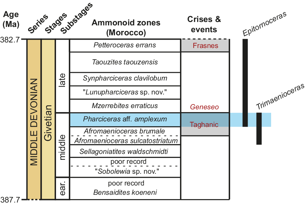
Despite being a typically late Givetian genus, Epitornoceras first occurs in the uppermost middle Givetian Pharciceras aff. P. amplexum zone of Morocco (Aboussalam and Becker, Reference Aboussalam and Becker2011; Allaire et al., Reference Allaire, Rustán, Korn, Vaccari, Ezpeleta and Balseiro2024, and references therein). Trimaenioceras, by contrast, is restricted to the upper middle Givetian of Morocco (Bockwinkel and Korn, Reference Bockwinkel and Korn2024). In turn, the Pharciceras aff. P. amplexum zone corresponds to the upper Taghanic Crisis interval (Aboussallam and Becker, Reference Aboussalam and Becker2011; Fig. 9).
Ammonoid succession for the Anti-Atlas of Morocco (modified from Becker et al., Reference Becker, El Hassani, Aboussalam, Hartenfels, Baidder, Hartenfels, Becker, El Hassani and Lüddecke2018) showing the range of the two genera, Epitornoceras and Trimaenioceras. Events and crises placed according to Aboussalam and Becker (Reference Aboussalam and Becker2011) and Becker et al. (Reference Becker, Königshof and Brett2016).

Figure 9. Long description
A stratigraphic table organized into six vertical columns.
1. Age (M a): The vertical axis ranges from 387.7 at the bottom to 382.7 at the top.
2. Series and Stages: The entire section is within the Middle Devonian series and the Givetian stage.
3. Substages: Divided into early (ear.), middle, and late.
4. Ammonoid zones (Morocco): From bottom to top, the zones are Bensaidites koeneni, poor record, Sobolewia sp. nov., poor record, Sellagoniatites waldschmidti, Afromaenioceras sulcatostriatum, Afromaenioceras brumale, Pharciceras aff. amplexum (highlighted in blue), Mzerrebites erraticus, Lunupharciceras sp. nov., Synpharciceras clavilobum, Taouzites taouzensis, and Petteroceras errans.
5. Crises and events: Three events are noted. At the top is Frasnes in red text on a gray background. In the middle is Geneseo in red text. Below that is Taghanic in red text on a gray background, which aligns with the blue-highlighted Pharciceras zone.
6. Genera Ranges: Represented by vertical black bars. The bar for Epitornoceras extends from the Taghanic event up to the top of the chart. The bar for Trimaenioceras is shorter, starting below the Taghanic event and ending within the blue-highlighted Pharciceras zone.
Therefore, Trimaenioceras and Epitornoceras co-occur only in the uppermost middle Givetian (see Becker et al., Reference Becker, Aboussalam, Bockwinkel, Ebbighausen, El Hassani and Nübel2004; Aboussalam and Becker, Reference Aboussalam and Becker2011; and Bockwinkel and Korn, Reference Bockwinkel and Korn2024, for synonymy). Hence, the Epitornoceras–Trimaenioceras assemblage, characterized by a worldwide distribution, lack of facies control, and short stratigraphic range, appears to represent a significant biostratigraphic marker for recognizing and correlating the Pharciceras aff. P. amplexum zone and, simultaneously, the Upper Taghanic Crisis interval.
This reasoning implies that the interval from the Sierra de Las Minitas, which records the Epitornoceras–Trimaenioceras assemblage, corresponds to the uppermost middle Givetian and correlates with the Pharciceras aff. P. amplexum zone of Morocco and the Upper Taghanic Crisis interval (Fig. 9). The ammonoid record from Argentina described herein thus represents the first undoubtedly biostratigraphically constrained occurrence of these cephalopods in extratropical basins, particularly within high-paleolatitude settings of Gondwana.
Regional biostratigraphic correlation of the Chigua Formation
The ammonoid-bearing stratigraphic intervals of the Sierra de Las Minitas typically consist of dark greenish to brownish, fine-grained, massive shales and siltstones with dispersed fossiliferous nodules (Fig. 3). Based on these lithostratigraphic characteristics, they can be correlated with the Chavela Member of the upper Chigua Formation in its type-area at Chigua, San Juan Province (located about 130 km to the SSW).
In addition, we documented the occurrence of typical biostratigraphic markers of the Chigua Formation, including fossil assemblages that are very similar: Epitornoceras baldisi occurs in association with trilobites dominated by the calmoniid Punillaspis argentina Baldis and Longobucco, Reference Baldis and Longobucco1977, and plant remains correspond to those reported as Haplostigma baldisii Gutiérrez and Archangelsky, Reference Gutiérrez and Archangelsky1997. Hence, in a general way, this fossil content supports a correlation of the ammonoid-bearing intervals from the Sierra de las Minitas (La Rioja Province) with the upper part of the Chavela Member of the Chigua Formation in its type area (San Juan Province). This conclusion agrees with the proposal by Carrizo and Azcuy (Reference Carrizo and Azcuy2015), although their suggestion lacked paleontological evidence beyond the mention of the lycopsid “Haplostigma”.
Despite intensive sampling, the type area of the Chigua Formation has yielded only Epitornoceras, suggesting a late Givetian age for the fossil-bearing layers (Leanza, Reference Leanza1968; Baldis and Sarudiansky, Reference Baldis and Sarudiansky1975; Allaire et al., Reference Allaire, Rustán, Korn, Vaccari, Ezpeleta and Balseiro2024). In contrast, the Sierra de Las Minitas records the Epitornoceras–Trimaenioceras assemblage, supporting a latest middle Givetian age. This observation raises questions regarding the precise biostratigraphic correlation between the two fossil localities. Since the absence of Trimaenioceras in the type area of the Chigua Formation is difficult to attribute solely to sampling bias, the ammonoid-bearing interval from the Sierra de las Minitas is interpreted as being older than the upper part of the Chigua Formation at its type area. The Chavela Member of the Chigua Formation at its type area, appears to be late Givetian in age, thus corresponding to a post-Taghanic stage (Fig. 9).
Regarding correlation towards the south, although the Codo Formation in San Juan Province has been correlated with the Chavela Member of the Chigua Formation (Amenábar and di Pasquo, Reference Amenábar and di Pasquo2008; Rustán et al., Reference Rustán, Rolan, Arnol and Uriz2025), the available evidence is too scarce and preliminary to provide further insights here.
In the context of remaining fossiliferous Middle Devonian units from the Central Andean Basin, mainly including Bolivia and Argentina, no more precise age than probable Givetian can be established. For example, the upper third of the Upper Member of the Belén Formation in the Bolivian Altiplano, which yielded ammonoids (Babin et al., Reference Babin, Racheboeuf, Le Hérissé and Riglos1991), has been successively considered from Lower to Upper Devonian (e.g., Racheboeuf et al., Reference Racheboeuf, Le Hérissé, Paris, Babin, Guillocheau and Truyols-Massoni1993; Blieck et al., Reference Blieck, Gagnier, Bigey, Edgecombe, Janvier, Loboziak, Rachebȩuf, Sempere and Steemans1996; Dalenz Farjat et al., Reference Dalenz Farjat, Riglos, Paulsen, Soruco, Leyton, Ortiz and Rios2024). A revision of the taxonomy and exact stratigraphic provenance of the ammonoids from this unit is needed, particularly in light of the recent report of Epitornoceras cf. E. baldisi from the overlying Sica Sica Formation (Zallés et al., Reference Zallés, Mardónez, Quiroz, Párraga Quiroga, Nava Camargo, Rojas Jaramillo, Paniagua Fuentes, Farjat and Cordero2025).
Biogeographic remarks
Trilobites, together with brachiopods, are the main groups defining the Malvinoxhosan Biogeographic Realm (Penn Clarke and Harper, Reference Penn-Clarke and Harper2021, Reference Penn-Clarke and Harper2023; Dowding et al., Reference Dowding, Ebach and Mavrodiev2022). The demise of this realm has classically been linked to an influx of warm waters or to different Middle Devonian biotic crises, particularly the Kačak or the Taghanic events (Penn-Clarke and Harper, Reference Penn-Clarke and Harper2023).
The presence of Devonian ammonoids at high paleolatitudes of Gondwana has been interpreted as indicating warmer “tropical” water influx or climatic “amelioration” (Babin et al., Reference Babin, Racheboeuf, Le Hérissé and Riglos1991), since ammonoids were known only from lower paleolatitudes, before the first South American records. This interpretation relies heavily on the assumption that high sea levels removed barriers, thereby allowing dispersal. Evidence from other indicators, such as the brachiopod Tropidoleptus carinatus (Conrad, Reference Conrad1839) (see discussions in Penn-Clarke and Harper, Reference Penn-Clarke and Harper2023, and Videira-Santos and Scheffler, Reference Videira-Santos and Scheffler2023), has also been used to support the collapse or dilution of the Malvinoxhosan Realm during the Middle Devonian. However, ammonoids in particular, were already present in southwestern Gondwana at least from the early Eifelian (Troth et al., Reference Troth, Marshall, Racey and Becker2011). This is significant since the Malvinoxhosan Bioregion is clearly recognizable during the Eifelian (Penn-Clarke and Harper, Reference Penn-Clarke and Harper2021, Reference Penn-Clarke and Harper2023). In fact, the Middle Devonian ammonoids reported from the upper member of the Belén Formation in the Bolivian Altiplano (Babin et al., Reference Babin, Racheboeuf, Le Hérissé and Riglos1991) occur in the context of the maximum development of Malvinoxhosan trilobite endemism at the genus level, particularly within the family Calmoniidae, which is entirely endemic to the Malvinoxhosan Realm (Lieberman et al., Reference Lieberman, Edgecombe and Eldredge1991; Lieberman,Reference Lieberman1993, and references therein). In turn, the presence of Tropidoleptus carinatus does not support warm ecological settings since it has been interpreted as an eurythermal taxon (Penn-Clarke and Harper, Reference Penn-Clarke and Harper2023, p. 25). Moreover, it would not be limited to a Middle Devonian immigration event, because it would have been present across Malvinoxhosan basins since the Early Devonian (Harper et al., Reference Harper, Alvarez, Boucot, Williams, Wright, Schemm-Gregory, Alvarez and Curry2010; Penn Clarke and Harper, Reference Penn-Clarke and Harper2021, Reference Penn-Clarke and Harper2023).
Although eustatic variations associated with transgressive events are well documented, no clear sedimentological indicators of warm-water conditions (e.g., limestones) have been reported from Devonian successions of southwestern Gondwanan basins (Boucot and Racheboeuf, Reference Boucot, Racheboeuf and Suárez-Soruco1993; Videira-Santos and Scheffler, Reference Videira-Santos and Scheffler2023, and references therein). Fossiliferous successions, including the Chigua Formation studied here, are clearly siliciclastic and consistent with cold-water settings. Therefore, in our view, there is currently no sedimentological evidence supporting the hypothesis of warm-water incursions into Malvinoxhosan basins. Indeed, several studies have documented a warming trend during the Givetian (e.g., Haq and Schutter, Reference Haq and Schutter2008; Joachimski et al., Reference Joachimski, Breisig, Buggisch, Talent, Mawson, Gereke, Morrow, Day and Weddige2009; Chen et al., Reference Chen, Ma, Mills, Qie, Joachimski, Shen, Wang, Xu and Wang2021; Scotese et al., Reference Scotese, Song, Mills and van der Meer2021). However, ammonoids were already present in southwestern Gondwana during the Eifelian (Troth et al., Reference Troth, Marshall, Racey and Becker2011), when climatic conditions were inferred to be cooler.
We therefore hypothesize that Middle Devonian ammonoids occurring in colder-water environments at the high paleolatitudes of southwestern Gondwana, as well as in warmer settings at lower paleolatitudes (i.e., Morocco; Fig. 1; Cocks and Torsvik, Reference Cocks, Torsvik, Gee and Stephenson2006; Torsvik et al., Reference Torsvik, Cocks, Domeier, Marcilly and Dowding2025), were simply tolerant of a wide range of water temperatures. There is no need to invoke warmer high-latitude waters to explain their broad geographic distribution and presence in circum-polar basins.
Our results also clearly dismiss the Kačak event (and likely also the Taghanic event) as a main cause of the disappearance of the Malvinoxhosan Realm, as they undoubtedly indicate that typical Malvinoxhosan faunas persisted at least until the late Givetian (see also Allaire et al., Reference Allaire, Rustán, Korn, Vaccari, Ezpeleta and Balseiro2024). This observation concurs with further evidence indicating that calmoniid trilobites were diverse during the entire Middle Devonian, becoming extinct in the earliest Frasnian (Rustán et al., Reference Rustán, Rolan, Arnol and Uriz2025). Moreover, calmoniids reported from the type locality of the Chigua Formation include Punillaspis argentina, Cryphaeoides rostratus (Kozłowski, Reference Kozłowski1923), and Bouleia dagincourti (Ulrich, Reference Ulrich1892) (Baldis, Reference Baldis and Oswald1967; Baldis and Sarudiansky, Reference Baldis and Sarudiansky1975; Baldis and Longobucco, Reference Baldis and Longobucco1977). No significant taxonomic turnover has been recorded during this interval, particularly with respect to the immigration of widely distributed taxa. Based on our new data, representing the first high-resolution global biostratigraphic markers from southwestern Gondwanan basins, we find no evidence for dilution, collapse, or demise of the Malvinoxhosan Realm during the Middle Devonian.
Conclusions
Based on lithological and biostratigraphic evidence (including the ammonoid Epitornoceras, the trilobite Punillaspis, and the lycopsid “Haplostigma” baldisii), we reliably identify the Chigua Formation in the Sierra de las Minitas, La Rioja Province (beyond its type area in San Juan Province to the south), in agreement with the preliminary proposals by Carrizo and Azcuy (Reference Carrizo and Azcuy2015).
Based on two new species of the middle Givetian ammonoid Trimaenioceras occurring in the same fossiliferous intervals (T. alegria n. sp. and T. ivanae n. sp.), we recognize the Epitornoceras–Trimaenioceras assemblage in the northernmost Argentine Precordillera Basin.
On the basis of well-defined biostratigraphy from Morocco, Epitornoceras and Trimaenioceras co-occur within the uppermost middle Givetian ammonoid biozone (i.e., the Pharciceras aff. P. amplexum zone; Becker et al., Reference Becker, Aboussalam, Bockwinkel, Ebbighausen, El Hassani and Nübel2004; Aboussalam and Becker, Reference Aboussalam and Becker2011; Bockwinkel and Korn, Reference Bockwinkel and Korn2024). Therefore, the ammonoid-bearing strata exposed in the Sierra de Las Minitas can be correlated with the Pharciceras aff. P. amplexum zone, allowing recognition of the upper Taghanic Crisis interval in southwestern Gondwana for the first time.
The Epitornoceras–Trimaenioceras assemblage appears to be a significant biostratigraphic indicator of the upper middle Givetian (upper part of the Taghanic Crisis interval) worldwide. Typical paleontological markers of the Chigua Formation can be clearly recognized in the Devonian of the Sierra de Las Minitas, however, based on the ammonoid assemblage, this succession might be older (i.e., uppermost middle Givetian) than the upper part of the Chavela Member of the Chigua Formation in its type area (i.e., upper Givetian).
The presence of ammonoids in the Middle Devonian of southwestern Gondwana has been classically interpreted as evidence of warm “tropical” water influxes and related to the collapse or dilution of the Malvinoxhosan Realm. However, because there is no clear sedimentological or faunal evidence for discrete episodes of warm-water influence, ecological tolerance to variations in water temperature may instead better explain the widespread distribution of ammonoids across a broad range of paleolatitudes during the Middle Devonian.
Acknowledgments
This work was part of a project funded by the PICT 3095-2017 grant to Juan J. Rustán (ANPCyT and SECyT UNLAR 530 00-05103/2019). N. Allaire further acknowledges CONICET for financial support through a postdoctoral fellowship. We thank D. Aquino, L. Eberhardt, E. Randolfe, G. Voldman, and I.S. Tapia for their valuable assistance during the fieldwork. We are also grateful to M. Yacobucci (Associate Editor, Bowling Green State University), K. De Baets (University of Warsaw), and an anonymous reviewer for their constructive comments, which helped improve the manuscript.
Competing interests
The authors declare none.
Appendix
Ontogenetic trajectories among Epitornoceras species. E. dennisoni is known only from its holotype, which is poorly preserved and allows only an incomplete set of measurements to be obtained; for this reason, the species is absent from the UWI, WER, and IZR plots. E. peracutum could not be included at all because it is represented solely by its holotype, a poorly preserved fragment of conch that prevents any measurements from being obtained.

Figure A1. Long description
A multi-panel figure containing five scatter plots, a cross-section diagram, and a list of indices. All plots use conch diameter in millimeters on the x-axis, ranging from 0.0 to 100.0.
* Top-left plot C W I (Conch Width Index): Values range from 0.00 to 0.90. Data shows a rapid exponential decay from 0.80 at small diameters, stabilizing between 0.20 and 0.40 as diameter increases.
* Top-right plot U W I (Umbilical Width Index): Values range from 0.00 to 0.25. Data points for E. transmeditaneum start high at 0.20 and drop sharply to near 0.00 by 20 millimeters. Other species remain near 0.00 throughout.
* Middle-left plot W E R (Whorl Expansion Rate): Values range from 1.5 to 3.1. Data shows a slight upward trend with high variance, clustering between 1.9 and 2.5.
* Middle-right plot W W I (Whorl Width Index): Values range from 0.00 to 2.00. Shows a steep power-law decrease from 1.80 at small diameters, leveling off to approximately 0.60 beyond 40 millimeters.
* Bottom-left plot I Z R (Imprint Zone Rate): Values range from 0.20 to 0.60. Data is widely scattered between 0.30 and 0.55 with no clear linear trend.
Bottom-center anatomical diagram: A cross-section of an ammonoid shell showing measurement parameters. Vertical anchors include conch diameter 1 (dm1), conch diameter 2 (dm2), apertural height (ah), and whorl height (wh). Horizontal anchors include whorl width (ww) and umbilical width (uw).
Bottom-right legend: Defines the indices as follows.
* Conch Width Index: C W I equals ww forward slash dm1.
* Whorl Width Index: W W I equals ww forward slash wh.
* Umbilical Width Index: U W I equals uw forward slash dm1.
* Whorl Expansion Rate: W E R equals (dm1 forward slash dm2) squared.
* Imprint Zone Rate: I Z R equals (wh minus ah) forward slash wh.
Ontogenetic trajectories of Trimaenioceras species.

Figure A2. Long description
A multi-panel figure containing five scatter plots, an anatomical cross-section, and a legend. All plots share a common x-axis: conch diameter in millimeters, ranging from 0.0 to 35.0. Data points represent six species: T. alegria n. sp. (blue diamond), T. ivanae n. sp. (red diamond), T. klugi (green triangle), T. eculeus (purple X), T. fuscina (orange circle), and T. paucum (yellow square).
* Top-left plot: C W I (Conch Width Index) on the y-axis from 0.00 to 0.80. Shows a general downward trend as conch diameter increases.
* Top-right plot: U W I (Umbilical Width Index) on the y-axis from 0.00 to 0.40. Shows a steep decline for most species as diameter increases.
* Middle-left plot: W E R (Whorl Expansion Rate) on the y-axis from 1.0 to 1.8. Data is scattered between 1.3 and 1.75 with no singular linear trend.
* Middle-right plot: W W I (Whorl Width Index) on the y-axis from 0.00 to 2.50. Shows a clear asymptotic decrease for all species.
* Bottom-left plot: I Z R (Imprint Zone Rate) on the y-axis from 0.00 to 0.80. Shows a general upward trend, leveling off around 0.50 to 0.60.
* Bottom-right panel: A cross-sectional diagram of a conch shell illustrating measurement parameters: conch diameter 1 (dm1), conch diameter 2 (dm2), whorl width (ww), whorl height (wh), umbilical width (uw), and apertural height (ah).
* Legend: Defines the indices as C W I = ww/dm1, W W I = ww/wh, U W I = uw/dm1, W E R = (dm1/dm2) squared, and I Z R = (wh-ah)/wh.