Introduction
Meningioma make up 18% of primary intracranical tumors though it is rarely identified as a primary middle-ear lesion without an intracranial component. The estimated incidence of meningioma occurring extracranially is 2 per cent, and its occurrence in the middle ear is extremely rare.Reference Friedman, Costantino, Teitelbaum, Berktold and Sisson1 Intracranial meningiomas can extend to the middle-ear cleft via the tegmen tympani, posterior fossa plate, internal auditory canal or jugular foramen. Most reported cases initially regarded as primary middle-ear meningiomas were eventually shown to have an intracranial origin.Reference Maniglia2–Reference D'Angelo, Marlowe, Marlowe and Mobini4 Most of these cases were reported before magnetic resonance imaging (MRI) with gadolinium enhancement became widely available.
This case report emphasises several important factors in the diagnosis and treatment of primary extracranial meningioma. The accompanying literature review also highlights salient features of this tumour.
Case report
A 55-year-old woman presented with a three-year history of decreased hearing and a sensation of fullness in the left ear. The symptoms were aggravated after alcohol intake or increased physical activity.
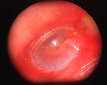
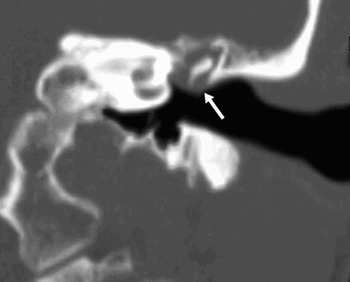
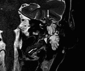
On otoscopy, the external auditory canal was found to be normal, but visualisation of the posterosuperior quadrant of the middle ear showed a mass behind an intact tympanic membrane (Figure 1). Audiometry showed a moderate conductive hearing loss. High resolution computed tomography (CT) scanning (Figure 2) and subsequent MRI with gadolinium enhancement (Figure 3) revealed a lesion in the epitympanum and mastoid antrum. The mass was confined to the mastoid antrum and epitympanum, with an intact ossicular chain, facial canal and otic capsule. The radiological appearances were non-specific, and a tentative diagnosis of schwannoma was made.
Otoscopic photograph showing a fullness (arrow) in the posterosuperior quadrant of the tympanic membrane.

Coronal, high resolution computed tomography scan of the left temporal bone, demonstrating a soft tissue mass (arrow) in the middle ear, with no evidence of bony erosion.

Coronal, T2-weighted magnetic resonance imaging scan with gadolinium enhancement, showing an isointense, extra-axial mass (arrow) within the petrous bone, inferior to the temporal basal dura. The lesion is in the superior aspect of the fluid-filled mastoid air cells, just lateral to the semicircular canal.

A transmastoid biopsy of the lesion was performed using a postaural approach. Histological examination of the biopsy specimen was consistent with meningioma.
After the biopsy, the patient's general condition was normal, and neurological examination showed no abnormality. Re-evaluation of the MRI confirmed no intracranial extension of the tumour.
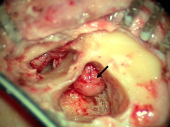
A combined approach tympanoplasty was performed. The tumour was removed in toto. The lesion was non-vascular and filled the attic and antrum (Figure 4). There was no evidence of infiltration of the ossicles or tympanic membrane by the tumour. The sinus plate, lateral semicircular canal and tegmen were intact. The head of the malleus and incus were removed to facilitate complete elimination of the tumour. Hearing reconstruction was achieved by refashioning the head of the malleus and replacing it over the stapes head.
Intra-operative photograph showing the meningioma in the epitympanum, aditus and antrum (arrow). No erosion of the tegmen or sinus plate was seen.

The patient did well after surgery, with no evidence of tumour recurrence at three months' follow up.
Histopathology
Macroscopic examination showed an encapsulated, 2 × 1 × 0.5 cm lesion. Histological examination showed a proliferation of bland, meningothelial-like cells forming diffuse sheets admixed with fibrous tissue. Nuclei were found with occasional pseudoinclusions and small nucleoli. No mitotic activity or necrosis was seen. Evidence of microcalcification was present. Periodic acid Schiff staining was negative.
Immunohistochemical analysis showed positive staining for vimentin, pancytokeratins (AE1AE3) and epithelial membrane antigen, but immunonegativity for S-100, smooth muscle actin and progesterone receptors. The MIB-1L1 labelling index was very low (less than 2 per cent).
The morphological and immunohistochemical characteristics of the tumour were consistent with a meningioma (World Health Organization (WHO) grade one) (Figure 5).
Photomicrograph of the meningioma showing proliferation of bland, meningothelial-like cells forming diffuse sheets admixed with fibrous tissue. Nuclei were found with occasional pseudoinclusions and small nucleoli. No mitotic activity or necrosis was seen (H&E; ×40).

Discussion
The occurrence of meningioma in the middle ear as a primary lesion is very rare, with few reported cases. Nager classified temporal bone meningiomas into two main types: type one (the commoner), representing extension of an intracranial meningioma; and type two, occurring without intracranial extension.Reference Nager5 The rare type two entity should be diagnosed only after confirmatory MRI. Most of the published case reports are from the pre-MRI era, with insufficient evidence to exclude the possibility of extension from a primary intracranial tumour. There are very few reported cases of primary middle-ear meningioma without intracranial extension.Reference Uppal, Kabbani, Reddy and Kaur6, Reference Kumar, Basu, Sen and Kamal7 Thompson et al. described 36 cases of temporal bone meningioma, but were not able to exclude intracranial extension due to the absence of proper radiological evaluation.Reference Thompson, Bouffard, Sandbeg and Mena8 An MRI with gadolinium enhancement is superior to plain X-ray or CT scan for differentiating peripheral extension of an intracranial meningioma from the less common primary extracranial meningioma (as encountered in our patient).Reference Parisier, Som, Shugar and Marovitz3, Reference Thompson, Bouffard, Sandbeg and Mena8
The differential diagnosis of a middle-ear mass includes schwannoma, glomus tumour, metastatic tumour and ceruminous gland tumour. Differentiation of middle-ear meningioma from facial nerve schwannoma is clinically difficult, due to these tumours' similar clinical presentations. Both tumours predominantly affect women aged 45–55 years, and present with symptoms of hearing loss and tinnitus. In addition, there are no characteristic radiological differences between the two tumours.Reference Hilton, Kaplan, Ang and Chen9 Therefore, the diagnosis can only be confirmed after histopathological examination, as the clinical symptoms and radiological signs are usually nonspecific, as in our patient.
Meningiomas are usually benign in nature, however, malignant transformation is possible in some patients. Histopathological examination of our patient's specimen revealed a mixed, grade one, benign meningioma (based on WHO classification). Other types include atypical (grade two) and anaplastic (grade three). The meningotheliomatous or transitional variant of grade one is more often associated with extracranial meningioma.Reference D'Angelo, Marlowe, Marlowe and Mobini4 The immunohistochemical profile of meningioma is quite characteristic, and is useful in differentiating these tumours from schwannoma. It has been noted that immunoreactivity for epithelial membrane antigen and keratin will be strongly positive in meningiomas, but negative or only weakly positive in schwannomas.Reference Borin, Okanda and Cruz10
• Meningiomas make up 18 per cent of primary intracranial tumours, whereas the estimated incidence of extracranial meningiomas is only 2 per cent. Meningioma in the middle ear without intracranial extension is extremely rare
• In cases of primary middle-ear meningioma, total excision of the lesion remains the mainstay of management
• Meningioma needs to be included in the differential diagnosis of middle-ear lesions
Complete surgical excision is the mainstay of management for primary middle-ear meningiomas. Surgery is difficult in the majority of cases due to the infiltrative nature of the tumour along the Haversian canals, and the absence of clear margins. However, complete excision of the lesion was possible in our patient, although removal of the head of the malleus and incus was temporarily required to facilitate tumour excision. We did not notice any erosion of the ossicles or tegmen plate.
As meningiomas are slow-growing tumours, long term follow up with MRI scanning is important to exclude recurrence. A change from conductive to sensorineural hearing loss may be the earliest sign of progressive disease; such a change may indicate erosion of the bony labyrinth, which is often spared in the early part of the disease.Reference O'Reilly, Kapadia and Kamerer11 In our patient, follow-up pure tone audiometry revealed improved hearing. The role of radiation therapy for residual tumours is unclear, due to the rare occurrence of these tumours.Reference Ferlito, Devaney and Rinaldo12
Conclusion
Primary middle-ear meningioma is a rare clinical entity, with nonspecific clinical and radiological features. Histopathological and immunohistochemical findings are helpful in differentiating it from other, more common middle-ear tumours, such as schwannoma and glomus tympanicum. An increased awareness among otolaryngologists is necessary in order to correctly diagnose this rare but well reported tumour. Long term follow up using MRI with gadolinium enhancement is necessary to exclude recurrence, which can occur even after complete surgical excision.