Introduction
A true ‘glomus tumour’ is an entirely different diagnosis from what otorhinolaryngologists know as ‘glomus tympanicojugulare’ (a paraganglioma of the skull base). Glomus tumours are mostly benign and are derived from cells of the neuromyoarterial glomus or glomus body, a structure most commonly present in the subungual region of the fingers, palm, wrist, forearm and foot.Reference Takahashi, Oizumi, Yanagawa and Sadahiro1, Reference Kayal, Hampton, Sheehan and Washington2 Malignant glomus tumours are extremely rare. Here, we describe the first reported case of malignant glomus tumour of the larynx.
Case report
A 37-year-old man presented with complaints of progressive hoarseness of voice for four months and dyspnoea for one month.
Fibre-optic nasolaryngoscopic examination revealed a smooth mass in the right supraglottic region causing narrowing of the airway.
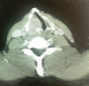
A computed tomography (CT) scan of the neck and chest was performed with intravenous contrast, which showed a heterogeneously enhancing lesion of the right hemilarynx, involving the whole of its length including the subglottis (Figure 1).
Axial computed tomography scan showing enhancing lesion of the right hemilarynx.

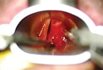
Microlaryngoscopy was performed and multiple deep biopsies were taken, after incising the overlying mucosa. The lesion involved the right false and true vocal folds (Figure 2), with some subglottic extension as well. It also caused a bulge into the medial wall of the right pyriform fossa.
Microlaryngoscopic view showing smooth mass of the right supraglottis.

Histopathological analysis of this biopsy was reported as ‘benign glomus tumour’.
Later, a tracheostomy had to be done due to increasing stridor. Laser ablation was attempted with a diode laser, but a second CT scan did not show much improvement in the size of the lesion.
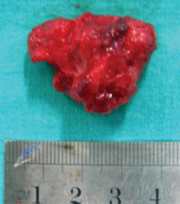
Due to the biopsy report of the ‘benign’ nature of the lesion, procedures such as total or even partial laryngectomy were considered unjustified at that time. Therefore, a wide local excision was performed via a suprahyoid pharyngotomy and laryngofissure approach. The tumour was adherent to the thyroid and cricoid cartilages. It was separated from these structures by stripping, and was also stripped off the pyriform mucosa. The tumour was removed in two to three pieces, the largest measuring 3.0 × 2.5 × 1.4 cm (Figure 3).
The largest piece of tumour removed during wide resection, measuring 3.0 × 2.5 × 1.4 cm.

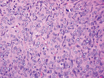
Histopathological analysis of this second specimen revealed a tumour composed of clusters and nests of polygonal cells with round nuclei, small nucleoli and fine chromatin. There was abundant eosinophilic cytoplasm. There was only mild cytological atypia but brisk mitotic activity, with up to 20 mitoses/50 high power fields (Figure 4). Necrosis and atypical mitotic figures were absent. The tumour also had an infiltrative growth pattern with focal vascular invasion. Given the large size of the tumour (approximately 4 cm), the presence of mitotic activity (20 mitoses/50 high power fields) and the deep visceral location, it was thought to be best characterised as a ‘malignant glomus tumour’, according to Folpe and colleagues' World Health Organization (WHO) classification.Reference Folpe, Fanburg-Smith, Miettinen and Weiss3, Reference Folpe, Fletcher, Unni and Mertens4
Photomicrograph of the tumour showing mitotic figures. (H&E; ×40)

Immunohistochemical analysis revealed diffusely positive cytoplasmic staining for smooth muscle actin, weakly positive staining for cluster of differentiation 34 glycoprotein, and positive staining for vimentin. In addition, there was also positive collagen type IV immunostaining surrounding individual tumour cells, further supporting the diagnosis. Staining for cytokeratin was negative. Staining for chromogranin was also negative, excluding the possibility of a paraganglioma or neuroendocrine tumour.
After the final biopsy report, the patient was investigated for metastases. An ultrasound scan of the abdomen and CT of the chest were normal. A bone scan did not show bone metastasis.
At this stage it was decided, after consultation with the radiation oncologist, that the patient should undergo total laryngectomy as a definitive, curative procedure, with the option of post-operative radiotherapy depending upon resection margins. However, the patient did not consent to surgery at that time. He was thus decannulated and kept under close follow up.
For a time, the patient remained well without any airway problems.
However, follow-up flexible nasolaryngoscopy performed six months after the excision revealed the presence of tumour, which was also evident on a magnetic resonance imaging (MRI) scan. The patient was counselled again, and finally consented to a total laryngectomy.
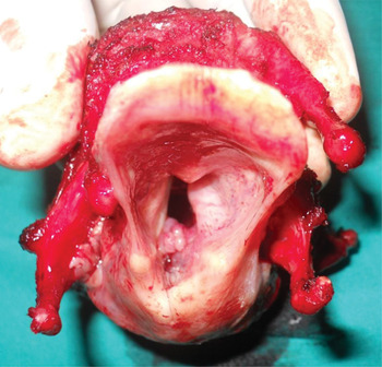
During the procedure, it was found that the tumour had now invaded the left side of the larynx as well as the posterior commissure (Figure 5). The thyroid cartilage was involved on the right side. The tumour was still confined to the larynx, but it had caused a more prominent bulge in the medial wall of the right pyriform fossa and the postcricoid area than before. Rather than being smooth and submucosal, the tumour now had a cobblestone, papillomatous appearance (Figure 5).
Laryngectomy specimen showing the right-sided tumour, which has crossed to the opposite side via the posterior commissure.

Histopathological analysis of the laryngectomy specimen was consistent with the previous report of ‘malignant glomus tumour’. It showed that the tumour had invaded the laryngeal cartilages and soft tissues posteriorly. It was 0.1 cm from the posterior margin.
Post-operatively, the radiation oncologist decided not to recommend radiotherapy for the patient. Thus, the patient was kept under close follow up. At the time of writing, he was doing well, without any complications.
Discussion
Glomus tumours are classically benign neoplasms. They typically consist of three components: glomus cells, vasculature and smooth muscle cells. Depending upon the relative amounts of these components, tumours are further categorised as (1) solid glomus tumour (glomus tumour proper), (2) glomangioma or (3) glomangiomyoma.Reference Cocker, Messaoudi, Waelput and Van Schil5
Glomus tumours are rare neoplasms which constitute approximately 1.6 per cent of all soft tissue tumours. The most common location is the subungual region of the fingers. Rarely, the tumour can also be encountered at other sites where the normal glomus body may be sparse or even absent, such as the mediastinum, respiratory tract, gastrointestinal tract, urinary tract and gynaecological regions.Reference Cullen and Hanna6–Reference Shim, Choi and Cho15
The malignant counterpart, or ‘glomangiosarcoma’, is very rarely reported. In 1990, Gould et al. Reference Gould, Manivel, Albores-Saavedra and Monforte16 proposed the following classification of malignant and atypical glomus tumours: (1) locally infiltrative glomus tumour, (2) glomangiosarcoma arising in a benign glomus tumour and (3) de novo glomangiosarcoma. As a group, these locally aggressive or potentially malignant glomus tumours were larger and more deeply located than conventional glomus tumours.
In 2001, Folpe et al. Reference Folpe, Fanburg-Smith, Miettinen and Weiss3 reviewed 52 cases of atypical glomus tumours from different body sites (none from the head and neck). Based on this large series, they identified the following histopathological criteria for malignancy in glomus tumours: deep location and a size of more than 2 cm; atypical mitotic figures; or a moderate to high nuclear grade and five or more mitotic figures/50 high power fields. Other types of atypical glomus tumours classified were glomus tumours with uncertain malignant potential, symplastic glomus tumours and glomangiomatosis. Using this classification scheme, Folpe et al. classified 32 of their 52 reviewed cases as malignant; furthermore, metastasis was observed in 38 per cent of them. This classification has now been accepted by the WHO.Reference Folpe, Fletcher, Unni and Mertens4
Searching the PubMed database, we found 22 more case reports of malignant glomus tumour, in addition to those reported by Folpe et al.,Reference Folpe, Fanburg-Smith, Miettinen and Weiss3 making a total number of 54 cases of malignant glomus tumour reported at the time of writing this paper.
We based our diagnosis upon the new WHO classification.Reference Folpe, Fanburg-Smith, Miettinen and Weiss3, Reference Folpe, Fletcher, Unni and Mertens4 We labelled our case as a ‘malignant glomus tumour’ based on the following features of this classification system: deep visceral location, large size (approximately 4 cm) and increased mitotic activity (20 mitoses/50 high power fields). In addition to these features, the tumour in our patient's laryngectomy specimen showed cartilage and soft tissue invasion, as well as focal vascular invasion and mild to moderate cytological atypia. These findings are consistent with case reports by De Chiara et al. Reference De Chiara, Apice, Mori, Silvestro, Losito and Botti17 and Matsumoto et al. Reference Matsumoto, Kakizaki, Yagihashi and Yagihashi18 Necrosis is only an occasional feature of malignant glomus tumours,Reference Terada, Fujimoto, Shirakashi, Kamo and Sugiura19 and no areas of necrosis were found in our case. Malignant glomus tumours or glomangiosarcomas usually do not metastasise,Reference Folpe, Fletcher, Unni and Mertens4 and represent a locally infiltrative malignancy.Reference Oh, Stephenson, Schnall, Fassola and Dinh20 In fact, the presence of mitotic figures, rather than metastasis, has been described by some authors as the most important diagnostic criterion for malignant glomus tumour or glomangiosarcoma.Reference Choi, Yang, Gang, Kim and Kim21 Hiruta et al. Reference Hiruta, Kameda, Tokudome, Tsuchiya, Nonaka and Hatori22 have described a case of malignant glomus tumour of the thigh in which the histological features were almost identical to those of our case. These authors also performed wide resection after open biopsy. According to Katabami et al.,Reference Katabami, Okamoto, Ito, Kimura and Kaji12 the presence of vascular invasion, as in our case, might be an important aspect of malignant behaviour of visceral glomus tumours, although this feature was not included in Folpe and colleagues'Reference Folpe, Fanburg-Smith, Miettinen and Weiss3 diagnostic criteria.
In the head and neck region, glomus tumour has been reported in the nose, paranasal sinuses, oral mucosa and eyelids.Reference Cocker, Messaoudi, Waelput and Van Schil5, Reference Keelawat, Hirunwiwatkul and Thanakit11 It has also been reported in the superior mediastinum,Reference Choi, Yang, Gang, Kim and Kim21 oesophagus,Reference Papla and Zieliński9 trachea and bronchi. To our knowledge, there have been 22 reports of glomus tumours arising in the lower respiratory tract (excluding the trachea)Reference Kayal, Hampton, Sheehan and Washington2 and 16 reports of glomus tumours arising in the trachea.Reference Altinok, Cakir, Gulhan and Tastepe10 Only one laryngeal case of benign glomus tumour (glomangiomyoma) has been reported in the English language literature.Reference Lee, Murthy, Gildea and Lorenz7 Ours is the first reported case of ‘malignant glomus tumour’ or glomangiosarcoma of the larynx.
• A case of laryngeal malignant glomus tumour in a 37-year-old man is reported
• The tumour was treated with total laryngectomy
• This tumour is very rare in visceral locations
• This is the first case reported in the larynx
In their previous report of a benign laryngeal glomangiomyoma, Lee et al. Reference Lee, Murthy, Gildea and Lorenz7 described a lesion with more or less similar macroscopic features to our own case. The lesion was submucosal and involved the left false vocal fold and aryepiglottic fold. It had a rubbery consistency and a cystic core, while our specimen was rubbery to hard in consistency throughout, including its core.
Immunohistochemical analysis of Lee and colleagues' case showed positive staining for cluster of differentiation 34 glycoprotein and smooth muscle actin. The various reported cases of benign and malignant glomus tumours have differed in their immunostaining pattern, but most, like ours, have shown positivity for smooth muscle actin, type IV collagen, cluster of differentiation 34 glycoprotein and vimentin.Reference Folpe, Fletcher, Unni and Mertens4, Reference Cullen and Hanna6, Reference Altinok, Cakir, Gulhan and Tastepe10, Reference Miettinen, Paal, Lasota and Sobin13, Reference De Chiara, Apice, Mori, Silvestro, Losito and Botti17–Reference Terada, Fujimoto, Shirakashi, Kamo and Sugiura19, Reference Calduch, Monteagudo, Martỉnez-Ruiz, Ramón, Pinazo and Cardá23–Reference Park, Oh, Yang and Kim25