Congenital complete tracheal rings are a rare tracheal anomaly, usually presenting in neonates with respiratory distress.Reference Tsai, Wong, Lien, Huang, Liao and Hsu 1 It is particularly associated with cardiac anomalies, especially pulmonary slings.Reference Chao, Peng, Lee, Lin and Chen 2 We report a rare association of congenital complete tracheal rings with hypoplastic left heart variant. She underwent a tracheal slide reconstruction at 2 months of age with multiple subsequent dilations. However, she returned to the emergency room 8 months post-procedure with sudden difficulty in breathing and died after resuscitation attempts.
Case report
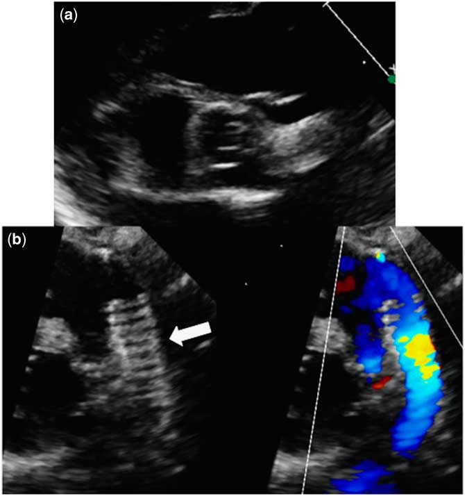
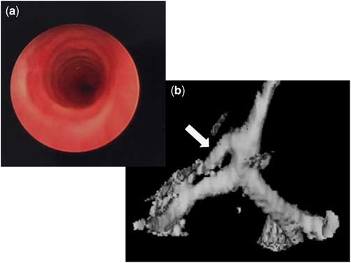
A full-term female infant, birth weight 2705 g, had an antenatal diagnosis of small left-sided structures with hypoplastic left heart variant. A postnatal echocardiogram confirmed the diagnosis of a hypoplastic left heart variant, including a mildly hypoplastic mitral valve with z-score of −2.3 and no gradient, unicuspid aortic valve (Fig 1a), a large perimembranous ventricular septal defect, moderately hypoplastic aortic arch with severe coarctation of the aorta, and large patent ductus arteriosus. She was started on prostaglandin infusion at 0.25 mg/kg/minute. In our patient, the hybrid procedure, which is a variant of the first-stage Norwood surgery, was performed in neonatal period to give the patient enough time to grow the left ventricle, with the hope to have a possible biventricular repair. At 13 days of life, she underwent a hybrid procedure (Fig 1b) consisting of ductus arteriosus stent placement and banding of bilateral branch pulmonary arteries. During the post-operative period, she failed extubation several times and had significant respiratory symptoms with each failed attempted extubation. Of note, in the immediate newborn period before hybrid procedure, she was noted to have significant tachypnoea, out of proportion to her underlying heart condition. A direct laryngo-bronchoscopy was performed, which revealed a diagnosis of complete tracheal rings of almost the entire trachea (Fig 2a). There was no subglottic stenosis and complete tracheal rings were visualised occupying approximately 80% of the trachea length with significantly tracheal stenosis around 1 cm above the carina. Surgeons were not able to pass the #2.5 bronchoscope through this ring at this time. She had a balloon dilation of the trachea performed because of evidence of tracheal stenosis. The diagnosis was confirmed with a CT scan, which showed the presence of a bronchus suis, serving the right upper lobe (Fig 2b). As she continued to have persistent desaturations, she underwent a tracheal slide reconstruction at 2 months of age. During the surgery complete tracheal rings were visualised all the way till 0.5 cm above the carina causing a funnel-like stenosis. The procedure was performed through a median sternotomy on cardiopulmonary bypass. A left atrial vent was placed to avoid pulmonary steal through the open ductus. The trachea and brochial suis were dissected. At the midpoint of the trachea, the bronchus suis and the right and left main stem bronchi were widely opened and the two ends were then anastomosed together. A posterior leak was noted on inflation of the endotracheal tube with pressure up to 50 cm of water, which was fixed. She was able to come off cardiopulmonary bypass without difficulty. Rigid bronchoscopy performed immediately after the procedure showed significantly enlarged lumen and intact suture lines. The airway was slightly narrow, approximately 1 cm above the carina. However, the #2.5 bronchoscope could be advanced with ease. The surgeons performed a surveillance direct laryngo-bronchoscopy with a planned extubation in the operation room on post-operative day 5 from the procedure, which was successful. Subsequently, two more surveillance direct laryngo-bronchoscopies were performed on post-operative days 13 and 26. However, she required multiple direct laryngo-bronchoscopies with dilations, that is a total of 11 balloon dilations over a span of 8 months post tracheoplasty. Some dilations required steroid or Kenalog injections during the dilatations. For the first 2 months, the dilations were performed every week. The next dilatation was performed 3 months post-surgery and after that the dilations were performed every 2 months.
( a ) A two-dimensional echocardiographic image of the aortic valve from parasternal short-axis view. The valve is visualised to be unicuspid with total fusion of all leaflets and small opening in the centre of the valve. ( b ) A two-dimensional echocardiographic colour-compare image of the stent (arrow) in the patent ductus arteriosus after the hybrid procedure (left-sided image) and colour (right-sided image).

( a ) A picture from the direct laryngoscopy showing the complete tracheal rings with absence of posterior membrane in the trachea of our patient. ( b ) A three-dimensional reconstruction of the trachea from the CT scan showing the bronchial suis (arrow), supplying the right upper lob directly from the trachea.

She was discharged home at 5 months of age but had a re-admission because of tracheal stenosis requiring balloon dilation and triamcinolone injections at the site of stenosis. She did have an outpatient cardiology visit where she was found to be doing well from a cardiac standpoint and was scheduled for a complete repair soon. However, she returned to the emergency room with sudden increase in work of breathing and rapid progression to unresponsiveness. She received cardio-pulmonary resuscitation for 12 minutes in the emergency room but was eventually declared dead due to failure of return of spontaneous circulation.
Comment
Congenital complete tracheal ring is a rare congenital malformation in neonates and often remains under-diagnosed. Most patients develop respiratory distress, cyanosis, and subsequent respiratory failure.Reference Tsai, Wong, Lien, Huang, Liao and Hsu 1 The most common pathologic finding is the absence of the normal posterior tracheal membrane, leading to significant stenosis of the lower trachea.Reference Chao, Peng, Lee, Lin and Chen 2 It is associated with cardiovascular anomalies in up to 70% of patients,Reference Butler, Speggiorin and Rijnberg 3 with the most common association being pulmonary artery sling. Other common cardiac associations include patent ductus arteriosus, atrial septal defect, and ventricular septal defect.Reference Chao, Peng, Lee, Lin and Chen 2 , Reference Loukanov, Sebening, Springer, Ulmer and Hagl 4 This is the first reported case of an association of congenital complete tracheal rings with hypoplastic left heart syndrome variant, where the patient had an unfavourable outcome. The complexity of the associated cardiac anomaly complicates the diagnosis of congenital complete tracheal rings with tracheal stenosis and increases the operative risks and results in poorer outcomes.Reference Loukanov, Sebening, Springer, Ulmer and Hagl 4
Complete cartilaginous rings occur in varying length, location, and severity. Conservative management is not successful in children with long-segment complete tracheal rings, like in our patient, especially with funnel-like stenosis.Reference Rossi, Moreddu, Mace, Triglia and Nicollas 5 The presence of stenosis can often lead to life-threatening respiratory insufficiency in children.Reference Hofferberth, Watters, Rahbar and Fynn-Thompson 6 Staged surgical intervention has been shown to have poor prognosis because of the post-operative complications due to the untreated lesion.Reference Loukanov, Sebening, Springer, Ulmer and Hagl 4 It is possible that the multiple reintubations and repeated instrumentation required for direct laryngoscopy and dilations of the stenosed trachea probably exacerbated the problem due to repeated trauma. However, the repeated dilatations are often required in these patients even after a successful surgery. We believe that in our patient, it was the combination of hypoplastic left heart syndrome variant and the respiratory insufficiency caused by the tracheal stenosis that led to her eventual demise.
Complete tracheal rings are often associated with abnormal bronchial branching pattern.Reference Loukanov, Sebening, Springer, Ulmer and Hagl 4 , Reference Hofferberth, Watters, Rahbar and Fynn-Thompson 6 Bronchus suis or pig bronchus is an anomalous origin of the right upper lobe bronchus from the right lateral wall of the trachea, which is also seen in our patient.Reference McLaughlin, Strieder, Harris, Vawter and Eraklis 7
The gold standard for definitive diagnosis of tracheal stenosis is rigid laryngo-bronchoscopy under general anaesthesia.Reference Hofferberth, Watters, Rahbar and Fynn-Thompson 6 Direct visualisation of the airway enables accurate assessment of the length and diameter of the stenosed tracheal segment. A timely diagnosis is imperative for prompt management and good outcomes. However, in our patient, we believe the diagnosis was delayed due to the unusual association, not reported in the literature previously as well as no upper respiratory symptom to suggest the need for an otolaryngology evaluation. She eventually had a laryngoscopy after multiple failed extubation attempts and the diagnosis was made. It is possible that the rigid stent in ductus could have caused compression of the airway. However, in our case, on direct laryngoscopy no discrete indentations into the airway were noted.
Indications for surgery are primarily based on functional status like in our case where respiratory failure resulted in failure of extubation.Reference Anton-Pacheco, Cano, Garcia, Martinez, Cuadros and Berchi 8 However, long-segment, funnel-shaped congenital tracheal stenosis associated in our patient with the complete tracheal rings is not effectively treated with conservative management.Reference Rossi, Moreddu, Mace, Triglia and Nicollas 5 In our patient with long-segment funnel-type stenosis, endoscopic dilation of the trachea did not result in resolution of symptoms, thus requiring surgical intervention. There are multiple surgical optionsReference Hofferberth, Watters, Rahbar and Fynn-Thompson 6 such as slide tracheoplasty, free tracheal homografts, and external stabilisation.Reference Loukanov, Sebening, Springer, Ulmer and Hagl 4 Our patient underwent a slide tracheoplasty, which has the advantages of a lower mortality and post-operative airway complication rates compared with other techniques.Reference Hofferberth, Watters, Rahbar and Fynn-Thompson 6 Our patient had recurrent stenosis due to scar tissue formation. In spite of multiple dilations, she had persistent respiratory distress and eventually had a poor outcome.
To our knowledge, this is the first case of the association of congenital complete tracheal rings with hypoplastic left heart variant. The presence of a significant heart lesion may delay the diagnosis of congenital complete tracheal rings. Therefore, in patients with failure of extubation post-cardiac surgery, a diagnosis of complete tracheal rings should be included in the differential.
Acknowledgements
None.
Financial Support
This research received no specific grant from any funding agency, or from commercial or not-for-profit sectors.
Conflicts of Interest
None.