INTRODUCTION
Episodic memory (EM) involves a widely distributed network that includes the mesial temporal lobe (MTL) as a central processing hub that communicates with the posterior cingulate, the diencephalic region, and cortical frontal lobe areas (Dickerson & Eichenbaum, Reference Dickerson and Eichenbaum2010; Mesulam, Reference Mesulam1990). Subtle changes in EM are commonly associated with normal aging (Oishi, Faria, van Zijl, & Mori Reference Oishi, Faria, van Zijl and Mori2011), but can also be the first and most prominent neuropsychological domain affected by Alzheimer’s disease (AD) (Albert, Moss, Tanzi, & Jones, Reference Albert, Moss, Tanzi and Jones2001; Backman, Small, & Fratiglioni, Reference Backman, Small and Fratiglioni2001; Bondi, Salmon, Galasko, Thomas, & Thal, Reference Bondi, Salmon, Galasko, Thomas and Thal1999). Successful EM requires efficient white matter (WM) connectivity, both within the MTL and between the MTL and other regions within the EM network.
Diffusion tensor imaging (DTI) provides a potentially important method for interrogating the integrity of MTL WM tracts that interconnect regions within the EM network. DTI studies have commonly found WM disruption in mild cognitive impairment (MCI) and AD patients in limbic tracts important for EM, including the cingulate-hippocampal connections and the perforant pathway to the hippocampus (Canu et al., Reference Canu, McLaren, Fitzgerald, Bendlin, Zoccatelli, Alessandrini and Frisoni2010; Salat et al., Reference Salat, Tuch, van der Kouwe, Greve, Pappu and Lee2010; Zhang et al., Reference Zhang, Schuff, Jahng, Bayne, Mori, Schad and Weiner2007). The fornix, the major WM projection tract from the hippocampus to a diencephalic-frontal network, is also affected in MCI and AD (Bennett & Madden, Reference Bennett and Madden2014; Geschwind, Reference Geschwind1965a, Reference Geschwind1965b; Wisse et al., Reference Wisse, Reijmer, ter Telgte, Kuijf, Leemans and Luijten2015), as is the uncinate fasciculus, which connects the anterior temporal lobe to lateral orbitofrontal regions (Alves et al., Reference Alves, O’Dwyer, Jurcoane, Oertel-Knochel, Knochel, Prvulovic and Laks2012; Canu et al., Reference Canu, McLaren, Fitzgerald, Bendlin, Zoccatelli, Alessandrini and Frisoni2010). Indeed, based on these DTI findings, some (Bartzokis et al., Reference Bartzokis, Sultzer, Lu, Nuechterlein, Mintz and Cummings2004) have suggested that AD pathology produces a “disconnection” between the network of regions subserving EM.
The purpose of the current longitudinal study was to determine if DTI measures are capable of predicting future EM changes in cognitively intact elders. We are aware of only one other such study. Zhuang et al. (Reference Zhuang, Sachdev, Trollor, Kochan, Reppermund, Brodaty and Wen2012) found lower baseline fractional anisotropy (FA) in the left parahippocampal cingulum, inferior temporal lobe, parahippocampal gyrus, and thalamus of cognitively intact, healthy elders who demonstrated verbal EM decline over the subsequent 2 years. Unlike the Zhuang et al. (Reference Zhuang, Sachdev, Trollor, Kochan, Reppermund, Brodaty and Wen2012) study, the current study recruited a disproportionately larger number of elders with a genetic risk of AD based on possession of an Apolipoprotein E (APOE) ε4 allele and/or a family history (FH) of dementia. This was done to increase the probability that some of our older participants would be in the preclinical stage of AD (Bondi et al., Reference Bondi, Salmon, Galasko, Thomas and Thal1999).
Change in EM was measured using the Rey Auditory Verbal Learning Test (RAVLT; Rey, Reference Rey1958), which was administerd twice: at study entry and again at the 3-year follow-up examination. The RAVLT measures learning over trials as well as immediate and delayed recall. Whereas changes in both learning and immediate/delayed recall have been associated with healthy aging (Balthazar, Yasuda, Cendes, & Damasceno, Reference Balthazar, Yasuda, Cendes and Damasceno2010; Vyhnalek et al., Reference Vyhnalek, Nikolai, Andel, Nedelska, Rubinova, Markova and Hort2014), we hypothesized that WM abnormalities would be more sensitive in predicting changes in delayed recall performance, which has been shown to be a strong predictor of conversion from healthy aging to MCI or AD (Albert et al., Reference Albert, Moss, Tanzi and Jones2001; Hamel et al., Reference Hamel, Kohler, Sistermans, Koene, Pijnenburg, van der Flier and Ramakers2015; Mistridis, Krumm, Monsch, Berres, & Taylor, Reference Mistridis, Krumm, Monsch, Berres and Taylor2015; Papp et al., Reference Papp, Amariglio, Mormino, Hedden, Dekhytar, Johnson and Rentz2015).
To minimize a potentially large number of statistical comparisons, we confined our DTI analysis to three MTL fiber tracts that are critical to EM function: the hippocampal projections of the cingulum, the fornix/stria terminalis, and the uncinate fasciculus. Left and right MTL structures were investigated separately based on findings of Zhuang et al (Reference Zhuang, Sachdev, Trollor, Kochan, Reppermund, Brodaty and Wen2012) that left but not right MTL structures predicted verbal EM decline. Additionally, left MTL structures correlate more strongly with verbal EM tasks in older adults than right MTL structures (Ezzati, Katz, Lipton, Zimmerman, & Lipton, Reference Ezzati, Katz, Lipton, Zimmerman and Lipton2016; Rosen et al., Reference Rosen, Prull, Gabrieli, Stoub, O’Hara, Friedman and deToledo-Morrell2003). Greater left MTL dysfunction is also observed in individuals with Alzheimer’s disease (Loewenstein et al., Reference Loewenstein, Barker, Chang, Apicella, Yoshii, Kothari and Duara1989; Thompson et al., Reference Thompson, Hayashi, de Zubicaray, Janke, Rose, Semple and Toga2003).
In addition to FA, we also report results from three diffusion measures: mean diffusivity (MD), axial diffusivity (DA), and radial diffusivity (DR). Diffusion measures have been shown to be sensitive in distinguishing healthy aging from MCI and AD (Acosta-Cabronero, Williams, Pengas, & Nestor, Reference Acosta-Cabronero, Williams, Pengas and Nestor2010; Agosta et al., Reference Agosta, Pievani, Sala, Geroldi, Galluzzi, Frisoni and Filippi2011; Nir et al., Reference Nir, Jahanshad, Villalon-Reina, Toga, Jack and Weiner2013). Whereas DA and DR are mathematically independent and are thought to reflect different types of WM pathology based on the direction of diffusion (Acosta-Cabronero et al., Reference Acosta-Cabronero, Williams, Pengas and Nestor2010; Song et al., Reference Song, Sun, Ju, Lin, Cross and Neufeld2003), MD is mathematically related to DA and DR, since MD represents an average of the two diffusion measures and reflects global alterations in diffusion rather than abnormalities in a specific diffusion direction.
At study entry, we also recorded other imaging and non-imaging measures that could potentially predict EM decline in this sample of elders with a disproportionately higher genetic risk for AD. From anatomical scans, we measured volumes of key MTL structures associated with EM: hippocampus, entorhinal cortex, and parahippocampal gyrus. Longitudinal studies conducted in cognitively intact elders indicate that MRI volumes of the hippocampus and entorhinal cortex (Cardenas et al., Reference Cardenas, Chao, Studholme, Yaffe, Miller, Madison and Weiner2011; den Heijer et al., Reference den Heijer, Geerlings, Hoebeek, Hofman, Koudstaal and Breteler2006; Golomb et al., Reference Golomb, Kluger, de Leon, Ferris, Mittelman, Cohen and George1996; Woodard et al., Reference Woodard, Seidenberg, Nielson, Smith, Antuono, Durgerian and Rao2010, Reference Woodard, Sugarman, Nielson, Smith, Seidenberg, Durgerian and Rao2012) predict EM decline in healthy elders. In addition, metabolic/vascular disorders are known preclinical risk factors for AD (Balthazar et al., Reference Balthazar, Yasuda, Cendes and Damasceno2010; Caselli et al., Reference Caselli, Reiman, Osborne, Hentz, Baxter, Hernandez and Alexander2004). Thus, we also examined whether the presence/absence of metabolic/vascular disorders, that is, diabetes, hypertension, hypercholesterolemia, and elevated triglycerides, predict future EM decline.
The primary goal of this study was to test the hypothesis that DTI variables from MTL fiber tracts, collected at study entry, could predict 3-year change in EM test performance in elders with a preponderant genetic risk for AD. Our secondary aim examined whether DTI measures provide unique information regarding the prediction of future changes in EM performance compared to other well-established AD risk factors, including possession of an APOE ε4 allele, FH of dementia, volume of MTL WM and gray matter (GM) structures, and pre-existing metabolic/vascular disorders.
METHODS
Participants
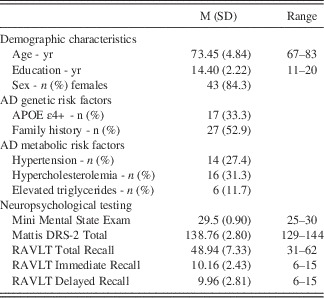
Fifty-one healthy, cognitively intact, older adults participated in a longitudinal study of genetic risk for Alzheimer’s disease funded by the National Institute on Aging (R01 AG022304). Demographic characteristics of the sample (age, education, and sex) are shown in Table 1. Participants were initially recruited from newspaper advertisements and were excluded if they were currently taking psychoactive medication or had a history of neurologic, psychiatric, or medical conditions that may affect brain functioning. Details of the recruitment process and inclusion/exclusion criteria are provided in a previous publication (Seidenberg et al., Reference Seidenberg, Guidotti, Nielson, Woodard, Durgerian, Antuono and Rao2009). The project was approved by the Medical College of Wisconsin Institutional Review Board; all participants provided written informed consent.
Baseline demographic characteristics, AD genetic risk factors, AD metabolic risk factors, and neuropsychological testing.

Note. AD=Alzheimer's disease; APOE=Apolipoprotein E; DRS-2=Dementia Rating Scale, 2nd Edition; RAVLT=Rey Auditory Verbal Learning Test.
To enrich the sample with individuals at genetic risk for AD, 52% (n=27) of the recruited sample had a FH of dementia (see Table 1). A positive FH was defined by either a clear clinical diagnosis of AD or a reported history of gradual decline in memory and other cognitive functions in the absence of a formal diagnosis of AD in a first-degree relative (i.e., parent or sibling). APOE genotype was then determined using a polymerase chain reaction method (Saunders et al., Reference Saunders, Hulette, Welsh-Bohmer, Schmechel, Crain and Burke1996); 33% (n=17) of the sample possessed one (n=15; 29.4%) or both (n=2; 3.9%) APOE ε4 alleles.
Participants completed a health questionnaire regarding their history of potential metabolic risk factors for AD (i.e., diabetes, hypertension, hypercholesterolemia, elevated triglycerides). Data were coded dichotomously (condition present/absent) and were based on participant self-report. Table 1 indicates the number of patients with each self-reported metabolic condition. Only one participant reported being treated for diabetes.
All participants were administered the following measures at study entry to ensure they were cognitively intact: Mini Mental State Examination (MMSE; Folstein, Folstein, & McHugh, Reference Folstein, Folstein and McHugh1975), RAVLT (Rey, Reference Rey1958), and Mattis Dementia Rating Scale, 2nd Edition (DRS-2; Jurica, Leittten, & Mattis, Reference Jurica, Leittten and Mattis2001). They were excluded if they scored below 25 on the MMSE or 1.5 standard deviations below age-appropriate means on RAVLT delayed recall (Woodard et al., Reference Woodard, Seidenberg, Nielson, Antuono, Guidotti, Durgerian and Rao2009). A score below 10 on the Geriatric Depression Scale (GDS; Yesavage et al., Reference Yesavage, Brink, Rose, Lum, Huang and Adey1983) was also required for inclusion in the study to rule out moderate to severe depressive symptoms, as was a score in the normal range on a measure of activities of daily living (Lawton Instrumental Activities of Daily Living Scale; Lawton & Brody, Reference Lawton and Brody1969). Results of baseline neuropsychological testing is presented in Table 1.
Longitudinal Episodic Memory Testing
Participants underwent repeat RAVLT testing at the 3-year follow-up examination. The RAVLT was chosen as the primary outcome measure based on findings that EM tests are the most sensitive to early cognitive decline in healthy, non-demented elders (Albert et al., Reference Albert, Moss, Tanzi and Jones2001; Backman et al., Reference Backman, Small and Fratiglioni2001; Blacker et al., Reference Blacker, Lee, Muzikansky, Martin, Tanzi, McArdle and Albert2007; Bondi et al., Reference Bondi, Salmon, Galasko, Thomas and Thal1999). From the RAVLT, three primary indices were investigated: total recall of trials 1–5, immediate recall, and delayed recall. Participants received alternate forms of the RAVLT, with a counterbalanced order of administration; no significant differences in performance accuracy were observed across the test forms.
MRI Scanning
At study entry participants underwent anatomical and DTI scans. Images were acquired on a General Electric (Waukesha, WI) Signa Excite 3.0 Tesla short bore scanner at Froedtert Memorial Lutheran Hospital (Milwaukee, WI). Foam padding was used to reduce head movement within the coil.
Diffusion tensor imaging
Whole brain (35 axial slices; 4 mm) diffusion tensor images were acquired with 25 encoding directions (b=1000 s/mm2; echo time [TE]=min (86.5 ms); repetition time [TR]=10, 000 ms; field of view [FOV]=240 mm, matrix=128 × 128; acquisitions=1). Images were smoothed at 6 mm full-width half-maximum to eliminate Gibbs ringing. Head movement and eddy currents were corrected and the Brain Extraction Tool (BET; Smith, Reference Smith2002) was applied to exclude non-brain voxels. Each diffusion-weighted image was registered to the corresponding B0 image (no diffusion encoding). FA, MD, DA, and DR images were created by fitting a tensor model to the raw diffusion data using the FSL Diffusion Toolbox (Behrens et al., Reference Behrens, Woolrich, Jenkinson, Johansen-Berg, Nunes and Clare2003).
DTI data were first processed using the Tract-Based Spatial Statistics (TBSS; Smith et al., Reference Smith, Jenkinson, Johansen-Berg, Ruckert, Nichols and Mackay2006) program found in FSL 4.0 image processing software (http://www.fmrib.ox.ac.uk/fsl/tbss/index.html) to create a mean WM skeleton. Specifically, all FA images were aligned to the FMRIB58 standard image in cubic 1 mm standard space. FA images were averaged and a skeleton created with an FA threshold of 0.2 representing tracts common to all participants. This method of “thinning” each WM tract perpendicular to the tract and thresholding FA helps minimize partial volume effects and areas of high inter-subject variability (Smith et al., Reference Smith, Jenkinson, Johansen-Berg, Ruckert, Nichols and Mackay2006). Each participant’s maximum FA value from the nearest relevant tract center was assigned to the corresponding skeleton voxel, which corrects for residual misalignments after nonlinear registration. Diffusivity measures from the same maximum FA voxels were also assigned to the corresponding skeleton voxels.
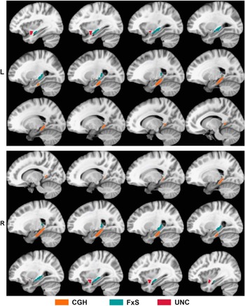
Three WM tracts of interest (TOIs) were defined because of their importance for EM: the bilateral cingulate-hippocampal tract (CGH), fornix/stria terminalis (FxS), and uncinate fasciculus (UNC). These TOIs (see Figure 1) were derived from the ICBM DTI-81 WM atlas (Mori et al., Reference Mori, Oishi, Jiang, Jiang, Li, Akhter and Mazziotta2008). Using the Analysis of Functional Neuroimages (AFNI) software package (Cox, Reference Cox1996), the TBSS-derived FA skeleton was overlaid onto the three DTI-81-defined TOIs. From this conjunction mask, mean FA, MD, DA, and DR values were calculated for each subject for each TOI. Figure 1 displays these tracts as represented in the ICBM DTI-81 atlas.
Three medial temporal white matter tracts investigated in this study: cingulate-hippocampal (CGH), fornix/stria-terminalis (FxS), and uncinate fasciculus (UNC). Tracts defined by the ICBM DTI-81 white matter atlas (Mori et al., Reference Mori, Oishi, Jiang, Jiang, Li, Akhter and Mazziotta2008).

Anatomical MRI
High-resolution anatomical scans were derived from a three-dimensional spoiled gradient-recalled at steady-state (SPGR) pulse sequence [TE=3.9 ms; TR=9.5 ms; inversion recovery preparation time=450 ms; flip angle=12 degrees; number of excitations=2; slice thickness=1.0 mm; FOV=24 cm; resolution=256 × 224; slices=144]. Bilateral volumes of selected GM (hippocampus, parahippocampal gyrus, entorhinal cortex) and WM (parahippocampal gyrus, entorhinal cortex) regions related to DTI tracts of interest as well as total intracranial volume (ICV) were calculated from SPGR images using FreeSurfer software version 5.1.0 (Fischl et al., Reference Fischl, Salat, Busa, Albert, Dieterich and Haselgrove2004). GM and WM volumes were then corrected for variations in head size by dividing by ICV.
Statistical Analyses
Prediction of change in EM performance was accomplished by correlating residualized 3-year change score outcomes on three EM measures (RAVLT total recall of trials 1–5, RAVLT immediate recall, and RAVLT delayed recall) with the following set of baseline (study entry) predictors: DTI (FA, MD, DA, DR), volumetric MRI, demographic characteristics, AD genetic risk factors (APOE, FH), and AD metabolic risk factors (hypertension, hypercholesterolemia, and elevated triclycerides). Residualized change scores were calculated by regressing baseline scores on 3-year follow-up scores; this technique adjusts for baseline performance, practice effects and regression to the mean (McSweeny, Naugle, Chelune, & Luders, Reference McSweeny, Naugle, Chelune and Luders1993; Tucker, Damarin, & Messick, Reference Tucker, Damarin and Messick1966). Pearson and point biserial correlations were calculated for continuous and dichotomous predictor variables, respectively. To control for multiple comparisons, a false discovery rate (FDR; Benjamini & Hochberg, Reference Benjamini and Hochberg1995) threshold was used to control family-wise error rate with significance established at q<.05. FDR thresholds were assessed separately for each class of DTI (FA, MD, DA, and DR), volumetric (GM and WM), and non-imaging variables. Multiple regression was performed when one or more of the correlations significantly predicted the same RAVLT outcome measure following FDR correction. We also report, but do not interpret, p<.05 correlations that did not survive FDR correction.
RESULTS
Over the 3-year retest interval, seven participants (14%) converted to a diagnosis of MCI based on our previously published criteria (see Woodard et al., Reference Woodard, Seidenberg, Nielson, Antuono, Guidotti, Durgerian and Rao2009 for details), five of whom (71%) were APOE ε4 carriers. A Fisher’s exact test indicated a significantly higher proportion of carriers amongst those who converted to MCI (p=.034), while no significant association was found between the presence/absence of FH of dementia and MCI conversion.
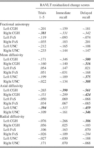
Table 2 summarizes correlations between baseline DTI tract measures and residualized change scores on the three EM measures: RAVLT total recall of trials 1–5, RAVLT immediate recall, and RAVLT delayed recall. Of the FA tract measures, only 1 of 18 correlations survived FDR correction: right CGH tract correlated negatively with 3-year change in RAVLT total recall of trials 1–5 (r=−.381; p=.006).
Pearson product-moment correlations between baseline DTI indices for each WM tract and residualized RAVLT change scores

Bold/italics indicate correlations surviving FDR correction for multiple comparisons. Italics indicate correlations significant at p<.05, but not surviving FDR correction. CGH=cingulate-hippocampal tract; FxS=fornix/stria terminalis tract; UNC=uncinate fasciculus tract; RAVLT =Rey Auditory Verbal Learning Test.
Significant negative correlations between MD and RAVLT delayed recall were observed bilaterally in the CGH and UNC tracts (Table 2). These results indicate that higher baseline MD was associated with decreased memory performance over time. Since MD represents an average of DA and DR, we then conducted separate analyses to determine if these significant MD correlations were being driven by DA, DR, or both.
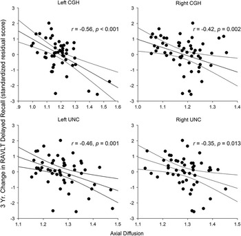
Similar to MD, baseline DA correlated negatively with changes in RAVLT delayed recall change scores in the bilateral CGH and UNC tracts (see Table 2; Figure 2). In addition, DA correlated negatively with RAVLT immediate recall in the left CGH and RAVLT total recall of trials 1–5 in the left UNC (Table 2).
Correlations between baseline axial diffusion and 3-year changes in Rey Auditory Verbal Learning Test (RAVLT) delayed recall scores. Regression line includes the 95% confidence interval. CGH=cingulate-hippocampal tract; UNC=uncinate fasciculus.

The relationship between baseline DR and changes in RAVLT test performance was less pronounced. Only 1 of 18 correlations was significant: the left CGH tract correlated negatively with changes in RAVLT delayed recall (Table 2). These results indicate that the significant correlations between MD and RAVLT delayed recall scores were being driven primarily by baseline axial rather than radial diffusion.
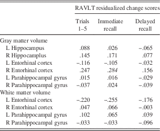
Table 3 summarizes correlations between baseline MTL GM and WM volumes and changes in RAVLT test performance. None of these correlations were significant after FDR correction. These results indicate that DTI measures of WM microstructure are better at predicting changes in EM test performance than measures of atrophy within MTL structures.
Pearson product-moment correlations between baseline mesial temporal lobe gray and white matter volumes and residualized RAVLT change scores

Bold/italics indicate correlations surviving FDR correction for multiple comparisons. Italics indicate correlations significant at p<.05, but not surviving FDR correction. RAVLT=Rey Auditory Verbal Learning Test.
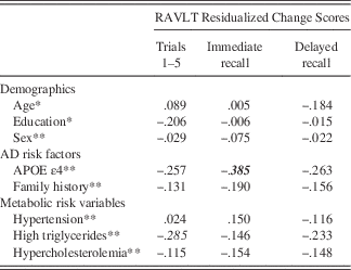
Table 4 reports correlations between 3-year change in RAVLT scores and demographic characteristics, genetic risk factors, and metabolic risk factors at baseline. Only one correlation was significant: change in RAVLT immediate recall correlated negatively with APOE status (i.e., carriers exhibited poorer performance over time).
Correlations between demographic, AD genetic risk, and metabolic risk variables and residualized RAVLT change scores

*Pearson corelations; **Point biserial correlations.
Bold/italics indicate correlations surviving FDR correction for multiple comparisons. Italics indicate correlations significant at p<.05, but not surviving FDR correction. RAVLT=Rey Auditory Verbal Learning Test.
Because both DA within the left CGH tract (Table 2) and APOE status (Table 4) correlated significantly with changes in RAVLT immediate recall, a multiple regression was performed with RAVLT immediate recall as the outcome and left CGH DA and APOE status as predictors. Both APOE status (β=−.334; t(50)=−2.609; p=.012) and left CGH DA (β=−.327; t(50)=−2.556, p=.014) remained significant predictors in the overall model (F(2,48)=8,242; p =.001; R2=.260), suggesting that each variable made independent contributions to the prediction of longitudinal EM changes.
DISCUSSION
Our results indicate that DTI diffusion measures (predominantly DA and MD) of mesial temporal lobe white matter tracts (cingulate-hippocampal and uncinate fasciculus) obtained from cognitively intact older adults predicted subsequent episodic memory decline over a 3-year interval. It is important to emphasize that these results were driven primarily by DA, since MD represents an average of DR and DA measures. Specifically, greater DA in these two critical EM-related WM tracts predicted a 3-year decline predominantly in delayed free recall on the RAVLT (Table 2, Figure 2). FA and DR in the cingulate-hippocampal tract also predicted EM change, although these results were less striking. Left hemisphere structures were more predictive of verbal EM decline overall, but most of the significant correlations between DTI measures and EM change involved bilateral WM tracts. Both APOE ε4 status and DA within the left CGH tract made unique and independent contributions to the prediction of change in RAVLT immediate recall. In contrast, volumes of MTL WM and GM structures did not predict 3-year EM change (Table 3). Likewise, demographic variables (age, education, and sex), FH of dementia, and metabolic/vascular risk factors (hypertension, hypercholesterolemia, and elevated triglycerides) did not predict long-term changes in EM functions (Table 4).
It is important to note that, in the current study, microstructural changes in WM, as reflected predominantly by increased DA within the CGH and UNC tracts, were able to predict EM changes, whereas atrophy of the MTL GM and WM structures was not. Abnormalities in DA are commonly associated with axonal disruption (Agosta et al., Reference Agosta, Pievani, Sala, Geroldi, Galluzzi, Frisoni and Filippi2011; O’Dwyer et al., Reference O’Dwyer, Lamberton, Bokde, Ewers, Faluyi, Tanner and Hampel2011; Song et al., Reference Song, Sun, Ju, Lin, Cross and Neufeld2003) that likely precede neuronal loss and atrophy as observed on anatomical MR scans, whereas DR has been associated with demyelination or myelin breakdown (Song et al., Reference Song, Sun, Ramsbottom, Chang, Russell and Cross2002). Recent studies, however, suggest this explanation may be overly simplistic (Pierpaoli et al., Reference Pierpaoli, Barnett, Pajevic, Chen, Penix, Virta and Basser2001; Wheeler-Kingshott & Cercignani, Reference Wheeler-Kingshott and Cercignani2009). Consistent with our findings, DA was found to be more sensitive than DR in studies of cognitive decline on a general cognitive screening measure in healthy older adults (Wang et al., Reference Wang, Fratiglioni, Laukka, Lovden, Kalpouzos, Keller and Qiu2015) and in conversion to MCI and AD in healthy elders (Fletcher et al., Reference Fletcher, Raman, Huebner, Liu, Mungas, Carmichael and DeCarli2013). DA also appears more sensitive than other DTI measures to WM changes along the symptomatic stage of the AD neurodegenerative continuum (Acosta-Cabronero et al., Reference Acosta-Cabronero, Williams, Pengas and Nestor2010; Agosta et al., Reference Agosta, Pievani, Sala, Geroldi, Galluzzi, Frisoni and Filippi2011). Thus DA changes may serve as an earlier and more sensitive biomarker than atrophy for predicting future EM decline, especially in cognitively intact, healthy elders.
DTI abnormalities in the CGH and UNC tracts have been observed in prior studies involving healthy elders and patients with MCI and AD. The CGH tract consists of posterior cingulate projections to the parahippocampal gyrus, the major input system to the hippocampus (Dickerson & Eichenbaum, Reference Dickerson and Eichenbaum2010). DTI measures from this tract have previously been shown to predict EM decline (Zhuang et al., Reference Zhuang, Sachdev, Trollor, Kochan, Reppermund, Brodaty and Wen2012) and general cognitive decline (Wang et al., Reference Wang, Fratiglioni, Laukka, Lovden, Kalpouzos, Keller and Qiu2015) in healthy elders. The UNC tract, which provides an important connection between MTL structures including the hippocampus, amygdala, and orbitofrontal cortex (Von Der Heide, Skipper, Klobusicky, & Olson, Reference Von Der Heide, Skipper, Klobusicky and Olson2013), has a demonstrated role in verbal EM performance (Christidi et al., Reference Christidi, Zalonis, Kyriazi, Rentzos, Karavasilis, Wilde and Evdokimidis2014; Schaeffer et al., Reference Schaeffer, Krafft, Schwarz, Chi, Rodrigue, Pierce and McDowell2014). Diffusivity measures (DA and DR) in both the CGH and UNC tracts are altered in MCI and AD (Canu et al., Reference Canu, McLaren, Fitzgerald, Bendlin, Zoccatelli, Alessandrini and Frisoni2010; Fu, Liu, Li, Chang, & Li, Reference Fu, Liu, Li, Chang and Li2014; Hiyoshi-Taniguchi et al., Reference Hiyoshi-Taniguchi, Oishi, Namiki, Miyata, Murai, Cichocki and Fukuyama2015; Salat et al., Reference Salat, Tuch, van der Kouwe, Greve, Pappu and Lee2010; Yasmin et al., Reference Yasmin, Nakata, Aoki, Abe, Sato, Nemoto and Ohtomo2008; Zhang et al., Reference Zhang, Schuff, Jahng, Bayne, Mori, Schad and Weiner2007), and have shown negative associations with EM in MCI (Li et al., Reference Li, Muftuler, Chen, Ward, Budde, Jones and Goveas2014; Metzler-Baddeley et al., Reference Metzler-Baddeley, Hunt, Jones, Leemans, Aggleton and O’Sullivan2012; Remy, Vayssiere, Saint-Aubert, Barbeau, & Pariente, Reference Remy, Vayssiere, Saint-Aubert, Barbeau and Pariente2015; Zhuang et al., Reference Zhuang, Sachdev, Trollor, Reppermund, Kochan, Brodaty and Wen2013).
Surprisingly, DTI measures derived from the fornix, the major WM output tract from the hippocampus, did not predict 3-year changes in EM. Metzler-Baddeley et al. (Reference Metzler-Baddeley, Hunt, Jones, Leemans, Aggleton and O’Sullivan2012) observed that whereas DTI measures correlated with EM performance in healthy older adults, DTI measures from the uncinate and parahippocampal tracts, but not the fornix, correlated with EM in MCI patients. They hypothesized that early fornix damage associated with the AD pathology may cause other associated WM tracts to become disproportionately involved in EM function. The finding by Ray et al. (Reference Ray, Metzler-Baddeley, Khondoker, Grothe, Teipel, Wright and O’Sullivan2015) that MCI participants’ performance on EM measures was more related to parahippocampal cortex than fornix volume also supports this hypothesis.
The current findings suggest that DTI of MTL tracts may provide important information for the detection of preclinical changes associated with AD (Sperling et al., Reference Sperling, Aisen, Beckett, Bennett, Craft, Fagan and Phelps2011). Our sample was enriched with individuals at increased genetic risk of AD by virtue of possessing at least one APOE ε4 allele and/or having FH of dementia. We focused our DTI analysis on fiber tracts within the MTL, since this region is affected early in the AD process and is less susceptible to the effects of normal aging (Raz & Rodrigue, Reference Raz and Rodrigue2006). In addition, as hypothesized, DTI measures were observed to be most effective in predicting changes in delayed recall, one of the earliest cognitive indicators of conversion to AD in asymptomatic elders (Albert et al., Reference Albert, Moss, Tanzi and Jones2001; Hamel et al., Reference Hamel, Kohler, Sistermans, Koene, Pijnenburg, van der Flier and Ramakers2015; Mistridis et al., Reference Mistridis, Krumm, Monsch, Berres and Taylor2015; Papp et al., Reference Papp, Amariglio, Mormino, Hedden, Dekhytar, Johnson and Rentz2015). We also note that a significantly higher proportion of our APOE ε4 carriers than non-carriers converted to MCI at the 3-year follow-up examination, suggesting that our recruitment procedure likely resulted in a higher proportion of persons in the preclinical stage of AD than the general population of similarly-aged adults. Post hoc supplementary moderated regression analyses were conducted to examine the interaction effects of WM microstructure and APOE ε4 status on EM change. These analyses were not significant, suggesting that APOE ε4 and early WM changes may be independent contributors to EM decline.
It is important to note that we did not directly measure amyloid β and tau derived from cerebrospinal fluid (CSF) nor did we conduct amyloid positron emission tomography (PET) imaging. Such measures are also able to predict subsequent cognitive decline in healthy elders (Ellis et al., Reference Ellis, Lim, Harrington, Ames, Bush and Darby2013; Fagan et al., Reference Fagan, Roe, Xiong, Mintun, Morris and Holtzman2007; Lim et al., Reference Lim, Pietrzak, Ellis, Jaeger, Harrington, Ashwood and Maruff2013; Mattsson et al., Reference Mattsson, Forsberg, Persson, Nyberg, Nilsson, Halldin and Farde2015; Moghekar et al., Reference Moghekar, Li, Lu, Li, Wang and Albert2013; Thai et al., Reference Thai, Lim, Villemagne, Laws, Ames and Ellis2015). It should also be noted, however, that not all healthy adults with evidence of amyloid β or abnormal tau demonstrate cognitive decline (Edmonds, Delano-Wood, Galasko, Salmon, & Bondi, Reference Edmonds, Delano-Wood, Galasko, Salmon and Bondi2015; Sperling et al., Reference Sperling, Aisen, Beckett, Bennett, Craft, Fagan and Phelps2011). A recent study of cognitively normal elders from the ADNI database (Edmonds et al., Reference Edmonds, Delano-Wood, Galasko, Salmon and Bondi2015) demonstrated that 17–22% of participants with evidence of cerebral amyloid accumulation on PET imaging converted to MCI or AD over a period of 12–96 months. Adding abnormal CSF tau to the presence of amyloidosis increased the prediction to 37%. These findings suggest that additional neuroimaging markers may be needed to improve the prediction of conversion to MCI and/or AD in healthy older adults. Future larger scale longitudinal studies are needed to compare the sensitivity of DTI measures to other imaging techniques (anatomical MRI, amyloid and tau PET) and CSF biomarkers in predicting EM decline and conversion from the cognitively healthy state to MCI and AD.
The current study is not without limitations. Despite enriching our sample with persons with a higher genetic risk for developing AD, only seven participants converted to MCI after the three year follow-up interval. This sample size is too small to draw conclusions about the accuracy of DTI to predict AD progression. Our tracts-of-interest were based on a WM skeleton created in TBSS using the ICBN DTI-81 atlas (Mori et al., Reference Mori, Oishi, Jiang, Jiang, Li, Akhter and Mazziotta2008). By focusing on the WM skeleton based on the highest FA values, we reduced, but possibly did not entirely eliminate, partial volume effects. Finally, metabolic/vascular risk factors were based on self-report.
To summarize, our findings suggest that DTI measures of axial diffusion within MTL WM tracts are capable of predicting EM decline in healthy, cognitively intact older adults. Such information may also help in characterizing the pathophysiological changes associated with the preclinical stage of AD. With data from future larger scale longitudinal validation studies, it is conceivable that DTI measures will prove useful, in combination with other imaging and non-imaging biomarkers, in the identification of healthy individuals at greatest risk for cognitive decline to serve in AD prevention studies and for monitoring the effects of interventions administered during the preclinical stage.
ACKNOWLEDGMENTS
We thank Alissa M. Butts, Kelli L. Douville, Christina M. Figueroa, Amelia Gander, Evan Gross, Leslie M. Guidotti-Breting, Nathan C. Hantke, Kathleen E. Hazlett, Emily Hoida, Cassandra Kandah, Christina D. Kay, Dana Kelly, Monica Matthews, Sarah K. Miller, Andria L. Norman, Katherine Reiter, Michael A. Sugarman, and Qi Zhang for their assistance. This work was supported by the National Institutes of Health Grants R01 AG022304 and M01 RR00058. The content is solely the responsibility of the authors and does not necessarily represent the official views of the National Institute on Aging or the National Institutes of Health. Conflict of interest: The authors declare no conflict of interest.