Introduction
The zoonotic significance of Toxocara canis larvae is of prime interest considering its strong tendency to cause ‘visceral larva migrans’ (VLM) in abnormal hosts, including humans, which act as paratenic hosts of T. canis. Toxocara canis larvae persist for months or years in a wide range of mammalian laboratory hosts (Pahari & Sasmal, Reference Pahari and Sasmal1990a) and it is presumed that they are transferred from animal to animal through paratenism in nature (Sprent, Reference Sprent1954; Pahari & Sasmal, Reference Pahari and Sasmal1990b). With regard to the role of invertebrates, Sprent (Reference Sprent1956), Okoshi & Usui (Reference Okoshi and Usui1968) and Pahari & Sasmal (Reference Pahari and Sasmal1991) reported successful infections of earthworms with T. cati and T. canis and the establishment of patent infections in the definitive host. In the present study the disseminating capacity of the cockroach Periplaneta americana and the establishment of patent infections of T. canis in pups with disseminated larvae or eggs from P. americana were investigated.
Materials and methods
Infective eggs
Bulk samples of embryonated eggs suspended in water were developed using the method described by Pahari & Sasmal (Reference Pahari and Sasmal1990a). Ten T. canis-infected pups, 5–7 weeks old, were treated with albendazole for collection of adult T. canis. The following morning the excreted worms were thoroughly washed with normal saline and mature females were separated. The gravid uteri of the worms were dissected out and teased on Petri dishes containing 0.5% formalin. The eggs in formalin were incubated at 27°C for 22 days for full embryonation. The number of embryonated eggs in bulk samples and the inoculum was determined by the method of Oshima (Reference Oshima1961) and larvae were recovered from tissues as described by Sprent (Reference Sprent1952).
Paratenic host (cockroaches, Periplaneta americana)
Sixty cockroaches, almost equal in size, were collected from domestic kitchen stores were reared in six glass jars at room temperature and fed with pieces of bread, sugar and minced potato throughout the experimental period. Before experimental use ten cockroaches were selected randomly, the tissues and intestines were squashed and digested in artificial gastric juice (pepsin, 0.5 g; HCl, 0.7 ml; 0.9% saline, 99.3 ml; pH adjusted to 1.0 with HCl) and were confirmed to be free from naturally acquired helminth infections. Faeces were also examined to ensure the absence of helminth eggs or larvae.
Definitive hosts
Five 6-week-old, locally bred pups of either sex were obtained from a local breeder and dewormed with albendazole (20 mg/kg body weight) for two consecutive days. Faeces of pups were examined for 3 days and on day 7 after medication all pups were found to be free of infection. Pups were maintained in sterilized individual iron cages and fed on bread, boiled rice, milk, sugar and glucose.
Experimental design
Experimental infection of cockroaches
Sixty cockroaches, almost equal in size were reared free of infection and their faeces examined to confirm the absence of a naturally acquired infection. Three groups – 1 and 2 (experimental) and 3 (control) – each of 16 cockroaches, were placed into three separate glass jars. Following starvation for a day, groups 1 and 2 were supplied with two pieces of sweetbread containing 1 ml suspensions of 3 × 10⁵ and 5 × 10⁵ embryonated eggs of T. canis, respectively. Cockroaches in group 3 were treated as uninfected controls.
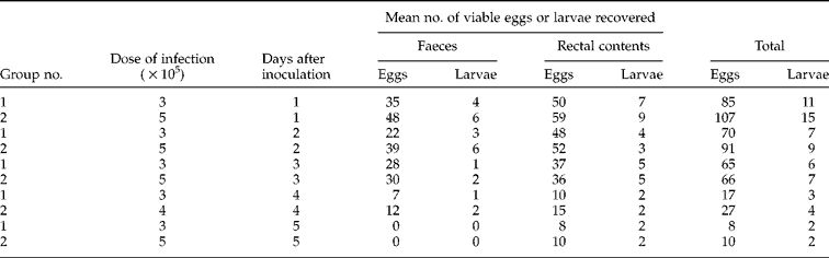
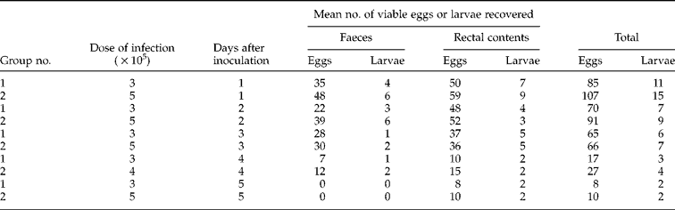
From 24 h post-infection, two cockroaches from each group, including the control, were killed 2, 3, 4, 5, 10, 15 and 20 days post-infection (DPI) for recovery of larvae. The entire alimentary tract was dissected out and the mid and hind gut contents examined separately by squashing. Tissues of the head, thorax and abdomen were minced finely and digested in artificial gastric juice and the sediment was examined for the presence of larvae. The pelleted faeces of groups 1, 2 and 3 were also collected on glass slides, soaked with normal saline and examined by the direct smear method for the presence of any eggs or larvae from 1 to 5 DPI at 24-h intervals. Five faecal samples from each group were examined and a mean count taken (table 1). Eggs and larvae were identified after Nichols (Reference Nichols1956) and Sprent (Reference Sprent1955) and percent inoculum was calculated following the formula of Pahari & Sasmal (Reference Pahari and Sasmal1990a).
Experimental infection of pups
One gram of infected faeces from each of group 1 and 2 cockroaches was examined by the squash method for calculation of total number of eggs or larvae present in 5 g of faeces used for infecting each pup. Five worm-free pups were divided into three groups: two pups in each of groups 1 and 2 and one pup in control group 3. Two pups in each of groups 1 and 2 were fed with 5 g of faeces from cockroaches infected with 3 × 10⁵ and 5 × 10⁵ eggs of T. canis, respectively. One pup from group 3 was fed with 5 g of uninfected faeces from control cockroaches. Any accidental T. canis infection from other sources was prevented by feeding pups with boiled food and water and maintaining strict hygienic measures. The faeces of each pup were examined at 24-h intervals from 20 to 38 DPI for the presence of T. canis eggs and expelled adult worms. Pups were sacrificed on day 38 PI by chloroform anaesthesia and post-mortem examination undertaken.
Results
Experimental infection of cockroaches
No mortality occurred in infected cockroaches within 20 DPI. No larvae were recovered from any tissues or organs using the direct or digestion method of examination. Daily examination of faeces and rectal contents of infected cockroaches revealed numerous viable eggs and only a few hatched larvae up to 5 DPI. The highest number of viable eggs (85 and 107) and larvae (11 and 15) were recovered from groups 1 and 2, respectively, on 1 DPI. This output decreased gradually up to 5 DPI. On day 5 faeces were found to be free of eggs and larvae; the rectal contents of groups 1 and 2 contained 8 and 10 eggs, plus 2 and 2 larvae, respectively (table 1). By 6 DPI no egg or larval output was observed. Neither eggs nor larvae were recovered from faeces or rectal contents of cockroaches in control group 3.
Recovery of viable eggs and larvae of Toxocara canis from the faeces and rectal contents of cockroaches inoculated with 3×10⁵ and 5×10⁵ infective eggs in food.
Experimental infection of pups
Regular faecal examination of pups in groups 1 and 2 from day 20 PI revealed the first appearance of immature T. canis worms entangled in stools on day 35 PI and Toxocara eggs on day 38 PI. The control pup remained free of infection. On day 38 PI, the worm burdens at necropsy ranged from 6 to 9 and 18 to 23 in groups 1 and 2, respectively. The majority of worms were slender and highly motile and located in the proximal part of the duodenum.
Discussion
The worm-free status of the control pup confirmed the contamination-free experimental conditions of both cockroaches and pups. Results on feeding of cockroaches with T. canis eggs indicate that the larvae cannot migrate in the tissues of cockroaches but infective embryonated eggs can remain viable in the gut of cockroaches and by 5 DPI they are disseminated with the faeces. Sprent (Reference Sprent1956) succeeded in recovering larvae from the tissues of cockroaches infected with T. cati eggs, but the present study revealed different results. Instead of a somatic migration, most of the viable eggs and a small number of actively motile hatched second-stage larvae of T. canis passed out with the faeces of infected cockroaches. This output increased with an increasing dose of embryonated eggs, as observed in cockroaches infected with 5 × 10⁵ eggs. A similar finding was also reported by Okoshi & Usui (Reference Okoshi and Usui1968) in cockroaches infected separately with T. canis, T. cati and Toxascaris leonina, although no larvae were recovered from the somatic tissues of the cockroaches, irrespective of ascarids used for infection. Bonfante et al. (Reference Bonfante, Faust and Giraldo1961) in an epidemiological survey in Cali, Columbia, detected many intestinal parasites of humans, including the eggs of Ascaris lumbricoides, in the intestinal contents of cockroaches (P. americana) and thus concluded that the cockroach is a potential mechanical disseminator of many natural parasites of humans. Thus cockroaches, as one of the commonest invaders of human dwellings, mainly in kitchens and store rooms, can be a potential source of ascarid infections to humans, especially where infected pups are reared with free access to kitchen areas. The danger of this infection for humans must be considered, but other than a disseminator of viable T. canis eggs, cockroaches cannot be considered as paratenic hosts.
In experimental infections of pups, the viability of eggs and larvae liberated with the faeces of cockroaches has been confirmed by the establishment of patent infections in the definitive host, the dog. The control pup showed no evidence of worm burdens at necropsy on 38 DPI, which confirmed the overall worm-free status of experimental pups.
The establishment of patent infections in worm-free pups has been reported following the feeding of migratory T. canis larvae from different abnormal hosts (Japanese quail (Pahari & Sasmal, Reference Pahari and Sasmal1990a, b) and piglets (Acharya et al., Reference Acharya, Sasmal, Jana and Roy2002)), but no such studies on the establishment of patent infections have been reported previously by feeding infected droppings of cockroaches to worm-free pups. The infection rate of pups with adult T. canis also varied with the dose of infected faeces fed to these pups. Therefore it can be concluded that, as in the case of other abnormal hosts, cockroaches cannot be considered as a paratenic host of T. canis, but because of their habitat and food habits, they can transmit the infection from infected faeces of dogs or from soil, and contaminate human food and water supplies. Cockroaches can also act as mechanical disseminators of viable ascarid and other eggs of zoonotic significance, resulting in various helminthic diseases.
Acknowledgements
The authors are grateful to the West Bengal University of Animal and Fishery Sciences, Kolkata and Bidhan Chandra Agricultural University, Mohanpur, Nadia, West Bengal, India, for providing financial assistance and facilities.