INTRODUCTION
Progression through the eukaryotic cell cycle is driven by a family of protein kinases known as cyclin-dependent kinases (CDKs). These proteins are highly conserved among eukaryotic organisms and are classified into two categories: essential and non-essential CDKs. Within the essential kinases, two of the most well characterized CDKs in lower eukaryotes are Cdc2 from Schizosaccharomyces pombe and Cdc28 from Saccharomyces cerevisiae. Both of these proteins act as the essential regulators of cell cycle progression in these organisms, and their inactivation affects cell viability (Nurse and Bissett, Reference Nurse and Bissett1981; Reed and Wittenberg, Reference Reed and Wittenberg1990). Several CDK proteins have been described in mammalian cells but only CDK1 and the interphase CDKs (CDK2, CDK3, CDK4 and CDK6) participate actively throughout the cell cycle (Malumbres and Barbacid, Reference Malumbres and Barbacid2005; Malumbres, Reference Malumbres2014). In mutant mouse embryos lacking interphase CDKs, CDK1 may bind to all cyclins resulting in the phosphorylation of many substrates, whereas embryos fail to develop in the absence of CDK1. These results indicate that CDK1 plays an essential role in cell cycle progression in mice (Santamaría et al. Reference Santamaría, Barrière, Cerqueira, Hunt, Tardy, Newton, Cáceres, Dubus, Malumbres and Barbacid2007; Enders, Reference Enders2012).
Saccharomyces cerevisiae also encode another protein kinase, Pho85, which is a non-essential CDK with 51% amino acid identity to Cdc28 (Toh-e et al. Reference Toh-e, Tanaka, Uesono and Wickner1988; Nishizawa et al. Reference Nishizawa, Suzuki, Fujino, Oguchi and Toh-e1999). Pho85 may function under special growing conditions such as nutrient starvation (Swinnen et al. Reference Swinnen, Wanke, Roosen, Smets, Dubouloz, Pedruzzi, Cameroni, De Virgilio and Winderickx2006). The homologue of S. cerevisiae Pho85 in mammalian cells is CDK5, which is not thought to be involved in controlling cell cycle progression. Instead, CDK5 plays a role in neuronal morphogenesis and migration in post-mitotic neurons (Huang et al. Reference Huang, Patrick, Moffat, Tsai and Andrews1999; Kawauchi et al. Reference Kawauchi, Shikanai and Kosodo2013).
CDK levels tend to remain constant during the cell cycle, and their activities are controlled mainly at the post-translational level by different mechanisms, including periodic expression of activating cyclins, repression caused by binding to inhibitory proteins [CKIs (cyclin-dependent kinase inhibitors)], a combination of both activation and inhibition by the phosphorylation of conserved amino acids, and the formation of interactions with a Cdc2/Cdc28 kinase subunit (CKS) (Horton and Templeton, Reference Horton and Templeton1997; Malumbres and Barbacid, Reference Malumbres and Barbacid2005; Piscopo and Hinds, Reference Piscopo and Hinds2008; Frontini et al. Reference Frontini, Kukalev, Leo, Ng, Cervantes, Cheng, Holic, Dormann, Tse, Pommier and Yu2012). Cyclins are positive regulatory subunits of CDKs and constitute multiprotein families in metazoans. Tight binding of a cyclin to a kinase and subsequent complete kinase activation requires CDK phosphorylation at a conserved residue (Thr-161 in human CDK1) by a CDK-activating kinase (CAK) (Brown et al. Reference Brown, Noble, Lawrie, Morris, Tunnah, Divita, Johnson and Endicott1999). In contrast, phosphorylation of residues in the ATP-binding site of the kinase (Thr-14 and Tyr-15 in human CDK1) inhibits kinase activity (Morgan, Reference Morgan1995). CKS proteins are evolutionarily conserved in eukaryotes and, among other functions, are essential components for cell cycle transition through regulation of CDK activity (McGrath et al. Reference McGrath, Balog, Kõivomägi, Lucena, Mai, Hirschi, Kellogg, Loog and Rubin2013). Mammalian cells contain two CKS paralogues (CKS1 and CKS2, which share over 80% sequence identity) (Richardson et al. Reference Richardson, Stueland, Thomas, Russell and Reed1990), but yeast and the protozoa Leishmania mexicana and Trypanosoma cruzi have only one reported CKS (Mottram and Grant, Reference Mottram and Grant1996; Muñoz et al. Reference Muñoz, Santori, Rojas, Gómez and Téllez-Iñón2006).
Cell cycle studies have been predominantly performed in yeast, animals and plants. In protozoa, the few studies of proteins involved in cell cycle control have focused mainly in members of the order trypanosomatida: CRKs and cyclins have been identified and characterized in T. cruzi, Trypanosoma brucei and L. mexicana (Mottram et al. Reference Mottram, Kinnaird, Shiels, Tait and Barry1993; Mottram and Smith, Reference Mottram and Smith1995; Gómez et al. Reference Gómez, Santorini, Laria, Engel, Swindle, Eisen, Szankasi and Téllez-Iñón2001; Hammarton et al. Reference Hammarton, Clark, Douglas, Boshart and Mottram2003; Ali et al. Reference Ali, Ibrahim, Grant and Mottram2010). In Trichomonas vaginalis, another early divergent protist, the presence of molecules similar to the Cdc2/28 protein kinase family was demonstrated, suggesting that they may represent the earliest ancestors of key cell division control molecules (Riley et al. Reference Riley, Campbell, Puolakkainen and Krieger1993). Trichomonas vaginalis is the causative agent of trichomoniasis in humans, and the World Health Organization has classified it as the most common non-viral sexually transmitted disease in developing countries (World Health Organization, 2011). Phylogenetic analyses based on the 16S-like ribosomal RNA gene have placed this parasite near the base of the eukaryotic tree (Sogin, Reference Sogin1991); however, the egalitarian model of evolution has challenged this proposal (Dacks et al. Reference Dacks, Walker and Field2008).
In this paper, we identified and characterized a panel of proteins potentially involved in T. vaginalis cell cycle control: three CDKs, designated as TvCRK1, TvCRK2 and TvCRK5 (parasitic CDKs are named CRKs, from Cdc2-related protein kinases), and two CKSs, designated TvCKS1 and TvCKS2. Interactions among these proteins were evaluated, and TvCRK1 and TvCRK5 showed kinase activation in the presence of a cyclin (TvCYC1). This work is the first reported characterization of proteins involved in cell cycle control in T. vaginalis.
MATERIALS AND METHODS
Parasites and culture conditions
The T. vaginalis isolate CNCD147 (Alvarez-Sánchez et al. Reference Alvarez-Sánchez, Avila-González, Becerril-García, Fattel-Facenda, Ortega-López and Arroyo2000) was used throughout the experimental work. Parasites were grown at 37 °C in TYM (trypticase-yeast extract-maltose) medium supplemented with 10% (v/v) heat-inactivated horse serum (GIBCO) as previously described (Diamond, Reference Diamond1957).
Gene identification
Gene sequences encoding CRKs and CKSs in the T. vaginalis genome (TrichDB, http://trichdb.org/trichdb/) were identified by BLAST analyses (Basic Local Alignment Search Tool) (https://blast.ncbi.nlm.nih.gov/Blast.cgi) using selected representative amino acid sequences from different organisms, including human, yeast and protozoa. A genomic sequence annotated as a putative cyclin B (herein referred to as TvCYC1) was used in this work.
Reverse-transcriptase-coupled PCR
Reverse-transcriptase-coupled PCR was used to assess the mRNA expression of the genes identified in this work. Total RNA was prepared from exponentially growing parasites using TRIzol reagent (Invitrogen) according to the manufacturer's instructions. cDNA was synthesized with a Superscript III RNase H-kit and the adapter primer according to the manufacturer's instructions (Invitrogen). To perform a semi-nested PCR, specific internal primers were used for each gene together with the AUAP-B primer (Invitrogen). The PCR conditions were as follows: 50 µL reactions were set up with 40 pmoles of each primer, 1× PCR buffer (Invitrogen), 10 mm of each dNTP, 3 mm MgCl2, 2·5 units of Taq DNA polymerase (recombinant, Invitrogen) and 1·5 µL of total cDNA. The amplification reactions consisted of 30 cycles of 94 °C for 1 min, 50 °C for 30 s, and 72 °C for 30 s, followed by a final 10-min incubation at 72 °C. The PCR products were cloned using a Zero Blunt TOPO PCR cloning system (Invitrogen) and the cDNA clones were automatically sequenced.
Bioinformatics analyses
Amino acid sequence identity and similarity values were calculated using the Sequence Identity And Similarity (SIAS) Tool from the Immunomedicine Group, Universidad Complutense, Madrid (http://imed.med.ucm.es/Tools/sias.html). Protein sequences were analysed using programs from the ExPASy Bioinformatics Resource Portal (https://www.expasy.org/), Clustal Omega from the EMBL–EBI server (http://www.ebi.ac.uk/Tools/msa/clustalo/), and the Align tool from UniProt (http://www.uniprot.org/); protein structure predictions were obtained using the RaptorX server (http://raptorx.uchicago.edu/StructurePrediction/predict/) (Källberg et al. Reference Källberg, Wang, Wang, Peng, Wang, Lu and Xu2012).
Cloning and expression of T. vaginalis genes encoding CRKs, CKSs and a cyclin
The identified open reading frames (ORFs) encoding CRKs, CKSs and a cyclin were amplified by PCR from T. vaginalis CNCD147 genomic DNA using specific primers with flanking restriction sites. The PCR products were cloned into a TOPO vector (Invitrogen, Carlsbad, CA, USA) and sub-cloned into an expression vector: the pQE-30 plasmid was used as an expression vector for the TvCRKs and TvCKS, which were modified to contain 6xHis tags at their N-termini; TvCYC1 was modified to contain a glutathione S-transferase (GST) tag at its N-terminus through cloning into a pGEX-4T-1 plasmid. All the plasmid constructs were automatically sequenced. The plasmids were transformed into Escherichia coli, and recombinant protein expression was induced with 1 mm IPTG (isopropyl-1-thiol-β-D-galactopyranoside). Recombinant proteins were purified under native conditions from bacterial lysates with the aid of Ni-NTA agarose beads (for the 6xHis-tagged proteins) or a glutathione Sepharose matrix (for the GST-tagged proteins) as described by the manufacturers (Qiagen and GE Healthcare, respectively) and were subsequently used for the kinase activity assays.
Complementation of a S. cerevisiae cdc28-knockout mutant
TvCRKs ORFs were cloned into the yeast expression vector pYES2 (Invitrogen), which contains a URA3 gene for transformant selection. The lithium acetate method was used to transform the S. cerevisiae cdc28-knockout heterozygous diploid strain BY4743 (MATa/α, his3Δ1/his3Δ1, leu2Δ0/leu2Δ0, LYS2/lys2Δ0, met15Δ0/MET15 ura3Δ0/ura3Δ, CDC28/cdc28Δ::KANMX) (Thermo Fischer Scientific cat. no. YSC1021-674028). Following the transformation, cells were plated on selection medium (synthetic dextrose minimal medium supplemented with histidine and leucine) and grown at 30 °C. The clones were then transferred to a pre-sporulation medium (0·8% yeast extract, 0·3% peptone, 10% anhydrous dextrose, 2% agar) and finally to minimal sporulation medium (1% potassium acetate, 5 mg L−1 histidine, 25 mg L−1 leucine, 2% agar). Complementation was determined by spore viability on YPGal medium (1% yeast extract, 2% peptone, 2% galactose) of meiotic clones obtained by tetrad dissection. The S. cerevisiae CDC28 gene was cloned into a pYES2 vector for use as a positive control. An empty pYES2 vector was used as a negative control. Expression of the recombinant TvCRKs in the S. cerevisiae knockout strain was corroborated by western blot, using an anti-PSTAIRE antibody that recognizes the TvCRKs and the endogenous S. cerevisiae Cdc28 and Pho85 (data not shown).
Assessment of binding between TvCRKs and a heterologous CKS (p13Suc1)
A commercial S. pombe p13Suc1 protein–agarose conjugate (Millipore) was used for heterologous binding assays. A total of 300 µg of crude protein extract from each 6xHis-tagged TvCRK recombinant protein or from bacteria transformed with the empty plasmid (negative control) was added to 100 µg of the p13Suc1–agarose conjugate or to control agarose beads and incubated for 2 h at 4 °C. The beads were washed five times with 500 µL of wash buffer (50 mm Tris–HCl pH 7·5, 250 mm NaCl, 5 mm NaF, 5 mm EDTA, 5 mm EGTA, 100 mm benzamidine and 0·1% NP-40). Bound protein was eluted by adding 25 µL of 2× Laemmli sample buffer, heating to 93 °C for 2 min, and recovering the supernatant after a brief spin in a microcentrifuge. The samples were electrophoresed on a SDS–12% PAGE (sodium dodecyl sulphate polyacrylamide gel electrophoresis) and transferred to a nitrocellulose membrane for western blotting with affinity HisProbe-HRP (Thermo Scientific). The western blots were developed with a chemiluminescent substrate (Pierce).
Two-hybrid system
To test for interactions between each of the three TvCRKs and TvCKS1, TvCKS2 and the cyclin TvCYC1, a two-hybrid assay was performed using the LexA system (Béranger et al. Reference Béranger, Aresta, De Gunzburg and Camonis1997; Ongay-Larios et al. Reference Ongay-Larios, Saviñon-Tejeda, Williamson, Durán-Avelar and Coria2000). The six ORFs were amplified by PCR from T. vaginalis genomic DNA. For the kinases, PCR products of the coding regions were cloned into a pEG202 plasmid to express the recombinant proteins as fusions with the LexA DNA-binding domain; this plasmid contains a HIS3 gene that allows for the selection of transformants. TvCKS1, TvCKS2 and TvCYC1 were expressed as recombinant proteins fused to the B42 activation domain (AD) using the pJG4-5 plasmid; this plasmid contains a TRP1 gene for the selection of transformants. All the plasmid constructs were confirmed by automated sequencing. The plasmids were transformed into the S. cerevisiae EGY48 strain [Mat α, trp1, ura3, his3, leu2 (LexAop-LEU2)] containing an integrated reporter gene (LEU2 under the LexAop). The pSH18-34 plasmid (containing the URA3 gene for transformant selection) was cotransformed and used as a second reporter to express lacZ under the LexAop. Yeast transformants were selected for tryptophan, uracil and histidine prototrophy in synthetic dextrose minimal medium supplemented with leucine. Interactions between the tested molecules were evaluated through two conditions: (1) by the ability of the transformed cells to grow in galactose minimal medium without leucine and (2) by the development of blue colour of the transformed cells due to β-galactosidase activity, in galactose minimal medium with leucine and X-gal (40 µg mL−1).
Pull-down assays
The recombinant GST–TvCYC1 fusion protein was bound to glutathione beads and incubated at room temperature for 30 min with E. coli protein extracts expressing the recombinant TvCRKs. The beads were then washed four times in 1× PBS before elution with 10 mm reduced glutathione. The eluted proteins were separated by SDS–12% PAGE and analysed by western blotting (data not shown). The TvCRK/cyclin complexes (either eluted or bound to the glutathione beads) were used for substrate phosphorylation in the kinase assay.
Kinase assays using pRB and histone HI
The recombinant 6xHis–TvCRKs were tested for their intrinsic kinase activity and for their kinase activity when bound to a cyclin. TvCRK/TvCYC complexes were obtained by pull-down assays as described above. The recombinant proteins or the complexes were maintained on ice during the assembly of the reaction, during which they were mixed with kinase assay buffer (50 mm MOPS [3-(N-morpholino)propanesulfonic acid] pH 7·2, 20 mm MgCl2, 2 mm dithiothreitol, 10 mm EGTA), followed by the addition of 4 µ m ATP, 1 µCi [gamma-32P]-ATP (3000 Ci/mmol) (Perkin Elmer), and 4 µg of either histone H1 (Calbiochem) or a purified recombinant GST-retinoblastoma protein (pRB) as phosphorylatable substrates. The reactions were then incubated for 30 min at 30 °C and stopped by the addition of SDS–PAGE loading buffer. The samples were boiled for 5 min and centrifuged, and the resultant supernatant was electrophoresed on a SDS–12% PAGE. The gel was dried under vacuum and submitted to autoradiography at −70 °C.
RESULTS
Trichomonas vaginalis CRKs
BLAST analysis of the T. vaginalis genome in the TrichDB database (http://trichdb.org/trichdb/) revealed three genes (accession numbers: TVAG_453900, TVAG_076000, and TVAG_166840) annotated as members of the CMGC family of protein kinases. This family comprise CDKs, mitogen-activated protein kinases, glycogen synthase kinases and CDK-like kinases. The encoded identified protein products are hereby called TvCRK1, TvCRK2 and TvCRK5, respectively. The mRNA expression of these genes was determined by reverse transcriptase-coupled PCR, cloning of the products and sequencing of the cDNA clones. All cDNA sequences contained the UAAA polyadenylation motif that is characteristic of T. vaginalis mRNA (Fuentes et al. Reference Fuentes, Barrera, Sánchez, Hernández and López-Villaseñor2012) (data not shown). An expressed sequence tag is also annotated in the TrichDB database for TvCRK5.
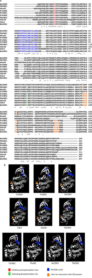
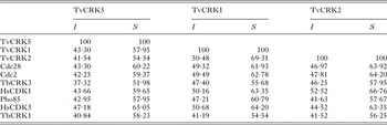
The overall sequence similarity among the three TvCRKs deduced protein sequences and their comparison with CDKs from other species are shown in Table 1. The TvCRKs were found to contain most elements involved in kinase activity that are found in the CDKs of other organisms (Fig. 1A). Human CDK1 is phosphorylated at three residues, Thr-14, Tyr-15 and Thr-161, which are highly conserved both within the CDK family and between species. The three TvCRKs were found to contain Thr and Tyr residues at positions equivalent to HsCDK1 Thr-14 and Tyr-15, respectively, which are located in the C-lobe and inhibit kinase activity when phosphorylated. In addition, two of the three TvCRKs have an activating phosphorylatable Thr at the position equivalent to HsCDK1 Thr-161 located within the region known as the T-loop (Malumbres, Reference Malumbres2014), while TvCRK5 has a Ser residue at this position. Another important element that is highly conserved among CDKs is the 16-amino-acid-long sequence EGVPSTAIREISLLKE, known as the PSTAIRE motif. This domain is important for cyclin binding and is located in the N-terminus lobe (Fig. 1A and B). TvCRK5 was the most conserved in this sequence among the three TvCRKs and showed only two substitutions compared to HsCDK1: an Ile for a Val and an Ala for a Ser. In contrast, TvCRK1 and TvCRK2 each showed seven substitutions. The GDSEID and (S/T)FPXW motifs, which are characteristic of the CDK1 subfamily and are important for interactions with proteins of the CKS family (Bourne et al. Reference Bourne, Watson, Hickey, Holmes, Rocque, Reed and Tainer1996), are conserved in TvCRK1 and TvCRK2. In contrast, ScPho85 and TvCRK5 lack these motifs (Fig. 1A and B). Predicted models of TvCRKs compared with CDKs from yeast, human and T. brucei are shown in Fig. 1B. The three TvCRKs have a similar general folded structure that includes a small amino-terminal lobe and a larger carboxy-terminal lobe. In this arrangement, the regulatory phosphorylatable amino acids are oriented outwards, towards the opening of the cleft, as has been described for other CDKs (Echalier et al. Reference Echalier, Endicott and Noble2010). The location of the PSTAIRE motif is conserved among all the compared CDKs.
Aligned sequences of T. vaginalis TvCRK1, TvCRK2 and TvCRK5 along with CDKs of diverse organisms (accession numbers are in brackets): Cdc28: S. cerevisiae CDK1 (CAA85119·1); Cdc2: S. pombe CDK1 (NP_595629·1); TbCRK3: T. brucei CRK3 (XP_822746·1); HsCDK1: Homo sapiens CDK1 (NP_001307847); Pho85: S. cerevisiae Pho85 (CAA68773·1); HsCDK5: H. sapiens CDK5 (NP_004926) and TbCRK1: T. brucei CRK1 (XP_822365·1). (A) Conserved motifs and regions among CDKs. Threonine and tyrosine inhibitory residues are shown in red, the PSTAIRE motif involved in cyclin binding is shown in blue, the threonine activating residue is shown in green, and the GDSEID and (S/T)FPXW motifs that form the CKS-binding site are shown in orange. (B) Predicted models of TvCRKs compared with CDKs fom yeast, human and T. brucei. Colour code is the same as in panel A.

Amino acid sequence identity (I) and similarity (S) among the TvCRKs and cyclin-dependent kinases from different organisms a

a Values are expressed in percentage.
Cdc28: S. cerevisiae CDK1 (CAA85119·1); Cdc2: S. pombe CDK1 (NP_595629·1); TbCRK3: T. brucei CRK3 (XP_822746·1); HsCDK1: H. sapiens CDK1 (NP_001307847); Pho85: S. cerevisiae Pho85 (CAA68773·1); HsCDK5: H. sapiens CDK5 (NP_004926) and TbCRK1: T. brucei CRK1 (XP_822365·1).
CRK complementation assay in S. cerevisiae
To evaluate the possible roles of the TvCRKs in cell cycle control, we tested whether these proteins could rescue a S. cerevisiae cdc28-knockout strain from lethality, since Cdc28 is an essential kinase that controls cell cycle in yeast (Reed and Wittenberg, Reference Reed and Wittenberg1990). Each of the three ORFs encoding the TvCRKs and the yeast CDC28 ORF were cloned into the pYES2 expression vector and individually transformed into the mutant yeast, as described in the Materials and Methods section. Tetrads were manually dissected, and their growth in galactose plates was monitored to assess complementation of Cdc28 function. Just two out of four spores were able to germinate when the cdc28-knockout strain was transformed with each of the TvCRK plasmid constructs. In contrast, when the cdc28 mutant was transformed with the CDC28 plasmid construct as the positive control, four spores were able to grow. These observations indicate that none of the three kinases from T. vaginalis could complement the lack of Cdc28 to restore the cell cycle progression of the yeast mutant (Fig. 2).
Complementation assay using the S. cerevisiae cdc28-knockout heterozygous diploid strain BY4743 and the TvCRKs analysed by tetrad dissection. None of the three T. vaginalis kinases tested could complement the lack of Cdc28 and restore cell cycle progression in the yeast mutants, as deduced by the growth of only two of the four spores. The S. cerevisiae CDC28 gene was cloned into the pYES2 vector and used as a positive control (+), and an empty pYES2 vector was used as a negative control (−). Only one of at least eight tetrad dissections for each complementation assay is shown.

Associations between the T. vaginalis CRK and CKS proteins
Mammalian CDK1 and CDK2 proteins, which are involved in cell cycle control, have been reported to associate with various CKSs including human CKS1 and CKS2, and S. pombe p13Suc1, with very high affinity in vitro. Because of this ability, p13Suc1-conjugated agarose beads are frequently used to identify CDKs belonging to the CDK1 subfamily from a wide variety of organisms (Brizuela et al. Reference Brizuela, Draetta and Beach1987; Dunphy et al. Reference Dunphy, Brizuela, Beach and Newport1988; Tang et al. Reference Tang, Pelech and Berger1994). Pull-down assays to assess the in vitro interactions formed by the three recombinant 6xHis-tagged TvCRK proteins with p13Suc1-agarose showed that both TvCRK1 and TvCRK2 formed interactions with the p13Suc1 protein. In contrast, TvCRK5, which does not have the GDSEID and (S/T)FPXW motifs known to mediate interactions with CKS proteins, did not bind to the p13Suc1-agarose beads (data not shown).
Because CKS proteins are important regulators of the cell cycle, we searched in the TrichDB database for the presence of the S. pombe p13Suc1 protein homologues. Two genes were identified (accession numbers TVAG_164620 and TVAG_142260), both of which were annotated as putative CDK regulatory proteins. The two encoded proteins are hereby called TvCKS1 and TvCKS2. The mRNA expression of both genes was assessed by reverse transcriptase-coupled PCR, and the corresponding cDNA clones were sequenced. Both genes contain the UAAA polyadenylation sequence that is characteristic of T. vaginalis mRNA (Fuentes et al. Reference Fuentes, Barrera, Sánchez, Hernández and López-Villaseñor2012) (data not shown). The two T. vaginalis CKS proteins are 82 amino acids long, with a molecular weight close to 10 kDa.
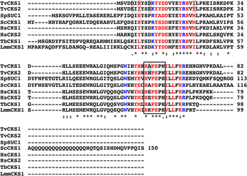
Amino acid sequence alignments of the TvCKSs with other CKSs showed that these proteins are conserved among eukaryotes, including the two CKSs that are present in humans (Richardson et al. Reference Richardson, Stueland, Thomas, Russell and Reed1990), the one in S. cerevisiae (Hadwiger et al. Reference Hadwiger, Wittenberg, Mendenhall and Reed1989) and those in the protists L. mexicana (Mottram and Grant, Reference Mottram and Grant1996) and T. brucei (Hammarton et al. Reference Hammarton, Clark, Douglas, Boshart and Mottram2003) (Fig. 3). The following key residues and motifs were observed: (1) the HxPEPH motif, which is involved in self-dimerization (Rousseau et al. Reference Rousseau, Schymkowitz, Wilkinson and Itzhaki2001), although the last His residue in this motif is changed to Asn in both T. vaginalis proteins; (2) residues involved in interactions with CDKs; and (3) residues that form anion-binding sites that are involved in interactions with several CDK substrates (Bourne et al. Reference Bourne, Watson, Hickey, Holmes, Rocque, Reed and Tainer1996). Five differences in amino acid sequence were identified between TvCKS1 and TvCKS2. Interestingly three of these five differences correspond to amino acids involved in the complex formation with CDKs.
Comparison of T. vaginalis CKSs and their homologues from several eukaryotic organisms. Sequence alignments of TvCKS1 and TvCKS2 with those from different species. SpSUC1: S. pombe CKS1 (CAA21308); ScCKS1: S. cerevisiae (NP_009693); HsCKS1 and HsCKS2: H. sapiens (NP_001817 and NP_001818); TbCKS1: T. brucei (CAD43044·1); LmmCKS1: L. mexicana (CAA90037). Amino acid residues involved in interactions with CDKs are shown in red, residues involved in interactions with CDK substrates are shown in blue, and the HxPEPH motif involved in self-dimerization is boxed. Amino acids that differ between TvCKS1 and TvCKS2 are shadowed.

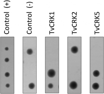
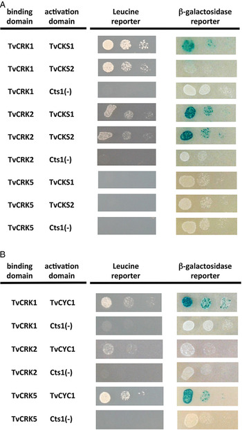
A second in vivo approach was used to identify interactions among the homologous TvCKSs and TvCRKs. A yeast two-hybrid assay confirmed the in vitro interactions observed with the p13Suc1-agarose beads, that is, both TvCKS1 and TvCKS2 interacted with TvCRK1 and TvCRK2, although a lower level of interaction was observed for TvCRK1 with TvCKS2. No interaction was detected between the TvCKSs and TvCRK5 (Fig. 4A). This result supports the proposal that TvCRK1 and TvCRK2 could belong to the CDK1 subfamily, and that TvCRK5 could be related to the CDK5 subfamily of CDKs.
Interactions between TvCRKs and TvCKSs (A) and between TvCRKs and TvCYC1 (B) as determined by yeast two-hybrid system. The binding domain corresponds to LexA-fused TvCRKs, and the activation domain (AD) corresponds to B42-fused TvCKSs or B42-fused TvCYC1, as indicated. Interaction between proteins is evidenced by the growth of the colonies in Leu-plates (Leucine reporter) and by the blue colour of the colonies grown in Leu+ plates with X-Gal (β-galactosidase reporter), plated in serial dilutions. S. cerevisiae endochitinase fused to the B-42 AD was used as a negative interaction control [Cts1(−)]. Pictures were taken after 72 h of incubation at 30 °C.

TvCRK interactions with a T. vaginalis cyclin
To identify a cyclin that can interact with and activate the TvCRKs, we searched for putative cyclins in the TrichDB database. A gene annotated as a putative cyclin B was identified under the accession number TVAG_121680, and the encoded protein is here named TvCYC1. The mRNA expression of TVAG_121680 was demonstrated by reverse transcriptase-coupled PCR and through the sequencing of cDNA clones. This mRNA contains the UAAA polyadenylation motif that is characteristic of T. vaginalis mRNA (Fuentes et al. Reference Fuentes, Barrera, Sánchez, Hernández and López-Villaseñor2012) (data not shown).
Interactions between the TvCRKs and TvCYC1 were assessed using an in vivo yeast two-hybrid assay, with the TvCRKs as the bait and TvCYC1 the as prey. TvCYC1 was found to interact with TvCRK1 and TvCRK5, but no interaction was detected with TvCRK2 (Fig. 4B). Pull-down assays were then performed using recombinant GST-TvCYC1 and the three 6xHis-TvCRKs, and the resulting complexes were used to test for activation of kinase activity by the cyclin.
Cyclin-mediated activation of the recombinant TvCRKs
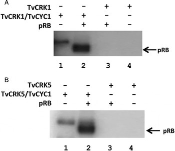
The kinase activities of the recombinant TvCRK1, TvCRK2 and TvCRK5 proteins were evaluated by an in vitro kinase assay using the pull-down complexes and the pRB as the phosphorylatable substrate. Figure 5 shows the activation of both TvCRK1 and TvCRK5 kinase activity by TvCYC1. Phosphorylation of the pRB substrate was observed with the CRK/CYC complexes, and phosphorylation of another protein that could correspond to the TvCRK was also detected in these in vitro assays. No kinase activity was observed in either case in the absence of the cyclin. In contrast, TvCRK2 showed no kinase activity either alone or in the presence of the cyclin (data not shown). This result was not surprising because no interaction was detected between TvCRK2 and TvCYC1 by the two-hybrid assay (Fig. 4B).
TvCYC1 activates the kinase activities of recombinant TvCRK1 and TvCRK5, as shown by an in vitro phosphorylation assay. A recombinant version of retinoblastoma protein (pRB) was used as the phosphorylatable substrate. (A) Kinase activity of the TvCRK1/TvCYC1 complex. Lane 1: Kinase activity of the TvCRK1/TvCYC1 complex. Phosphorylation of a protein with a molecular weight similar to TvCRK1 is detected. Lane 2: phosphorylation of pRB by the TvCRK1/TvCYC1 complex. Lanes 3 and 4: no kinase activity of the TvCRK1 recombinant protein is detected in the absence of TvCYC1. (B) Kinase activity of the TvCRK5/TvCYC1 complex. Lane 1: Kinase activity of the TvCRK5/TvCYC1 complex. Phosphorylation of a protein with a molecular weight similar to TvCRK5 is detected. Lane 2: phosphorylation of pRB by the TvCRK5/TvCYC1 complex. Lanes 3 and 4: no kinase activity of the TvCRK5 recombinant protein is detected in the absence of TvCYC1. Histone HI was also used as the phosphorylatable substrate and produced similar results to those obtained with pRB (data not shown).

DISCUSSION
Cell cycle control has been widely studied in higher eukaryotes; however, studies of the components involved in cell cycle control in parasitic protozoa are limited. The present work represents the first attempt to characterize some proteins involved in cell cycle regulation in T. vaginalis, an early divergent protist of both biological and medical relevance.
A general search for proteins involved in T. vaginalis cell cycle control identified three Cdc2-related protein kinases, designated TvCRK1, TvCRK2 and TvCRK5 (parasite CDKs are called CRKs, from Cdc2-related protein kinases). These TvCRKs contain sequence and structural features that place them in the CDK family. Their deduced amino acid sequences revealed the presence of key residues and motifs that led us to propose that these kinases belong to two different subfamilies. The first is the CDK1 subfamily, which TvCRK1 and TvCRK2 appear to be closely related to, similarly to Cdc28 from S. cerevisiae, Cdc2 from S. pombe and CRK3 from T. brucei. Moreover, TvCRK1 and TvCRK2 possess characteristic CKS-binding motifs, supporting the suggestion that these kinases belong to the CDK1 subfamily and may be directly involved in cell cycle control in T. vaginalis. The second is the CDK5 subfamily, in which the CKS-binding motifs are absent, and the activating phosphorylatable Thr residue is not required (Malumbres, Reference Malumbres2014). Regarding these features, TvCRK5 shares these characteristics with Pho85 from S. cerevisiae and the human CDK5. Therefore, we consider TvCRK5 a putative member of the CDK5 subfamily.
Complementation of yeast mutants has been a useful method of characterizing the functionality of heterologous proteins. When tested, none of the three kinases from T. vaginalis could complement the absence of Cdc28 in a S. cerevisiae cdc28-knockout mutant to restore cell cycle progression. This lack of complementation suggests that the TvCRKs are not able to phosphorylate Cdc28 substrates in yeast, or that the TvCRKs cannot interact with yeast regulatory proteins such as cyclins, phosphatases, activating kinases, inhibitor kinases or other regulators. Similar complementation assays have been reported for CDKs from L. mexicana, T. cruzi and Plasmodium (Mottram et al. Reference Mottram, Kinnaird, Shiels, Tait and Barry1993; Ross-MacDonald et al. Reference Ross-MacDonald, Graeser, Kappes, Franklin and Williamson1994; Gómez et al. Reference Gómez, Kornblihtt and Téllez-Iñón1998; Grant et al. Reference Grant, Hassan, Anderson and Mottram1998; Vinkenoog et al. Reference Vinkenoog, Sperança, Ramesar, Thomas, Portillo, Janse and Waters1998; Sperança et al. Reference Sperança, Vinkenoog, Ocampos, Fischer, Janse, Waters and del Portillo2001); in these cases, only the Leishmania major CRK3 kinase rescued the growth defect of a S. pombe cdc2-33ts mutant when integrated into the genome and expressed at the optimal level (Wang et al. Reference Wang, Dimitrov, Garrity, Sazer and Beverley1998). Therefore, our results do not rule out a regulatory role for the TvCRKs in the cell cycle of T. vaginalis.
Members of the CDK1 subfamily of CDK proteins have been reported to associate with CKSs. Indeed, CDKs and CKSs may form complexes with very high affinity in vitro (Bourne et al. Reference Bourne, Watson, Hickey, Holmes, Rocque, Reed and Tainer1996). Here we identified two CKSs in T. vaginalis (TvCKS1 and TvCKS2), which is an unusual finding among protists since only one has been reported in those protists where CKSs have been studied (Mottram and Grant, Reference Mottram and Grant1996; Hammarton et al. Reference Hammarton, Clark, Douglas, Boshart and Mottram2003). Few eukaryotes possess two CKSs. In humans, CKS1 and CKS2 (81% identical) have specific functions: only HsCKS1 can bind the SKP2 protein, which is part of the SCF (Skp1, Cul1, F-box-protein) ubiquitin ligase complex and is involved in the degradation of the CDK inhibitor p27, a key step in G1/S progression (Spruck et al. Reference Spruck, Strohmaier, Watson, Smith, Ryan, Krek and Reed2001; Frontini et al. Reference Frontini, Kukalev, Leo, Ng, Cervantes, Cheng, Holic, Dormann, Tse, Pommier and Yu2012). In mice, CKS2 is required in meiosis for the first metaphase to anaphase transition, and mice lacking CKS2 are viable but sterile in both sexes (Spruck et al. Reference Spruck, de Miguel, Smith, Ryan, Stein, Schultz, Lincoln, Donovan and Reed2003).
TvCKS1 and TvCKS2 are 94% identical, possessing only 5 amino acid differences between them. Preliminary results from our group showed TvCKS dimers in polyacrylamide gels during the preparation of recombinant TvCRK proteins. Because CKS proteins contain a self-dimerization motif (Rousseau et al. Reference Rousseau, Schymkowitz, Wilkinson and Itzhaki2001; Seeliger et al. Reference Seeliger, Spichty, Kelly, Bycroft, Freund, Karplus and Itzhaki2005), we speculate that TvCKSs fluctuate between at least two different states: a monomer able to interact with kinase proteins and a dimer that cannot interact with target proteins. In a two-hybrid assay, both TvCKSs interacted with TvCRK1 and TvCRK2 but not with TvCRK5. This result further supports that TvCRK1 and TvCRK2 belong to the CDK1 subfamily of CDK kinases. The differences in the interactions among the TvCRKs and the TvCKSs may be related to the amino acid changes between both TvCKSs, which are located in relevant regions for the interaction with CDKs, and therefore might play different roles in T. vaginalis cellular processes.
Kinase activity was evaluated for the three TvCRKs on pRB as the phosphorylatable substrate. TvCYC1 (a putative T. vaginalis-type B cyclin) binds to and activates the kinase activities of both TvCRK1 and TvCRK5. This result provides strong evidence that TvCRK1 and TvCRK5 are true CDKs. Since no interaction or kinase activity was observed for TvCRK2 with TvCYC1, it is possible that TvCRK2 associates with a cyclin other than TvCYC1 to display its activity. In fact, several sequences are annotated as putative cyclins in the TrichDB database that could interact with the TvCRKs. It can thus be speculated that a complex CRK-cyclin signalling network exists in T. vaginalis that may be similar to those found in other eukaryotic organisms.
The results presented here represent the first characterization of proteins involved in cell cycle control in T. vaginalis. Additional studies are needed to characterize the physiological substrates of TvCRK/TvCYC complexes to understand their precise roles in regulating the cell cycle of this parasitic protist.
ACKNOWLEDGEMENTS
The authors acknowledge preliminary work by Olivia Arizmendi regarding the identification of T. vaginalis CRKs. We thank Susana Kolb and Katya Frank from the Academic Writing Office at the UNAM's Coordinación de Estudios de Posgrado for their help revising an original version of the manuscript.
FINANCIAL SUPPORT
This work was supported by grant IN205813 from Programa de Apoyo a Proyectos de Investigación e Innovación Tecnológica (PAPIIT), Universidad Nacional Autónoma de México. Scholarships were awarded from Consejo Nacional de Ciencia y Tecnología (CONACyT), Mexico, to Erick Amador and Nataly Morales as graduate students from the Programa de Maestría y Doctorado en Ciencias Bioquímicas, UNAM.