Hughes–Stovin syndrome is a very rare clinical disorder of unknown aetiology, characterised by the combination of vascular aneurysms and thromboses, and is considered to be a variant of Behcet’s disease.Reference Durieux, Bletry, Huchon, Wechsler, Chretien and Godeau 1 – Reference Emad, Ragab, Shawki, Gheita, El-Marakbi and Salama 3 We report a paediatric case of Hughes–Stovin syndrome with a challenging differential diagnosis because of the absence of haemoptysis or respiratory complaints at onset, who was initially diagnosed as having Lemierre’s syndrome.
Case report
A 12-year-old, previously healthy, Albanian boy born to non-consanguineous parents was admitted to our department with the diagnosis of endocarditis.
His history began 4 months earlier in Albania with headache, and he was treated for acute sinusitis with antibiotics. Owing to persistent vomiting and diplopia, a brain MRI was performed, which showed thrombosis of the left sigmoid and transverse sinuses. Cerebral angiography through catheterisation revealed no vascular malformations; his coagulation tests were normal. The boy was discharged on antiplatelet therapy. He soon developed persistent fever, nausea, and right abdominal and leg pain. Appendectomy was performed without benefit. CT angiography revealed an aneurysm of the right femoral artery at the site of vascular access of the previous catheterisation. Echocardiography showed a mural mass in the right atrium attached to the posterolateral wall and floating through the tricuspid valve. His three consecutive blood cultures and serology for hepatitis A, B, and C, parvovirus B19, enterovirus, and Epstein–Barr virus showed negative results. Broad-spectrum antibiotic therapy with ceftriaxone, vancomycin, and amikacin was administered for 2 weeks, but high fever with chills persisted; thus, the therapy was switched to meropenem, linezolid, and fluconazole, and he was transferred to our unit.
On admission to our unit, he was febrile with chills, ill-appearing, and pale; he had reduced visual acuity. Abdominal pain in the right lower quadrant and a pulsatile right inguinal mass (3.5×3 cm) were present; no mouth or genital ulcers were present.
Laboratory tests showed increased inflammatory parameters (C-reactive protein 23 mg/dl, erythrocyte sedimentation rate 50 mm/hour), microcytic anaemia (haemoglobin 7.6 g/dl, mean cell volume 71 fl), and normal renal and hepatic functions. Serial blood cultures were sterile on antibiotics; the Quantiferon-TB test was negative. Echocardiography confirmed a mural endocardial mass (30×40 mm) in the right atrium. Ophthalmology evaluation was normal without ocular inflammation.
Cerebral CT showed thrombosis of the left sigmoid and transverse sinuses and both internal jugular veins. Pulmonary CT and CT angiography demonstrated pulmonary nodules and bilateral iliac vein thromboses. We suspected Lemierre’s syndrome because of the presence of jugular vein thrombosis and previous mastoiditis. Antibiotic treatment with daptomycin and levofloxacin was administered with no laboratory or clinical benefit.
Considering the lack of response to broad-spectrum antimicrobial therapy, the right atrial vegetation was surgically removed via sternotomy, but new vegetations, postoperative endocardial masses, rapidly formed in the right atrium and ventricle. Subcutaneous low-molecular weight heparin was started.
After 3 weeks, he underwent a resection of a ruptured right femoral artery aneurysm. Immediately, he developed peripheral thrombophlebitis and an aneurysm of the right brachial artery at the site of a previous arterial catheter placed before heart surgery. This aneurysm was surgically repaired. Caspofungin was added without benefit, whereas all operative specimens – endocardial mass, femoral, and humeral vascular tissue – and blood cultures were sterile. Viral serology for Brucella, Borrelia, parvovirus B19, enterovirus, Epstein–Barr virus, cytomegalovirus, immunodeficiency virus, and hepatitis B and C, antinuclear antibodies, anti-double-stranded DNA antibodies, circulating immune complexes, antimitochondrial antibodies, anti-smooth muscle antibodies, anti-liver kidney microsomal autoantibodies, anti-cardiolipins, and thrombophilic screening showed negative results; C-reactive protein level was 17 mg/dl.
Total body 18F-fluorodeoxyglucose positron emission tomography/CT did not identify areas of vascular inflammation: intense uptake in the bone marrow and spleen, a tiny area of uptake in the right lower lobe of the lung, and an area of uptake in the right upper lobe at a site of vascular bifurcation attributed to non-specific vascular activity. No vascular areas of active inflammation were identified.
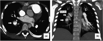
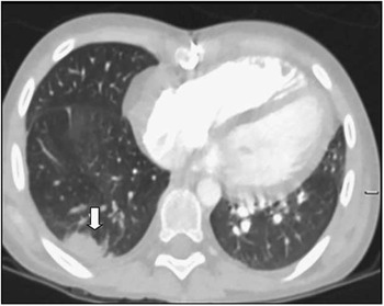
We hypothesised an inflammatory disease after exclusion of an infectious condition, given the absence of clinical and laboratory response and negative operative specimens and blood cultures, high-dose intravenous steroid pulses were started with defervescence and dramatic improvement of the clinical condition, but dry cough and chest pain developed. Chest X-ray showed a right round opacity of the upper lobe, and a thoracic CT showed new evidence of multiple pulmonary artery aneurysms originating from the artery to the right lower lobe, which were not present 2 months earlier: two were partially thrombosed and one was leaking; previously detected pulmonary nodules were not cavitated (Figs 1 and 2). With these findings, arterial aneurysms, venous thromboses, including the right-sided cardiac mass, and normalisation of inflammatory markers after steroid treatment, Hughes–Stovin syndrome was diagnosed. Skin pathergy test and HLA B51 were positive. Methylprednisolone was switched to oral prednisone after 5 days, and cyclophosphamide was added; anticoagulant therapy was stopped because of the risk of pulmonary bleeding.
Contrast-CT chest: axial ( a ) and posteroanterior ( b ) views showing aneurysms originating from the right lower lobe segmental artery: one aneurysm is completely obstructed and one is partially circumferentially obstructed by organised thrombus. The white arrows point out the aneurysms.

CT chest axial projection showing right lower lobe pulmonary infarction at the level of the posterobasal segment (white arrow).

After 20 days, repeat thoracic CT showed a slight decrease in the size of aneurysms, and the previously leaking lesion was partially thrombosed. Echocardiography showed normal pulmonary pressure and decreased size of the endocardial mass.
Discussion
Hughes–Stovin syndrome is an exceedingly rare vasculitis of unknown aetiology, consisting of multiple pulmonary artery aneurysms and deep venous thromboses. Since its first description in 1959,Reference Hughes and Stovin 4 there have been about 40 reports of its occurrence: therefore, there is still a lack of formal diagnostic criteria or laboratory investigation procedures, making its diagnosis a challenge.
Aneurysms usually involve the pulmonary and/or bronchial arteries, but they can also occur anywhere in the arterial circulation. Recurrent episodes of phlebitis commonly involve large veins leading to thrombus formation and thromboembolism, involving the jugular vein, the iliac and femoral veins, dural sinuses, caval veins, and cardiac chambers.
Patients, mostly males, aged 12–40 years usually present with cough, dyspnoea, chest pain, haemoptysis, and signs of pulmonary hypertension. Other associated non-specific features include fever and elevated intracranial pressure.Reference Chalazonitis, Lachanis and Mitseas 5
Many authors propose that Hughes–Stovin syndrome is a variant of Bechet’s disease, sharing clinical, radiological, and histopathological findingsReference Durieux, Bletry, Huchon, Wechsler, Chretien and Godeau 1 – Reference Emad, Ragab, Shawki, Gheita, El-Marakbi and Salama 3 and in particular the association of pulmonary aneurysms and venous thromboses. Therefore, when a patient presents with these findings and distinctive Bechet’s features – recurrent mouth and/or genital ulcers and iritis – are absent, as in the reported case, a diagnosis of Hughes–Stovin syndrome can be made. Histological examination of lesions can be useful for diagnosis,Reference Hughes and Stovin 4 , Reference Kopp and Green 6 but in the case presented all available specimens were evaluated only by culture for suspected infection.
Several hypotheses have been proposed regarding the pathogenesis of Hughes–Stovin syndrome, including infections, angiodysplasia, thrombophilia, and presence of HLA B51, but the current consensus is that vasculitis is the primary pathological process.Reference Khalid and Saleem 7 All of our findings – that is, femoral and humeral aneurysms, spontaneous thrombophlebitis, extended deep venous thrombosis, pulmonary aneurysms, with negative cultures, and clinical response to steroids – support primary vascular involvement.
In the medical literature, there are very few paediatric cases describedReference Hughes and Stovin 4 , Reference Roberts, Jimenez and Golladay 8 , Reference de Vries, Koppelman, Roofthooft, Bootsma, Leijsma and Armbrust 9 and they are all boys, as was in our case. By reviewing the clinical presentation in this age group, we suggest that it may be represented by the association of pulmonary artery aneurysms and cerebral venous sinus thromboses, rather than thrombosis in other systemic sites with persistent fever and chills, as in our patient in whom pulmonary symptoms appeared only later.
Jugular thrombophlebitis with pulmonary abscesses is typical in Lemierre’s syndrome, explaining clinical overlap with Hughes–Stovin syndrome that we underline in this report. Lemierre’s syndrome is characterised by jugular vein thrombophlebitis with metastatic septic pulmonary embolisation in the setting of oropharyngeal, sinus, or ear infections, usually caused by Fusobacterium necrophorum.Reference Righini, Karkas and Tourniaire 10 It is treated with antibiotics, whereas the use of anticoagulants is controversial.Reference Righini, Karkas and Tourniaire 10 , Reference Eilbert and Sigla 11 In our patient, pulmonary nodules detected on the initial chest CT were interpreted as septic emboli from the jugular vein. Moreover, various antibiotic regimens were ineffective and all cultures were negative.
Once pulmonary artery aneurysms are suspected, conventional angiography is the standard for diagnosis, but multi-detector row helical CT angiography has the advantage in that it is non-invasive and offers precise visualisation of the vascular lumen and wall, and shows mural thrombus.Reference Ketchum, Zamanian and Fleischmann 12 , Reference Fischer, Korman and West 13 Moreover, it enabled us to rule out the likelihood of Lemierre’s syndrome because pulmonary nodules did not cavitate over time, thus making infection much less likely. We decided not to perform percutaneous angiography because the child had no haemoptysis and because of the potential vascular complications connected with the procedure itself that were further increased by active vasculitis of Hughes–Stovin syndrome.
The therapeutical approach aims to reduce inflammation. Although there are no controlled trials and no standard treatment guidelines, the recommended therapy is a combination of cyclophosphamide and glucocorticoids.Reference Bowman and Honey 14 , Reference Lee, Noh and Hwang 15 Other agents variably used include colchicine, cyclosporine, and azathioprine.Reference Erkan, Yazici, Sanders, Trost and Yazici 2 , Reference Khalid and Saleem 7 Immunosuppression has the potential to stabilise small pulmonary aneurysms, which in some cases may regress. In our patient, immunosuppressive therapy was clinically effective without significant side-effects, but we do not know the long-term outcomes as he has been lost to follow-up. Despite thrombosis, anticoagulation and platelet inhibitors appear to be non-indicated as the risk of pulmonary haemorrhage may outweigh the risk of pulmonary embolism. The prognosis of Hughes–Stovin syndrome is usually poor related to rupture of pulmonary aneurysms. Surgical resection and/or transcatheter arterial embolisation are generally reserved for severe, acute cases because of the high associated morbidity and mortality.
Conclusion
Hughes–Stovin syndrome is an extremely rare condition, especially in children, which shares similar clinical, radiological, and histopathological findings with Behcet’s disease; however, differential diagnoses include other disorders such as polyarteritis nodosa and Lemierre’s syndrome and may be difficult because of the overlapping clinical features. We described the case of a 12-year-old boy with the association of cerebral venous thrombosis, endocardial mass with persistent fever, and recurrent arterial aneurysms, with only late pulmonary artery aneurysms. The initial diagnosis was Lemierre’s syndrome.
Investigating the genetic basis, establishing diagnostic criteria, and formulating management guidelines for Hughes–Stovin syndrome are needed to achieve early diagnosis in order to the improve prognosis of this very rare but very serious clinical entity. Earlier diagnosis may in some cases prevent the development of potentially life-threatening pulmonary artery aneurysms.
Acknowledgement
The authors thank Professor Marco Bonvicini, Professor Gaetano Gargiulo, and Dr Luca Ragni.
Financial Support
This research received no specific grant from any funding agency or from commercial or not-for-profit sectors.
Conflicts of Interest
None.