INTRODUCTION
The Apicomplexa are a diverse group of unicellular parasites that infect humans and animals. Plasmodium falciparum and Toxoplasma gondii are among the best-known Apicomplexa being among the leading causes of morbidity and mortality worldwide (Tenter et al. Reference Tenter, Heckeroth and Weiss2000; Guinovart et al. Reference Guinovart, Navia, Tanner and Alonso2006). Malaria is endemic in 104 countries putting an estimated 3·4 billion people at risk, with 207 million reported cases and 627 000 deaths globally in 2012 (WHO 2013). Other pathogens of importance include Cryptosporidium, Babesia, Eimeria and Neospora caninum which collectively can cause devastating and debilitating encephalitis, diarrhoea and foetal abortion in the host. Treatment options, which rely heavily on chemotherapeutics, are limited and an issue of immediate importance due to increasing resistance of many apicomplexans to available therapeutic agents (Nair et al. Reference Nair, Brooks, Goodman, Strurm, McFadden, Sundriyal, Anglin, Song, Moreno and Striepen2011). Currently available therapies include antifolate and macrolide antimicrobials such as sulfadiazine and pyrimethamine or clindamycin, spiramycin and imidocarb. All of these treatments present obstacles by exhibiting poor pharmacokinetic properties, toxicity, teratogenicity, tolerability and allergenicity (Norrby, Reference Norrby1978; Reeves and Wilkinson, Reference Reeves and Wilkinson1979; Luft et al. Reference Luft, Hafner, Korzun, Leport, Antoniskis, Bosler, Bourland, Uttamchandani, Fuhrer and Jacobson1993; Katlama et al. Reference Katlama, De Wit, O'Doherty, Van Glabeke and Clumeck1996).
Most apicomplexan parasites are obligate intracellular pathogens that must invade a host cell to grow and replicate (Morrissette and Sibley, Reference Morrissette and Sibley2002). They have distinctive structural and phenotypic characteristics that can be targeted for the development of safe and effective antimicrobial therapies. Recent studies of apicomplexan kinase biology have described the transmission of calcium signals under the control of calcium-dependent protein kinases (CDPKs) to inter-related pathways as essential in facilitating host cell invasion, namely gliding motility and microneme secretion (Lovett et al. Reference Lovett, Marchesini, Moreno and Sibley2002). Each CDPK has defined regulatory functions at distinct stages of the apicomplexan life cycle, which are integrated into various key phosphorylation-associated signalling events (Ojo et al. Reference Ojo, Merritt, Maly, Van Voorhis, Doerig, Späth and Wiese2013). Hence, expression levels and specific functions in each organism may vary based on the specific life cycle requirements, which are not completely understood at present. However, we have previously shown that interruption of these enzymes’ ability to transmit signals by the application of specific inhibitors resulted in inhibition of parasite cell replication and growth in in vitro and in vivo models for T. gondii (Ojo et al. Reference Ojo, Larson, Keyloun, Castaneda, Derocher, Inampudi, Kim, Arakaki, Murphy, Zhang, Napuli, Maly, Verlinde, Buckner, Parsons, Hol, Merritt and Van Voorhis2010; Johnson et al. Reference Johnson, Murphy, Geiger, DeRocher, Zhang, Ojo, Larson, Perera, Dale, He, Reid, Fox, Mueller, Merritt, Fan, Parsons, Van Voorhis and Maly2012; Doggett et al. Reference Doggett, Ojo, Fan, Maly and Van Voorhis2014), Cryptosporidium parvum (Murphy et al. Reference Murphy, Ojo, Larson, Castellanos-Gonzalez, Perera, Keyloun, Kim, Bhandari, Muller, Verlinde, White, Merritt, Van Voorhis and Maly2010; Castellanos-Gonzalez et al. Reference Castellanos-Gonzalez, White, Ojo, Vidadala, Zhang, Reid, Fox, Keyloun, Rivas, Irani, Dann, Fan, Maly and Van Voorhis2013) and P. falciparum (Ojo et al. Reference Ojo, Pfander, Mueller, Burstroem, Larson, Bryan, Fox, Reid, Johnson, Murphy, Kennedy, Mann, Leibly, Hewitt, Verlinde, Kappe, Merritt, Maly, Billker and Van Voorhis2012, Reference Ojo, Eastman, Vidadala, Zhang, Rivas, Choi, Lutz, Reid, Fox, Hulverson, Kennedy, Isoherranen, Kim, Comess, Kempf, Verlinde, Su, Kappe, Maly, Fan and Van Voorhis2014; Vidadala et al. Reference Vidadala, Ojo, Johnson, Zhang, Leonard, Mitra, Choi, Reid, Keyloun, Fox, Kennedy, Silver-Brace, Hume, Kappe, Verlinde, Fan, Merritt, Van Voorhis and Maly2014) without harmful effects to the hosts. The inhibitors’ library structure-activity relationships (SARs) for these parasites are similar. A structure-activity relationship is an iterative study that defines the contribution of each molecular group within an inhibitor's chemical structure to the overall biological activity associated with it (inhibitor). Specific inhibition of CDPKs was achieved using an adenosine-like pyrazolopyrimidine scaffold which binds to the hinge region of the active site, similar to the binding motif of ATP, but with an added bulky aromatic R1-substituent at the C3-position – the so-called ‘bump’ of bumped kinase inhibitors (BKIs) (Ojo et al. Reference Ojo, Larson, Keyloun, Castaneda, Derocher, Inampudi, Kim, Arakaki, Murphy, Zhang, Napuli, Maly, Verlinde, Buckner, Parsons, Hol, Merritt and Van Voorhis2010). Topologically, the aromatic R1-group projects into a hydrophobic pocket adjacent to the gatekeeper position of the ATP-binding site (the gatekeeper pocket), a feature exclusive to protein kinases with small gatekeepers, such as many apicomplexan CDPKs. Most mammalian kinases typically have larger gatekeeper residues that exclude the ‘bump’ of BKIs, although a few mammalian protein kinases like SRC do contain relatively smaller gatekeeper residues (threonine). Therefore, great care must be taken to maintain selectivity towards apicomplexan CDPKs over such human kinases. Fortunately, preliminary results also suggest that slight topographical deviations of the ATP-binding region of apicomplexan CDPKs from SRC, namely, the ribose binding pocket, also confer alternative selectivity to BKIs. Larson et al. (Reference Larson, Ojo, Murphy, Johnson, Zhang, Kim, Leibly, Fox, Reid, Dale, Perera, Kim, Hewitt, Hol, Verlinde, Fan, Van Voorhis, Maly and Merritt2012) previously demonstrated that mammalian kinases such as SRC have shallow ribose pockets as compared with apicomplexan CDPKs. Co-crystal structures of BKIs in complex with TgCDPK1 have revealed a hydrogen bond between residue E135, a conserved glutamic acid found within the ribose binding pocket and shared among other apicomplexan CDPKs, and inhibitors with a 4-piperidinylmethylene R2 group, an interaction which further enhances selectivity. Together, the gatekeeper residue and the differing topology of the ribose binding pocket allow selectivity for broad-spectrum anti-apicomplexan inhibitors with specificity for CDPKs over human kinases.
Furthering our work with apicomplexan CDPKs, this study reports the analysis of sensitivity of additional apicomplexan CDPK orthologues to a compound library of 333 BKIs. Using purified CDPK orthologues of veterinary pathogens Eimeria tenella, N. caninum and Babesia bovis, we investigate the impact of BKIs on several pathogens of great relevance to food security and veterinary medicine and further define potential structural constraints for consideration in the development of broad-spectrum apicomplexan CDPK inhibitors.
MATERIALS AND METHODS
Compound library
Chemical synthesis of BKIs used in this study was previously described (Murphy et al. Reference Murphy, Ojo, Larson, Castellanos-Gonzalez, Perera, Keyloun, Kim, Bhandari, Muller, Verlinde, White, Merritt, Van Voorhis and Maly2010; Johnson et al. Reference Johnson, Murphy, Geiger, DeRocher, Zhang, Ojo, Larson, Perera, Dale, He, Reid, Fox, Mueller, Merritt, Fan, Parsons, Van Voorhis and Maly2012; Zhang et al. Reference Zhang, Ojo, Johnson, Larson, He, Geiger, Castellanos-Gonzalez, White, Parsons, Merritt, Maly, Verlinde, Van Voorhis and Fan2012). The library is comprised of 53 benzoylbenzimidazole; 14 imidazo [1, 5-a] pyrazine analogues, 259 pyrazolo [3,4-d] pyrimidine analogues, with 7 other bumped inhibitors. The purity of all compounds (>98%) was confirmed by reverse-phase HPLC and 1H-NMR. The <2% impurity should have an insignificant effect on inhibitory activity at the final reaction concentration of 3 μ m.
Molecular cloning, protein expression and purification of parasite CDPK enzymes
The complete coding region of wild-type apicomplexan calcium-dependent protein kinase enzymes TgCDPK1 (T. gondii ME49), CpCDPK1 (C. parvum Iowa II), NcCDPK1 (N. caninum Liverpool), PfCDPK1 (P. falciparum 3d7), PfCDPK4 (P. falciparum 3d7), EtCDPK1 (E. tenella) and BbCDPK4 (B. bovis T2Bo) were PCR amplified from the cDNA library of the respective parasite strain in parentheses. Since the ATP binding sites of apicomplexan CDPKs seem to be highly conserved even between genera, we do not anticipate any structural differences or response to inhibitors between strains of the same species as shown by previous studies in T. gondii (Ojo et al. Reference Ojo, Larson, Keyloun, Castaneda, Derocher, Inampudi, Kim, Arakaki, Murphy, Zhang, Napuli, Maly, Verlinde, Buckner, Parsons, Hol, Merritt and Van Voorhis2010; Doggett et al. Reference Doggett, Ojo, Fan, Maly and Van Voorhis2014). The amplicons were cloned into the ligation-independent cloning (LIC) site of expression vector AVA0421 or the maltose-binding protein variant (AVA0421_MBP) (Alexandrov et al. Reference Alexandrov, Vignali, LaCount, Quartley, de Vries, De Rosa, Babulski, Mitchell, Schoenfeld, Fields, Hol, Dumont, Phizicky and Grayhack2004; Mehlin et al. Reference Mehlin, Boni, Buckner, Engel, Feist, Gelb, Haji, Kim, Liu, Mueller, Myler, Reddy, Sampson, Subramanian, Van Voorhis, Worthey, Zucker and Hol2006). The inserts were sequenced and confirmed to correspond with GenBank entries for their respective gene. Site-directed mutants of TgCDPK1, in which the gatekeeper amino acid was substituted for a serine or threonine residue (TgG128SCDPK1 and TgG128TCDPK1 respectively), were also included to further our understanding of the gatekeeper residue influence. Recombinant expression was in Rosetta® 2(DE3) competent Escherichia coli cells (Novagen EMD, Billerica, MA) using Studier auto-induction protocols at 20 °C (Studier, Reference Studier2005). Soluble enzymes were purified by immobilized metal-affinity chromatography (IMAC) in a Ni2+–NTA (Qiagen, Valencia, CA) column as earlier described (Ojo et al. Reference Ojo, Arakaki, Napuli, Inampudi, Keyloun, Zhang, Hol, Verlinde, Merritt and Van Voorhis2011). The binding buffer is composed of 20 mm HEPES pH 7·25, 500 mm NaCl, 5% glycerol, 30 mm imidazole, 0·5% CHAPS and 1 mm TCEP. Purified proteins were eluted with the same buffer supplemented with imidazole up to 250 mm. Human kinase SRC was included as a control to examine off-target liabilities of threonine gatekeeper kinases. SRC was expressed and purified according to an established protocol (Seeliger et al. Reference Seeliger, Young, Henderson, Pellicena, King, Falick and Kuriyan2005).
Apicomplexan CDPK enzyme activity assay
Compound screening included 333 compounds at 3 μ m final concentration in the experimental reaction. Each screen for a given enzyme was performed in three replicates. Inhibition concentration to give 50% reduction in enzyme activity (IC50) was determined in eight serial dilutions of compounds from 2 μ m to 0·0001 μ m using the non-radioactive Kinaseglo© luciferase assay (Promega, Madison, WI). Kinaseglo© is a reagent containing the enzyme luciferase, which utilizes ATP to give off light. Phosphorylation of peptide substrate (PLARTLSVAGLPGKK) (American Peptide Company, Inc. Sunnyvale, CA) was indirectly assessed as previously described (Ojo et al. Reference Ojo, Larson, Keyloun, Castaneda, Derocher, Inampudi, Kim, Arakaki, Murphy, Zhang, Napuli, Maly, Verlinde, Buckner, Parsons, Hol, Merritt and Van Voorhis2010). Enzyme activity assays in the presence of 20 μ m peptide substrate; 2·11 nm TgCDPK1; 0·80 nm NcCDPK1; 2·0 nm CpCDPK1; 4·06 nm EtCDPK1; 1·84 nm TgG128SCDPK1; 146·5 nm PfCDPK4; 8·80 nm PfCDPK1; 18·72 nm BbCDPK4; or 7·32 nm TgG128TCDPK1 were performed in a buffered solution containing 1 mm EGTA (pH 7·2), 10 mm MgCl2, 20 mm HEPES pH 7·5 (KOH), 0·1% BSA and enzyme activation reagent containing 2 mm CaCl2. The PfCDPK4 recombinant enzyme is marginally soluble even as a maltose-binding protein fusion, hence the necessity for a higher concentration per reaction. The reaction was stopped after 90 min incubation at 30 °C with the addition of 5 mm EGTA. Each experiment included reactions without peptide substrate as controls and no ATP consumption was noted without substrate.
SRC enzyme activity assay
Similarly, for the human SRC enzyme inhibition assay, Kinaseglo© luciferase assay was used to generate inhibition data with the compound library. Final concentration of components of assay buffer included 40 mm Tris-HCl (pH 7·5), 20 mm MgCl2, 1 mm MnCl2, 1 mm DTT and 0·1% BSA. SRC enzyme was used at a final concentration of 1·97 nm (25 μL final reaction volume), while SRC substrate sequence Ac-EIYGEFKKK (GenScript, Piscataway, NJ) was used at a final concentration of 61 μ m. SRC substrate was purified on a C-18 reverse phase prep column on a Varian Prostar® HPLC with an acetonitrile/water gradient containing 0·05% TFA. A final concentration of 10 μ m ATP was added per well to initiate the reaction. The assay was allowed to proceed for 90 min at 30 °C. Enzyme inhibition was obtained indirectly by assessing the percentage of unused ATP in the reaction via luminescence with Kinaseglo® and comparing it with SRC wells with no inhibitor (in the presence of 1% DMSO to account for the DMSO in experimental wells). This assay also included reactions without peptide substrate as controls. Luminescence was read on a MicroBeta2® plate reader (PerkinElmer, Waltham, MA) after addition of Kinaseglo®.
Human cell toxicity assay
Cell viability assays were performed to determine potential toxicity of compounds against mammalian cells. CRL-8155 human lymphocytic cells (ATCC, WIL2-NS) cultured in RPMI-1640 growth medium supplemented with 10% heat inactivated foetal bovine serum (FBS), 10 mm HEPES, 1 mm sodium pyruvate and 1 mm L-glutamine, were incubated in the presence of compounds. The Alamar Blue® assay (Life Technologies, Grand Island, NY), which measures general cellular metabolism, was used to quantify cell growth. Mid-log cells were seeded in 96-well flat-bottom plates at a density of 3×10⁵ cells mL−1 containing compounds (20 mm stock solutions dissolved in DMSO) at six concentrations (40, 20, 10, 5, 2·5 and 1·25 μ m) in quadruplicate and grown for 48 h at 37 °C in a 5% CO2 humidified incubator. 1/10th volume of Alamar Blue® developing reagent was added to each well and incubated for an additional 3 h and fluorescence was measured at the respective excitation and emission wavelengths of 560 and 590 nm in a FLx800 microplate reader (Biotek, Winooski, VT). Per cent growth inhibitions by test compounds were calculated based on DMSO vehicle and negative (50 μ m Quinacrine) controls, which represent 0 and 100% growth inhibition, respectively. Additionally, Quinacrine was used as a positive control in each assay for quality assurance.
Data analysis
Luminescence data were analysed and equated to per cent ATP usage. The results were then converted to per cent inhibition produced by each compound, subsequent IC50 analysis was performed using Graphpad® Prism software (GraphPad Software, San Diego, CA). Statistical analysis of data using Pearson's Correlation test was performed using Microsoft® Excel analysis package. For each enzyme's reaction plate, the Z′-factor, a coefficient that defines assay signal dynamic range and data variation associated with signal measurements were determined as earlier described (Zhang et al. Reference Zhang, Chung and Oldenburg1999). The Z′-factor was calculated as an intra-assay assessment of the quality of the assay using the equation below (Zhang et al. Reference Zhang, Chung and Oldenburg1999).
 $$Z{\rm \prime - {\rm Factor = {1 - {\displaystyle{\eqalign{ & 3{\rm \times} ({\rm Standard}\,{\rm deviation}\,{\rm of}\,{\rm negative}\,{\rm control})} \cr & + 3 {\rm \times} ({\rm Standard}\,{\rm deviation}\,{\rm of}\,{\rm positive}\,{\rm control)}} \over {{\rm Mean}\,{\rm of}\,{\rm negative}\,{\rm control} - {\rm Mean}\,{\rm of}\,{\rm postive}\,{\rm control}}$$
RESULTS
Overall inhibition of CDPKs and SRC from the BKI library
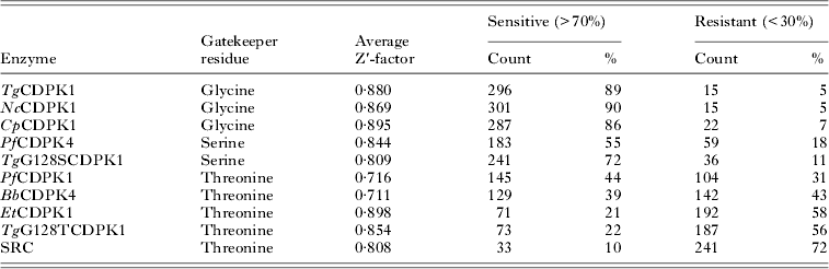
In order to establish the structure activity relationships (SAR) of BKIs against the CDPKs, we assayed the entire BKI library for inhibition of the recombinant apicomplexan CDPKs at a concentration of 3 μ m. Sensitivity to a compound at this final reaction concentration was defined as the inhibition of enzyme kinase activity by greater than 70% relative to the no inhibitor full reaction (positive control). Resistance was defined as the inhibition of an enzyme kinase activity by less than 30% relative to the positive control. The average Z′-factor of the compound reaction plate for enzymes screened ranged from 0·7112 for BbCDPK4 to 0·8949 for CpCDPK1 (Table 1). A high Z′-factor reflects two characteristics of the assay. It reflects a wide separation between the maximum signals of the assay (in this case, highest amount of luminescence in zero ATP consumption found in negative controls) and the lowest signal (lowest amount of luminescence found in positive enzyme reactions with no inhibitors causing a reduction of the initial ATP concentration). It also shows that standard deviations are low when assessing positive and negative controls. A Z′-factor value of greater than 0·5 implies a quality assay, and a perfect assay will approach 1·0 (Zhang et al. Reference Zhang, Chung and Oldenburg1999). The most sensitive CDPK orthologues to our BKI compound library were wild-type NcCDPK1, TgCDPK1 and CpCDPK1, where 90% (301 compounds), 89% (296) and 86% (287) of inhibitors exhibited over 70% to absolute inhibition of kinase activity, respectively (Table 1). These three enzymes share a glycine gatekeeper residue. These results were followed by PfCDPK4 with 55% (183 compounds), PfCDPK1 with 44% (145), BbCDPK4 with 39% (129), EtCDPK1 with 21% (71) and SRC with 10% (33) effective inhibitors. PfCDPK4 has a serine gatekeeper, while PfCDPK1, BbCDPK4, EtCDPK1 and the human enzyme SRC all share a threonine gatekeeper. The level of resistance to BKIs was much higher for EtCDPK1 and SRC compared with other kinases (Table 1).
Recombinant enzymes tested, gatekeeper residues, evaluation of reproducibility of assays (Z′-factor) and % BKIs that effectively inhibited the various enzymes

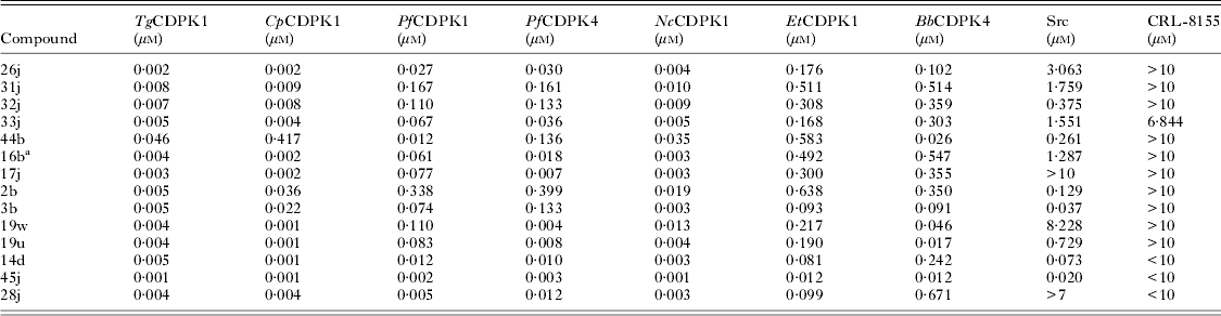
The selectivity of the BKIs’ inhibition towards the different protein kinases tested was variable. Fourteen of the 333 compounds have greater than 90% inhibition of kinase activity in all of the wild-type apicomplexan CDPK enzymes tested. Of these 14 compounds, five were completely non-selective as they also inhibited the human kinase SRC activity by greater than 90%. Forty compounds have >80% inhibition of all CDPKs with 11 of these 40 compounds also inhibiting SRC by greater than 80%. Sixty of the 333 compounds caused >70% inhibition of CDPKs where 17 also inhibited SRC by greater than 70%. Of these 60 compounds, none are benzoylbenzimidazole analogues, four are imidazo [1,5-a] pyrazine analogues and 56 are pyrazolo [3,4-d] pyrimidine analogues. Thus, when BKIs inhibit all of the CDPK orthologues, similar inhibition of SRC occurs at a ratio of one compound in approximately four (17 out of 60 compounds). Human SRC was chosen as a control because it contains a threonine gatekeeper. When the 14 compounds with >90% inhibition of all apicomplexan CDPK enzymes tested were assayed for cytotoxicity in human cells, four compounds achieved significant inhibition in the <10 μ m range in vitro against the human lymphotic cell line CRL-8155 (Table 2). Hence, the data suggest that the anti-proliferative effects of off-target BKIs on mammalian cells are likely not mediated by the inhibition of SRC.
Experimental enzymes IC50 and human cells CRL-8155 EC50 values of compounds with >90% inhibition of kinase activity at single dose of 3 μ m

a Imidazo [1, 5-a] pyrazine analogues.
Correlation relative to identity of the protein kinase domain
The apicomplexan CDPKs tested shared highly conserved ATP-binding domains. Multiple alignments of the amino acid residues proposed to interact with BKIs, as well as a matrix portraying the identity of the protein kinase domain between orthologues as a percentage are shown in Fig. 1 and Table 3. TgCDPK1 and EtCDPK1 share 94% amino acid identity within their ATP binding sites as defined by TgCDPK1 residues K52 to L198 (accession number XP_002371666). This is an important pair to compare because EtCDPK1 is the least-sensitive CDPK homologue to the BKI library, despite its close identity to TgCDPK1, the enzyme against which the focused BKI library was modelled. For comparison, TgCDPK1 and EtCDPK1 also share identities to the ATP binding sites of PfCDPK4 (77%), CpCDPK1 (74 and 73% respectively), BbCDPK4 (71 and 72%, respectively) and PfCDPK1 (54 and 55%, respectively). Amino acid identity information can sometimes be correlated to drug screening results. In theory, the greater the identities of the orthologous CDPKs, the more similar their drug inhibition profiles. A good example is illustrated by the comparison between TgCDPK1 and NcCDPK1 where in addition to 99% shared residues within the ATP-binding site, their activities with BKIs are similar. On the contrary, Cp and TgCDPK1 are very similar with respect to inhibitor binding, and yet are only 74% identical at the ATP-binding site whereas Tg and EtCDPK1 are more similar at the ATP binding site (94%) but are the most different in terms of inhibitor binding.
Multiple alignments of the amino acid residues proposed to interact with BKIs. Regions contributing to the active site as seen in crystal structures of several TgCDPK1 orthologues are shown. Numbering was based on TgCDPK1 protein database accession number XP_002371666. Specific residues that may confer differential sensitivity to some BKI are shaded yellow. Residues that are shaded grey are conserved between all the enzymes. The gatekeeper residues are shown in red. Weighted identity score (WS) of differences in amino acids, compared with Tg/NcCDPK1 (note, Tg identical to Nc in the active site) proposed to directly interact with BKIs in each enzyme is shown on the far right. The gatekeepers were excluded from this analysis since the influence of these residues on BKI inhibition of CDPKs is well defined.

Comparison of amino acid identities (%) of CDPK orthologues’ ATP-binding sites as defined by TgCDPK1 residues K52 to L198 (accession number XP_002371666). The smallest sum probability by BLASTP search is shown in parentheses

As orthologues, the apicomplexan CDPKs collectively share a positive correlation amongst one another against the BKI library. However, when assessing an individual compound amongst the enzymes, the level of sensitivity to the library varies (Table 2). This may be reflective of each enzyme's active site variations for BKI inhibition (Table 2). The shared concordance could be correlated by Pearson's R value (Table 4). Relative to other enzyme pairings, TgCDPK1, with glycine gatekeeper residue, and EtCDPK1, with threonine as the gatekeeper residue, have only fair correlation (Table 4), despite high protein kinase active site identity (Fig. 1 and Table 3). CpCDPK1's inhibition spectrum towards BKIs is very similar to TgCDPK1 and NcCDPK1 (Table 4), and distinct from the BKIs that inhibited EtCDPK1 (Pearson R value = 0·4252). PfCDPK4, the only CDPK studied with a serine gatekeeper, had fair to poor correlation when compared with other enzymes, achieving the most correlation with TgCDPK1 (Pearson R = 0·6028), CpCDPK1 (Pearson R = 0·6921) and NcCDPK1 (Pearson R = 0·5851 (Table 4).
Pearson correlation R values: Statistical comparative analysis of inhibition data for each enzyme relative to the other using Pearson's correlation test. Higher correlations among CDPKs with similar gatekeeper residues support their influence on BKI inhibition. The data showed relatively lower inhibition correlation of most CDPK enzymes with human SRC kinases

Effects of a gatekeeper residue mutation on sensitivity and characteristics of inhibitory compounds
Mutants were generated to explore the effect of the gatekeeper residue on the inhibition pattern. A TgCDPK1 threonine mutant, TgG128TCDPK1, was shown to have an almost perfect correlation and BKI compound sensitivity compared with EtCDPK1 (Tables 1 and 4). The compelling similarity between EtCDPK1 and the mutant TgG128TCDPK1 under the same conditions, as well as their relative resistance to compounds compared with the other orthologues with smaller gatekeeper residues further supports the hypothesis that gatekeeper residues are key drivers of compound binding. However, when comparing TgG128TCDPK1's SAR to BbCDPK4 and PfCDPK1, sensitivities to the compound library showed discordance (37% for BbCDPK4 and 44% for PfCDPK1 versus 21% for TgG128TCDPK1 or EtCDPK1, respectively). Thus, EtCDPK1 and TgG128TCDPK1 are more resistant to the library than BbCDPK4 and PfCDPK1.
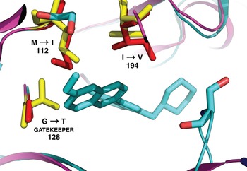
Weighted identity scores ([WS] in Fig. 1) of conserved differences in amino acids proposed to directly interact with BKIs were derived by assigning a unitless value of 1 for every property difference, based on well-established properties of charge, polarity and size as previously described (Livingstone and Barton, CABIOS, Reference Livingstone and Barton1993). The gatekeepers were excluded from this analysis since the influence of these residues on BKI inhibition of CDPKs is well defined. By this measurement, PfCDPK1 and CpCDPK1 have the highest divergence from the TgCDPK1 ATP binding site with a score of 15 for PfCDPK1 and 9 for CpCDPK1 (Fig. 1). However, CpCDPK1 showed identical SAR profile and with high sensitivity towards the library, much like TgCDPK1, probably due to the higher influence of the shared glycine gatekeeper residue. Conversely, the SAR profiles of Nc/TgCDPK1 versus those of PfCDPK1, BbCDPK4 and EtCDPK1 make it challenging to attribute all the inhibitor library observed characteristics to the gatekeeper size alone. In the latter case, we can partially attribute higher sensitivity to increased divergence from the model of TgCDPK1 on which compound design was based. However, it is likely that a combinatory effect resulting from differences in types and chemical properties of amino acids within the active sites of apicomplexa CDPKs can produce a variety of BKI inhibition SARs. Analysis of amino acid differences between PfCDPK1 and TgCDPK1 demonstrated deviations capable of promoting the differential sensitivity observed. Through modelling based on previous crystal structures of TgCDPK1 and CpCDPK1 co-crystallized with BKI inhibitors, probable structural models were created to simulate binding motifs of other CDPK enzymes. PfCDPK1 was compared to this effect with TgCDPK1 (Fig. 2). Three possible differences from the sequences of PfCDPK1 and TgCDPK1 were noted that may influence the binding of BKIs to PfCDPK1. These include TgCDPK1 F134V (Residue 117 in PfCDPK1), M112I (Residue 129 in PfCDPK1) and I194V (Residue 221 in PfCDPK1). Perhaps the most influential variation is at the position of methionine 112 in TgCDPK1 (isoleucine 129 in PfCDPK1). The larger (glycine->threonine) gatekeeper residue on one side of the binding pocket is paired with a complementary smaller residue (methionine->isoleucine) on the opposite side of the binding pocket (Fig. 2). This allows the BKI binding pose to shift slightly away from the gatekeeper, something that is otherwise sterically restricted in TgCDPK1 and EtCDPK1. Thus the effect of the larger threonine gatekeeper in PfCDPK1 is partially offset by the presence of a complementary amino acid variation elsewhere in the active site, which may explain its increased BKI sensitivity relative to EtCDPK1 (Fig. 2).
BKI interactions in the ATP-binding domains of PfCDPK1 and TgCDPK1. Model of PfCDPK1 based on previous crystal structure of TgCDPK1 in complex with inhibitor 19j (Larson et al. Reference Larson, Ojo, Murphy, Johnson, Zhang, Kim, Leibly, Fox, Reid, Dale, Perera, Kim, Hewitt, Hol, Verlinde, Fan, Van Voorhis, Maly and Merritt2012; PDB 3sx9). Gatekeeper residues and other residues postulated to affect inhibition are shown as red (TgCDPK1) and yellow (PfCDPK1) sticks. The larger G128M (Residue 145 in PfCDPK1) gatekeeper residue is paired with a complementary smaller residue M112I (Residue 129 in PfCDPK1), and I194V (Residue 221 in PfCDPK1) in PfCDPK1. This partially offset the effect of the larger gatekeeper in PfCDPK1 by the presence of a complementary amino acid variation elsewhere in the active site, which may explain its increased BKI sensitivity relative to EtCDPK1.

Within the TgG128TCDPK1 inhibitor SAR, patterns of compound characteristics were identified. In general, inhibition of TgG128TCDPK1 and its highly similar threonine partner, EtCDPK1, requires relatively small substituents at the C3 position (R1 group), such as an alkoxybenzyl group, 4-chlorophenyl, or naphthalen-2-yl (Fig. 3). Presumably, larger substituents exhibit steric clash with the threonine gatekeeper, disfavouring binding. However, a few compounds with larger R1 groups, such as 23j, 24j, 31j, 32j, 33j, 26a, 24a, 25j, 30a, 39a, 27a, 34a, 6j, did cause >70% inhibition of EtCDPK1 kinase activity, irrespective of the R2 groups size. Such constraints on inhibitors were not as obvious with the other threonine enzymes BbCDPK4 and PfCDPK1. A wider range of compounds were able to achieve inhibition for these orthologues (BbCDPK4 and PfCDPK1) relative to EtCDPK1, including compounds with larger R1 moieties (6-alkoxy- and 6-benzyloxy-naphthol) without the need of a smaller R2 group that may be associated with the spatial constraints of enzymes with larger gatekeeper residues.
R1 and R2 substructures of compounds.

Contrary to the near-identical sensitivity and correlation seen with EtCDPK1 and TgG128TCDPK1, the serine mutant TgG128SCDPK1 was sensitive to 241 (72%) of compounds while its serine gatekeeper partner PfCDPK4 was sensitive to 183 (55%). Although this enzyme pair shared the same gatekeeper residue, there was a difference in sensitivity patterns to compounds and change in correlation (Table 4: Pearson R value 0·7714) that was not dependent on the gatekeeper. A series of compounds was isolated which differentially inhibited TgG128SCDPK1 over PfCDPK4. This series shifted inhibition towards TgG128SCDPK1 by greater than 30% over PfCDPK4. Compounds with this pattern include 15k, 4a, 13a, 31a, 32a, 36a, 35a, 37a, 30a, 41b, 43b, 40b, 25b, 6b, 12b, 10b.
PfCDPK4 seems to accommodate R1 groups such as a naphthyl ring connected in an ortho configuration, while the larger threonine gatekeeper residue enzyme, PfCDPK1, was able to accommodate quite large and extended groups at this position, such as 6-alkoxy- and 6-benzyloxynaphthol substituents. In fact, a group of compounds with extended moieties inhibited PfCDPK1, yet against PfCDPK4 with a serine gatekeeper residue, did not achieve the same level of inhibition. These compounds include 35a, 7a, 8a, 9a, 13a, 36a, 35a, 11j, 10b (Fig. 3).
DISCUSSION
A comparative analysis of the structural activity relationship of veterinary pathogens N. caninum, E. tenella and B. bovis; zoonotic pathogens C. parvum and T. gondii CDPKs and those of human pathogen P. falciparum CDPKs was determined by screening against 333 BKI analogues. The degree of sensitivity or resistance of CDPK enzymes to BKIs was earlier postulated to be due to the size and characteristic of the gatekeeper residue and the adjacent pocket. Indeed, correlation of screening data was best among enzymes with the same gatekeeper residue, which corroborates the gatekeeper hypothesis (Table 4). However, sensitivity to compounds among enzymes with the same gatekeeper residue does not correlate perfectly, suggesting alternative modes of CDPK–drug interaction. We explored this by constructing TgCDPK1 mutants that substituted serine or threonine for the wild-type glycine at the gatekeeper position, and comparing their BKI sensitivity profile with that of the homologues with the corresponding wild-type gatekeeper residue; i.e. TgCDPK1G128S can be compared to PfCDPK4, and TgCDPK1G128T can be compared to PfCDPK1, EtCDPK1 and BbCDPK4 (Tables 3 and 4). Although EtCDPK1 has the closest sequence identity to wild-type Tg/NcCDPK1 in the ATP binding pocket (Table 3: 94%), it nonetheless proved to be the least sensitive enzyme to BKIs, with poor correlation to the sensitivity profile of wild-type TgCDPK1. However, the sensitivity of EtCDPK1 correlates very well with that of TgCDPK1G128T. The BKI sensitivity of two other enzymes with a threonine gatekeeper, BbCDPK4 and PfCDPK1, is also more similar to that of the T. gondii G128T mutant than to that of the wild-type TgCDPK1. However, these orthologues share less sequence identity with the T. gondii enzyme and have weaker correlation with the sensitivity of the TgCDPK1G128T mutant. TgCDPK1 and NcCDPK1 are also similar in their ATP binding domain identity (>99%) and predictably share similar sensitivity to compounds. Yet, CpCDPK1 is not as similar in identity to TgCDPK1 as EtCDPK1 (94% identity versus 74%) or NcCDPK1 (>99% versus 74%) (Table 3). However, the fact that Tg/NcCDPK1 and CpCDPK1 are the most sensitive enzymes to compounds, yet share less sequence similarity (74% identity) than that of EtCDPK1, returns us to the enormous role played by the size of the gatekeeper residue in determining the level of sensitivity to BKIs. This assertion was supported with experimental evidence showing excellent correlation and characteristics of structure activity relationship between EtCDPK1 and TgG128TCDPK1, completely reversing susceptibility of TgCDPK1.
Although the similarity of a homologue to TgCDPK1's ATP-binding site sequence is an attractive marker for determining sensitivity to BKIs for enzymes whose full binding pocket is uncharacterized, the binding pocket size, charge and hydrophobicity are all factors that ultimately determine sensitivity to BKIs. A combination of these factors contributes directly or indirectly to the sensitivity of CDPKs to BKIs. Further research is necessary to elucidate the relative impact of each factor to CDPK sensitivity to BKIs. Crystal structures of these new homologues may help understand the complex nature of the binding pocket. With this new information, we conclude that a large gatekeeper residue does not necessarily mean complete resistance to BKIs. In the example of our threonine gatekeeper enzymes, low nanomolar inhibition was achieved, even by BKIs designed to target small gatekeepers.
It is important that future drug development focuses on exploiting other characteristics of the drug-binding pocket in addition to the gatekeeper. In this respect, compounds could be optimized to target the conserved apicomplexan ribose-binding pocket. Similarly, targeting the calcium-binding regulatory domain could prevent activation of CDPK enzymes with a resultant disruption of downstream process. Compounds that inhibit EtCDPK1 but not SRC are important as lead compounds for the development of broad-spectrum antimicrobials. Because of its threonine gatekeeper and unaccommodating binding pocket towards BKIs, if potent compounds to target EtCDPK1 are synthesized, then clinically, these compounds can be useful as monotherapy with low likelihood of easily conferring resistance across CDPKs of the apicomplexa. Within our BKI library, 34 compounds effectively inhibited EtCDPK1 by >70% and inhibit SRC kinase activity by <30%. The structure and characteristics of these compounds will be the focus of future studies. These compounds include 19a, 19j, 22j, 23j, 26j, 24j, 21j, 31j, 33j, 25j, 3j, 38a, 30a, 42b, 19b, 10j, 17j, 6j, 25l, 19e, 16j, 19q, 19r, 19t, 19v, 19w, 20j, 5j, 5l, 19s, 20a, 28j.
CONCLUSION
It is apparent that the highly conserved nature of the ATP-binding domain shared by apicomplexan CDPK homologues could be exploited to some extent in development of potential broad-spectrum inhibitors if potential structural constraints are well defined and understood. A combination of these constraints concerning the gatekeeper residue, the structure and characteristic of the binding pocket, and differences among compounds, all contribute subtle distinctions in sensitivity to compounds among these highly conserved CDPKs. Further research into the structural constraints for the development of broad-spectrum, potent inhibitors of apicomplexan CDPKs is warranted.
ACKNOWLEDGEMENTS
The authors wish to express our gratitude to Steve M. Johnson, Ryan C. Murphy and Edward J. Dale for technical and valuable support.
FINANCIAL SUPPORT
This work is supported by the National Institutes of Health grants R01AI089441 (E.A.M. and W.C.V.V.) and 5 R01 AI080625 (W.C.V.V.). The Maly Lab was supported by NIH grant R01GM086858. K.R.K. was supported by a training scholarship from the University of Washington Plein Endowment for Geriatric Pharmacy Research.