Introduction
The manipulation of oocytes enclosed in preantral follicles (MOEPF) biotechnique, also known as artificial ovaries, has as a main objective the recovery of oocytes included in the preantral follicles and their in vitro culture until complete maturation. Preantral follicles represent about 95% of the entire follicular population and normally do not develop until the preovulatory follicle stage, as they enter a physiological process known as atresia (Araújo et al., Reference Araújo, Gastal, Figueiredo and Gastal2014).
To date, the success of MOEPF has been reported in mice, with the birth of healthy mice from primordial follicles grown, matured and fertilized in vitro (O’Brien et al., Reference O’Brien, Pendola and Eppig2003), as well as after cryopreservation followed by in vitro culture of ovarian tissue, also resulting in the gestation and birth of live offspring (Wang et al., Reference Wang, Catt, Pangestu and Temple-Smith2011). It was previously observed that, in the case of goats (Magalhães et al., Reference Magalhães, Duarte, Araújo, Brito, Soares, Lima, Lopes, Campello, Rodrigues and Figueiredo2011), sheep (Arunakumari et al., Reference Arunakumari, Shanmugasundaram and Rao2010) and buffaloes (Gupta et al. Reference Gupta, Ramesh, Manjunatha, Nandi and Ravindra2008), in vitro culture of preantral follicles resulted in the production of mature oocytes, fertilized in vitro, giving rise to embryos, but not gestated. In humans (Telfer et al., Reference Telfer, McLaughlin, Ding and Thong2008), cattle (Araújo et al., Reference Araújo, Gastal, Figueiredo and Gastal2014) and dogs (Serafim et al., Reference Serafim, Duarte, Silva, Souza, Magalhães-Padilha, Moura, Silva, Campello and Figueiredo2015), the maximum archived was the development of primordial follicles up to the antral stage.
Co-culture of oocytes or embryos with somatic cells has been strongly suggested in the attempt to simulate what occurs naturally in vivo, as follicular development is regulated by paracrine and autocrine factors of the surrounding somatic cells (Araújo et al., Reference Araújo, Gastal, Figueiredo and Gastal2014). Mesenchymal stem cells (MSCs) are somatic cells with greater potential for differentiation, which under appropriate conditions are able to establish a regenerative microenvironment through the secretion of bioactive factors, trophic and antiapoptotic molecules. In addition, they are able to remove toxic components and reactive oxygen species (ROS), thereby helping the survival and proliferation of other cell types (Moshkdanian et al., Reference Moshkdanian, Nematollahi-Mahani, Pouya and Nematollahi-Mahani2011). These benefits have already been observed in the co-culture of MSC with cattle (Miranda et al., Reference Miranda, Nascimento and Costa2016) and mice embryos (Moshkdanian et al., Reference Moshkdanian, Nematollahi-Mahani, Pouya and Nematollahi-Mahani2011), as well as in co-culture with preantral follicles in cattle (Itoh and Hoshi, Reference Itoh and Hoshi2000), mice (Green et al, Reference Green, Zhou and Shikanov2016) and humans (Xia et al., Reference Xia, Wang, Yin, Yan, Yan, Lu, Zhao, Li, Zhang, Jin, Zhu, Liu, Li and Qiao2015).
Although Saraiva et al. (Reference Saraiva, Rossetto, Brito, Celestino, Silva, Faustino, Almeida, Bruno, Magalhães, Matos, Campello and Figueiredo2010) and Magalhães et al. (Reference Magalhães, Duarte, Araújo, Brito, Soares, Lima, Lopes, Campello, Rodrigues and Figueiredo2011) have been able to obtain embryos from goat preantral follicles, which is considered an expressive advance, both recommended new studies for the definition of an appropriate medium for the complete in vitro development of primordial goat follicles, because in these two experiments the rate of oocyte maturation was suboptimal, therefore preventing subsequent large-scale production of oocytes and embryos developed in vitro.
This work aims to develop an in vitro co-culture system of goat preantral follicles with mesenchymal bone marrow stem cells, evaluating their influence on follicular growth, rate of activation and survival of morphologically normal follicles.
Materials and methods
Ovarian tissue preparation and experimental design
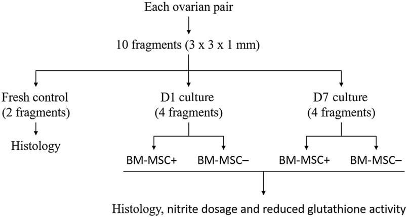
Five pairs of ovaries (n = 5) were collected from five adult cross-breed goats (Capra hircus) at a local slaughterhouse. Immediately postmortem, the ovaries were washed once in 70% ethanol for 10 s and then washed twice in HEPES-buffered minimum essential medium (MEM) supplemented with 100 μg/ml penicillin and 100 μg/ml streptomycin. Ovaries were then transported to the laboratory in MEM at 4°C within 1 h after collection. At the laboratory, ovaries were stripped of the surrounding fat and fibrous tissue and the ovarian cortex from each ovarian pair was cut into 10 (n = 10) small fragments (3 × 3 × 1 mm) using a scalpel blade under sterile conditions. Part of the fragments were randomly distributed as fresh control samples (n = 2) and the remaining fragments (n = 8) were cultured for 1 or 7 days with the presence (BM-MSC+) or absence (BM-MSC−) of MSC of the goat’s bone marrow (BM-MSC). Experimental design was performed as depicted in Fig. 1.
Experimental design. Ovarian fragments were divided into five groups: Fresh control and in vitro cultured for 1 or 7 days with the presence of BM-MSC (BM-MSC+) or absence of BM-MSC (BM-MSC−). Comprehensive histological analysis was performed in all study groups; and the nitrite dosage and reduced glutathione activity were performed after 1 or 7 days of culture.

Acquisition and preparation of mesenchymal stem cells from the bone marrow of goats (BM-MSC)
BM-MSC were obtained from thawing of cryopreserved cells in the stem cell bank of the Integrated Nucleus of Morphology and Research with Stem Cells (NUPCelt) of the Federal University of Piauí, located in the city of Teresina-PI. Cells were isolated, characterized and cryopreserved at the third passage during a previous study using healthy adult goats from the centre of Agricultural Sciences of the Federal University of Piauí.
After thawing in a water bath at 37°C, the BM-MSCs were transferred to 15 ml polypropylene tubes containing twice the volume of α-MEM growth medium (Invitrogen®, Carlsbad, CA, USA). They were then centrifuged at 1000 rpm for 5 min. The supernatant was discarded and the pellet resuspended in 1 ml of the same medium, this process was repeated twice; at the end the resulting solution was deposited in a 25 cm² culture bottle already containing 2 ml of the culture medium. The bottle was maintained in a humid atmosphere oven at 37°C and 5% CO2 and the culture medium was changed every 72 h until the cells reached 80% confluency. Adherence and cell growth were monitored by observation with an inverted microscope. After confluence, cells were harvested to 24-well culture plates. To detach adherent cells, 3 ml of trypsin–EDTA solution (0.25%) and pH 7.2 (Sigma-Aldrich, St. Louis, MO, USA) was added to the bottle, which was incubated in an oven at 37°C and 5% CO2 for 5 min. After cell detachment, double the culture medium was added to neutralize the trypsin, totalling a solution of 9 ml, which was centrifuged at 1500 rpm for 10 min. The supernatant was discarded and the pellet resuspended in 1 ml of medium to perform counting of cells in Neubauer’s chamber, so that each well received 1 × 10³ cells. After cell adhesion, which occurred generally after 24 h, the ovarian fragments were deposited with these for co-culture (one fragment per well).
Co-culture of goats preantral follicles in situ associated with BM-MSC
Co-culture started only when the BM-MSC showed 80% confluency in the culture wells. The culture medium for both treatments, BM-MSC+ and BM-MSC−, consisted of 1 ml of MEM (Invitrogen®, Carlsbad, CA, USA) culture medium supplemented with ITS (6.25 μg/ml insulin, 6.25 mg transferrin), 20 IU/ml penicillin, 2 mM glutamine, 2 mM hypoxanthine, 1.25 mg/ml bovine serum albumin (BSA; Gibco BRL, Rockville, MD, USA), streptomycin and 0.005 g of ascorbic acid. Incubation was carried out in a sterile environment in an atmosphere of 5% CO2, 95% relative humidity at 37°C. The follicles included in ovarian tissue were cultured for up to 7 days, with exchanges of medium performed every 48 h. During culture, on days D1 and D7, fragments were collected and sent for histological processing to evaluate the morphology, growth and rates of survival, and follicular activation. In addition to the fragments, culture medium was also collected on the same day for nitrite dosage and reduced glutathione activity.
Histological processing
All fragments collected during the experiment (fresh control and in vitro post-culture) were fixed in Carnoy’s solution for 4 h, dehydrated using a 70, 80, 90 and 100% alcohol sequence, diaphanized in xylol and paraffin embedded at 60°C. The paraffin-embedded fragments were sequentially sectioned at 7 μm thickness for assembly of the slides, which were stained using haematoxylin and eosin.
Morphological analysis
Overall, 150 follicles were evaluated for each treatment (30 follicles per treatment in 1 repetition × 5 repetitions = 150 follicles). Only preantral ovarian follicles with oocytes showing an evident nucleus in the observed section were part of the analysis. According to stage of development cells were defined as: primordial (oocyte surrounded by cells of the pre-granulosa of paved form) or in development (oocyte surrounded by one or more layers of cells of the pre-granulosa of paved form and at least one cell of the granulosa of cubic format). Preantral follicles were morphologically classified in normal (follicles containing oocytes and intact granulosa cells) or degenerate follicles (with pyknotic nucleus or presence of pyknotic bodies, cytoplasmic retraction and granulosa cell disorganization).
Rate of morphologically normal follicles and follicular activation
The rate of morphologically normal follicles was based on morphological classification as previously described (normal or degenerate) and the proportion of normal follicles in relation to the total analysed follicles was calculated. The proportion averages were calculated in each treatment and in each culture period and then compared.
To evaluate follicular activation, the proportions of primordial and growing follicles were calculated for the fresh control group and after 1 or 7 days of culture under various culture conditions. Follicular activation rate (%) was calculated as follows: number of primordial follicles or for growth after day 1 or 7 of culture divided by number of normal follicles evaluated × 100.
Evaluation of follicular development
Oocyte and follicular diameter of preantral follicles classified as normal were obtained from the average of two perpendicular measurements (90º). All histological sections were examined using a trinocular optical microscope (Nova Optical Systems®, São Paulo, Brazil), equipped with a digital camera (TOPCAM ™ 5 Megapixel) for photographic recording of images. All measurements were performed using ToupView® 3.7 software.
Determination of nitrite content and reduced glutathione activity (GSH)
The determination of nitrite content was based on the Griess reaction (Green et al., Reference Green, Tannenbaum and Goldman1978) in which 500 μl of the Griess reagent plus 500 μl of the culture medium without culture (White) were combined in a white tube. In another test tube, 500 μl of the Griess reagent plus 500 μl of the culture medium (50 mM sodium phosphate buffer, pH 7.4) (Test) were combined. The spectrophotometer was read at 560 nm. Results were expressed as μM/mg protein.
Determination of the GSH activity was based on the Ellman reaction (5,5′-dithiobis(2-nitrobenzoic acid)) with some modifications of the technique as described by Sedlak and Lindsay (Reference Sedlak and Lindsay1968). In a tube, 400 μl of culture medium, 320 μl of distilled water and 80 μl of 10% trichloroacetic acid were combined. The material was shaken and centrifuged at 3000 rpm for 15 min, then 400 μl of supernatant was combined with 800 μl of 0.4 M Tris–HCl buffer, pH 8.9 and a further 20 μl of 0.01 M DTNB and, after 1 min of reaction, the spectrophotometer was read at 412 nm. The concentration was expressed in mg/g haemoglobin.
Statistical analysis
The diameters, activation rate and morphologically normal follicular rate were obtained and analysed using the statistical package SAS (Statistical Analysis System®, version 9.2). The Shapiro−Wilk test was used to test normality. However, the diameter of oocytes and follicles in the various treatments did not follow a normal distribution. Therefore, a generalized linear model was used instead of non-parametric tests. The Gama model was the one that demonstrated possible distributions to be adjusted, being, therefore, the chosen one. The Wald test was used for comparison of means. For the follicular activation rate, the data had normal distribution, being submitted to analysis of variance (ANOVA) followed by the Tukey test to compare differences between the treatments. For the rate of morphologically normal follicles, the data were analysed by chi-squared test. Data on oxidative stress were also evaluated using ANOVA followed by the Neuman–Keuls test. Data were analysed using GraphPad Prism 6.0 software (San Diego, CA, USA). Results were expressed as mean ± standard error of the mean (SEM). Values were considered statistically when P-values were < 0.05.
Results
Percentage of morphologically normal goat preantral follicles
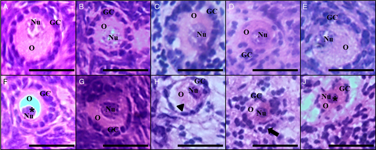
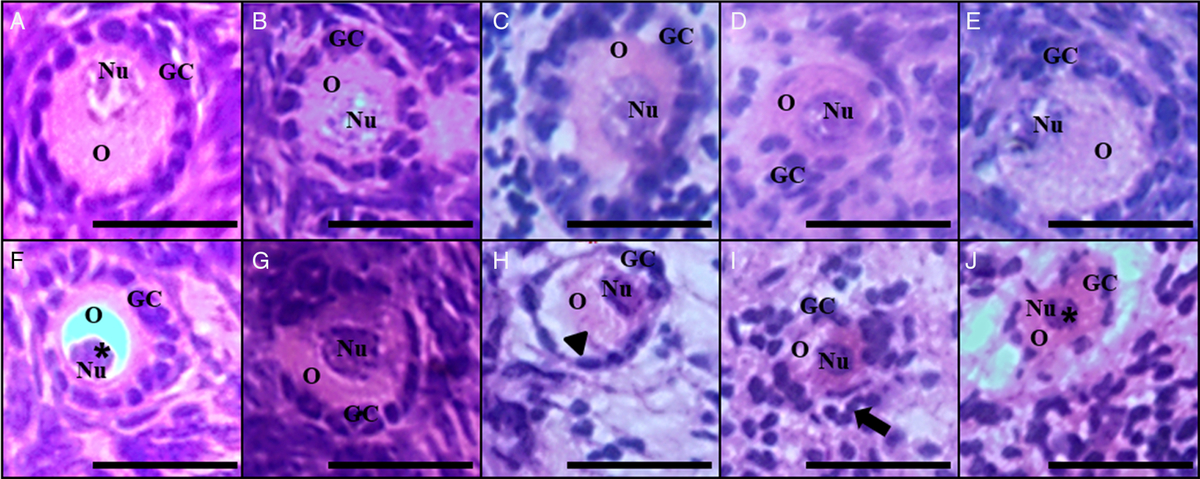
Preantral follicles included in the ovarian tissue were considered morphologically normal when the oocyte was spherical or slightly elongated, with a nucleus without evidence of pyknose and with well organized granulosa cells surrounding the oocyte. Those with cytoplasmic retraction, pyknotic oocyte nucleus or disorganized granulosa cells were considered to be degenerate. Normal and atresic follicles were found in the fresh control, as well as after 1 or 7 days of culture, in both treatments (Fig. 2).
Histological section of the ovarian cortex (×400 magnification) demonstrating the morphology of goat preantral ovarian follicles stained with haematoxylin and eosin. Normal follicles with intact oocyte and nucleus are presented in the fresh control (A), 1 day of co-culture with presence of BM-MSC (B), 1 day of culture with absence of BM-MSC (C), 7 days of co-culture with presence of BM-MSC (D), 7 days of culture with absence of BM-MSC (E). Degenerate follicles are shown in the fresh control (F), 1 day of co-culture with presence of BM-MSC (G), 1 day of culture with absence of BM-MSC (H), 7 days of co-culture with presence of BM-MSC (I), 7 days of culture with absence BM-MSC (J). Presence of pyknotic nucleus (*), cytoplasmic retraction (arrowhead) and disorganization of granulosa cells (arrow). O: oocyte; Nu: oocyte nucleus; GC: granulosa cells. Scale bars represent 25 μm.
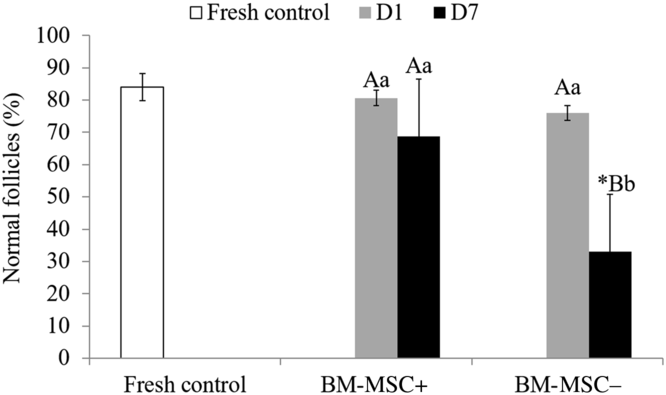
The percentage of morphologically normal follicles found in the fresh control, as well as after 1 or 7 days of in vitro culture in BM-MSC+ and BM-MSC− is represented (Fig. 3). It was observed that there was a reduction (P < 0.05) in this percentage compared with the fresh control only after 7 days of culture following BM-MSC− treatment. In treated BM-MSC+ no reduction (P > 0.05) was observed in the percentage of morphologically normal follicles between days 1 and 7 of culture. However, following BM-MSC− treatment the percentage of morphologically normal follicles in 7 days of culture was lower compared with day 1 (P < 0.05). When comparing treatments BM-MSC+ versus BM-MSC−, on day 1 there was no difference (P > 0.05) in the percentage of morphologically normal follicles, but on day 7 the number of morphologically normal follicles found for BM-MSC+ treatment was higher than BM-MSC− treatment (P < 0.05).
Percentage (mean ± SEM) of preantral follicles morphologically normal in fresh (uncultivated) control and after in vitro culture for 1 (D1) or 7 (D7) days with the presence of BM-MSC (BM-MSC+) and absence of BM-MSC (BM-MSC−). *Statistically different from control (P < 0.05). Different uppercase letters indicate differences in the same treatment on different days of culture (P < 0.05). Different lowercase letters indicate difference between treatments on the same day of culture (P < 0.05).

Follicular development
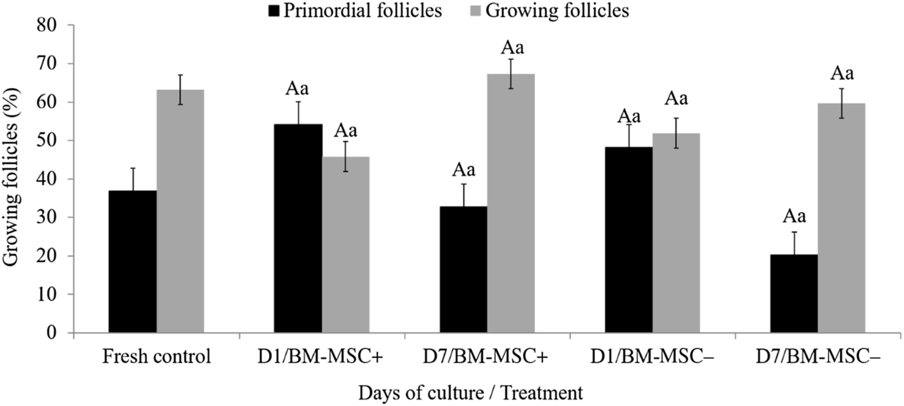
The percentage of primordial and developing follicles observed at all times is shown in Fig. 4. After 1 or 7 days of culture, there was no effect of the presence of BM-MSC on follicular activation, i.e. in the percentage of primordial and growing follicles (P > 0.05).
Percentage (mean ± SD) of primordial goat preantral ovarian follicles under development in fresh (uncultivated) control and after in vitro culture for 1 (D1) or 7 (D7) days with the presence of BM-MSC (BM-MSC+) and absence of BM-MSC (BM-MSC−). *Statistically different from control (P < 0.05). Different uppercase letters indicate difference in the same treatment on different days of culture (P < 0.05); distinct lowercase letters indicate difference between treatments on the same day of culture (P < 0.05).

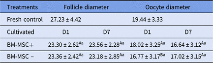
When comparing the treatments tested, as well as with the fresh control, on different days of culture, no differences were observed for follicular diameter (P > 0.05). However, on day 1 of culture, oocyte diameter was higher (P < 0.05) after BM-MSC+ treatment in relation to BM-MSC− treatment. After 7 days of culture, both treatments presented similar diameters (P > 0.05) and, when compared with the fresh control, also no differences were observed (Table 1).
Follicular, oocyte and nuclear (μm) diameter (mean ± SD) in fresh (uncultivated) control and after in vitro culture of preantral follicles for 1 or 7 days with presence of BM-MSC (BM-MSC+) and absence of BM-MSC (BM-MSC−)

Different uppercase letters indicate differences between treatments on the same day of culture (P < 0.05); distinct lowercase letters indicate differences between the same treatment on different days of culture (P < 0.05).
Concentration of nitrite and reduced glutathione
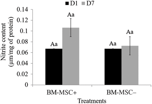
Figure 5 shows nitrite production after in vitro culture of preantral follicles for 1 or 7 days in the absence (BM-MSC−) or presence (BM-MSC+) of MSCs from the bone marrow of goats. There was no difference between the treatments tested, regardless of the day of culture (P > 0.05).
Concentration of nitrite (mean ± SEM) in the culture medium of preantral follicles after culture in vitro for 1 (D1) or 7 (D7) days with the presence of BM-MSC (BM-MSC+) and absence of BM-MSC (BM-MSC−). Different uppercase letters indicate difference in the same treatment on different days of culture (P < 0.05); distinct lowercase letters indicate difference between treatments on the same day of culture (P < 0.05).

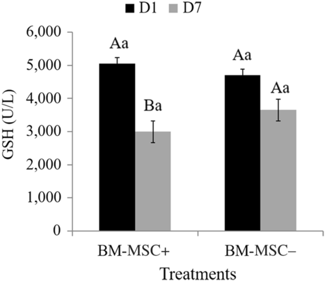
The levels of reduced glutathione are expressed in Fig. 6, in which a decrease (P < 0.05) in glutathione concentration from day 1 to day 7 was observed following BM-MSC+ treatment; whereas, in the BM-MSC− culture there was no statistical difference during the days of culture (P > 0.05). Although reduced glutathione levels decreased on day 7 of BM-MSC+ treatment, there was no difference (P > 0.05) from levels found on the same day for BM-MSC− treatment.
Concentration of reduced glutathione (GSH) in the culture medium of preantral follicles after culture in vitro for 1 (D1) or 7 (D7) days with presence of BM-MSC (BM-MSC+) and absence of BM-MSC (BM-MSC−). Different uppercase letters indicate differences in the same treatment on different days of culture (P < 0.05); distinct lowercase letters indicate differences between treatments on the same day of culture (P < 0.05).

Discussion
The aim of this study was to develop a co-culture system of mesenchymal bone marrow stem cells with preantral follicles of goats, to determine if BM-MSCs would be able to offer better developmental conditions in vitro for these follicles. It was observed in the BM-MSC+ treatment that the percentage of morphologically normal follicles was maintained throughout the growing period. Conversely, after 7 days of culture in BM-MSC− treatment, it was verified that only 33% of follicles had preserved morphology, differing (P < 0.05) from BM-MSC+ treatment on the same day of culture, in which 68% of preantral follicles were morphologically normal. These results are in agreement with those described by Xia et al. (Reference Xia, Wang, Yin, Yan, Yan, Lu, Zhao, Li, Zhang, Jin, Zhu, Liu, Li and Qiao2015), in which after 8 days of culture in the absence of MSC there were lower survival rates (30%) in relation to co-culture with high (0.5 × 10⁵) and low (0.5 × 10³) density of MSC, in which rates of 58.8% and 42.9% for follicular survival were reported, respectively.
The co-culture of preantral follicles with BM-MSC developed in this work can be considered as low MSC density, as the amount of cells present in the co-culture wells was 1 × 10³. This may explain, at least in part, the maintenance of the rate of morphologically normal follicles in the co-culture over 7 days compared with the fresh control, with no difference between the treatments tested, BM-MSC+ versus BM-MSC−, at the end of the period of culture. Therefore, under our experimental conditions strong evidence ws observed that BM-MSCs may provide adequate support for the survival of goat preantral follicles.
During the development of this study, an attempt was initially made to co-culture using 1 × 10⁵ BM-MSC per well. However, it was observed that this cell density was very high and made it impossible to develop the experiment, as cells reached 100% confluence in the 1.91 cm2 wells before 7 days of culture. After full confluency, BM-MSCs began to die, damaging the main goal of the study, which was to keep preantral follicles co-cultured with undifferentiated live cells in short-term culture.
The factors involved in the death of BM-MSCs are unknown, but it is believed that the lack of area for expansion, coupled with the lack of ideal conditions for differentiation, may cause increased oxidative stress of these cells predisposing to apoptosis (Park et al., Reference Park, Kim, Xia and Sung2011). It is believed that an alternative to this protocol would be to exchange the fragment for a new well containing BM-MSC still in cell expansion, when the former reached 100% confluence. However, this hypothesis was not tested in this experiment and should be analysed in the future.
The mechanisms by which MSCs act to maintain the survival rate of morphologically normal preantral follicles in co-culture are not yet fully elucidated. However, some studies have shown that MSCs are able to secrete factors that help maintain viability and follicular development, such as fibroblast growth factor and members of the superfamily of the transforming growth factor-β (TGF-β) (Kim et al., Reference Kim, Park and Sung2009; Parte et al., Reference Parte, Bhartiya, Manjramkar, Chauhan and Joshi2013). These growth factors have been shown to be fundamental for maintaining the ultrastructure of the ovarian follicle, maintaining not only its morphology but also its function (Rodrigues et al., Reference Rodrigues, Lima, Chaves, Rossetto, Costa, Castro, Barros, Matos, Lopes, Báo, Campello and Figueiredo2014; Kim et al., Reference Kim, Yun, Shin, Lee, Lee, Yu, Seo, Lee, Kang, Choi, Seo and Kang2015). Among members of this superfamily produced by MSCs with proven efficacy on follicular development are activin A (Park et al., Reference Park, Lee, Chang, Kim, Sung, Na and Chang2016) and bone morphogenetic protein (BMP) (Araújo et al., Reference Araújo, Silva, Magalhães, Silva, Báo, Silva, Figueiredo and Rodrigues2010; Xia et al., Reference Xia, Wang, Yin, Yan, Yan, Lu, Zhao, Li, Zhang, Jin, Zhu, Liu, Li and Qiao2015).
In this study, we chose to co-culture with MSCs derived from bone marrow precisely because of the possibility of this type of cell secreting BMP, as these are molecules synthesized by osteoprogenitor cells, as well as BM-MSCs. To date, 15 types of BMPs have been described and seven of these (BMP-2, -3, -3b, -4, -6, -7 and -15) have already been localized in mammalian ovaries (Glister et al., Reference Glister, Kemp and Knight2004; Fatehi et al., Reference Fatehi, Van Den Hurk, Colenbrander, Daemen, Van Tol, Monteiro, Roelen and Bevers2005; Persani et al., Reference Persani, Rossetti, Di Pasquale, Cacciatore and Fabre2014). These proteins play an essential role in the development of various cell types in various tissues, acting on cell proliferation and differentiation, organogenesis, embryonic development, apoptosis and repair of a wide variety of tissues (Carreira et al., Reference Carreira, Alves, Zambuzzi, Sogayar and Granjeiro2014). In addition, the use of BMPs as beneficial factors for the development of in vitro cultured preantral follicles has been recurrent (Araújo et al., Reference Araújo, Silva, Magalhães, Silva, Báo, Silva, Figueiredo and Rodrigues2010). These observations justify the high survival rate of morphologically normal follicles found in the present study after 7 days of BM-MSC co-culture. Moreover, it is important to note that the base culture medium used in this experiment did not receive exogenous supplementation of hormones and/or growth factors, as the objective was to evaluate the unique and exclusive influence of BM-MSC.
Co-culture with BM-MSC had no influence on the rate of development, activation or follicular and oocyte growth, except on the first day of culture, when oocyte diameter was higher in BM-MSC co-culture. However, it is possible that this difference was not caused by BM-MSC, as at the end of co-culture no difference was detected between the treatments tested. In a previous study, Rossetto et al. (Reference Rossetto, Matos, Celestino, Bruno, Silva and Faustino2010) also found no difference in the growth of preantral ovarian follicles after 7 days of culture with culture medium containing follicle stimulating hormone (FSH) and androstenedione. However, there was follicular activation, which can be explained by the possibility that the primordial follicles do not increase in diameter, but are activated, that is they develop from primordial to transitional or primary follicles as evidenced by a change in granulosa cell morphology, which are no longer paved to become cubic. This phenomenon often does not imply the diameter of the oocyte or the follicle, but represents follicular development, indicating activation.
To evaluate oxidative stress, nitrite and reduced glutathione were measured in the culture medium collected during the experiment. Bruno et al. (Reference Bruno, Lima-Verde, Martins, Matos, Lopes, Maia, Báo, Nobre Junior, Maia, Pessoa, Moraes, Silva, Figueiredo and Rodrigues2008) reported that nitrite levels in the culture medium can be used as indicators of granulosa cell viability of the preantral follicles of goat. However, in our experimental conditions there was no difference between treatments on the same day of culture, as well as in the same treatment in different periods. In relation to the concentration of reduced glutathione, there was a difference between days 1 and 7 in the co-culture, with the latter presenting lower levels; whereas, among the treatments there was no difference. These results demonstrated the possibility of higher oxidative stress on the seventh day of BM-MSC co-culture when compared with day 1. Generally, oxidative stress intensity is measured by the levels of ROS (Aguiar et al., Reference Aguiar, Lunardi, Lima, Rocha, Bruno, Magalhães-Padilha, Cibin, Nunes-Pinheiro, Gastal, Rodrigues, Apgar, Gastal and Figueiredo2016) and not by the direct concentration of reduced glutathione as demonstrated here. However, both methods can be used as parameters to determine the presence of free radicals that are deleterious to cells in culture (Crocomo et al., Reference Crocomo, Marques Fillho, Landin-Alvarenga and Bicudo2012).
In conclusion, co-culture of fragments of the ovarian cortex with BM-MSC had no influence on follicular and oocyte development. However, it was critical to maintain the survival of preantral follicles during 7 days of culture. In addition, it has been clearly shown that preantral follicle culture is possible simultaneously with BM-MSC.
Financial support
This research was supported by the National Council for Scientific and Technological Development (CNPq, Brazil, grant number: 407300/2013-9 – RENORBIO). Camila Arrivabene Neves is a recipient of a grant from the Coordination for the Improvement of Higher Education Personnel (CAPES). Maria Acelina Martins de Carvalho is supported by a grant from CNPq.
Conflicts of interest
None
Ethical standards
This study was approved by the Ethics Committee on Use (CEUA) of the Federal University of Piauí (Permit Number 117/15).