Introduction
Uranyl carbonate minerals are abundant products of hydration–oxidation weathering of uraninite (Plášil, Reference Plášil2014) in the presence of solutions with dissolved CO2 that can originate from various sources (e.g. atmospheric or dissolved gangue carbonates). Due to the potentially high mobility of uranium in carbonate-bearing groundwaters as uranyl-carbonate complexes, which are thermodynamically stable (Langmuir, Reference Langmuir1978), those minerals are of high environmental concern. Thus, a good knowledge of the crystal chemistry and behaviour of uranyl carbonate minerals is of great importance. Currently, 44 uranyl carbonates are known from nature and recognised by the International Mineralogical Association (IMA) as minerals. Pendevilleite-(Y) is a new mineral from the Kamoto-East open-cut in Lualaba (formerly Shaba) province, Democratic Republic of Congo, Africa. It is named after Jean-Marie Pendeville (1936–2002), a school teacher and an avid mineral lover and collector. He started with mineral collecting in 1967 and expanded it during his 28-year stay in Katanga (Democratic Republic of Congo, DRC). Pendeville had a curiosity for science and a profound gift for writing. He prospected during the 1970s and 1980s on most of the local deposits together with Gilbert Gauthier, another great connoisseur of the minerals from DRC. All samples collected by J. M. Pendeville are perfectly labelled and now stored in the collections of the Musée d’Histoire Naturelle in Luxembourg. This Congolese collection is internationally recognised as one of the finest. The mineral pocket, with subsequently described new minerals kamotoite-(Y), shabaite-(Nd) and astrocyanite-(Ce), was discovered by Pendeville in the early 1980s. The new mineral pendevilleite-(Y) was found within one of the specimens he collected and labelled as an “unknown new mineral”. The mineral and its name (symbol Pnv-Y) have been approved by the Commission on New Minerals, Nomenclature and Classification of the IMA (IMA 2022-054, Plášil et al., Reference Plášil, Steciuk, Škoda, Philippo and Guennou2022). The Levinson suffix modifier ‘-(Y)’ is in line with the dominance of Y over the other REEs in pendevilleite. The holotype specimen is deposited in the collections of the Musée d’Histoire Naturelle, Luxembourg, specimen no.VP230. Here, we report on its description, including crystal structure refinement from the 3D electron diffraction data.
Occurrence
Pendevilleite-(Y) was identified in the specimens from Kamoto-East Cu-Co deposit, exploited by the open-cut. More detailed information about the locality is given in the papers by Michel Deliens and Paul Piret, including descriptions of the new minerals kamotoite-(Y) (Deliens and Piret, Reference Deliens and Piret1986), françoisite-(Nd) (Piret et al., Reference Piret, Deliens and Piret-Meunier1988), shabaite-(Nd) (Deliens and Piret, Reference Deliens and Piret1989) and astrocyanite-(Ce) (Deliens and Piret, Reference Deliens and Piret1990). Pendevilleite-(Y) has been found in association with other uranyl carbonates that contain yttrium or lanthanoids (Ln): kamotoite-(Y), astrocyanite-(Ce) and shabaite-(Nd). Among non-REE-containing minerals, the type specimen we investigated contained uranophane and sklodowskite; the matrix consists of massive blackish pitchblende.
Physical and optical properties of pendevilleite-(Y)
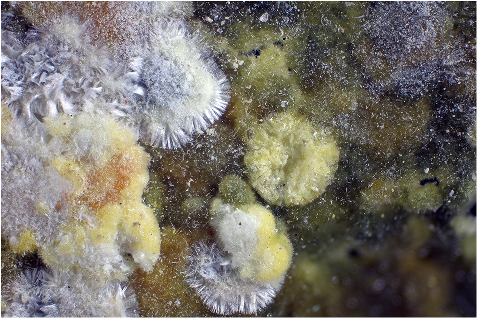
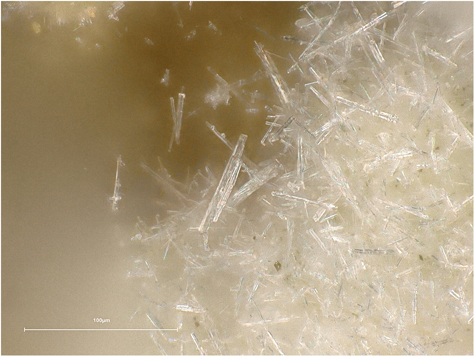
Pendevilleite-(Y) occurs as extremely thin blades up to ∼0.08–0.10 mm in length and only ∼1 μm thick. It often forms lichen-like aggregates and crusts (Fig. 1). Crystals are whitish or greyish-white, locally pale-bluish white in colour (Fig. 2). Pendevilleite-(Y) is non-fluorescent in both SW and LW ultraviolet light. The Mohs hardness is estimated at 2 based on tests when crystals are broken. Pendevilleite-(Y) is brittle; it has an irregular fracture. Cleavage is perfect on {001}. An experimental density could not be determined due to a lack of available pure material. A density of 2.51 g·cm–3 is calculated for the empirical formula and 2.42 g·cm–3 for the ideal formula. No optical properties were determined due to the minimal size of the crystals and complicated intergrowths. A mean index of refraction, calculated based on Gladston-Dale equations, is n GD = 1.47.
Whitish pendevilleite-(Y) growing along with the yellowish new unnamed mineral (most likely the yttrium analogue of shabaite-(Nd)) on uranophane (greenish crystalline crust). Part of the holotype specimen used for electron diffraction experiment. Kamoto-East open pit. FOV is 2.5 mm (Photo by P. Škácha).

Detail of the pendevilleite-(Y) crystalline aggregate from the holotype specimen showing parallel intergrowths of the long-prismatic crystals. (Photo by S. Philippo).

Chemical composition of pendevilleite-(Y)
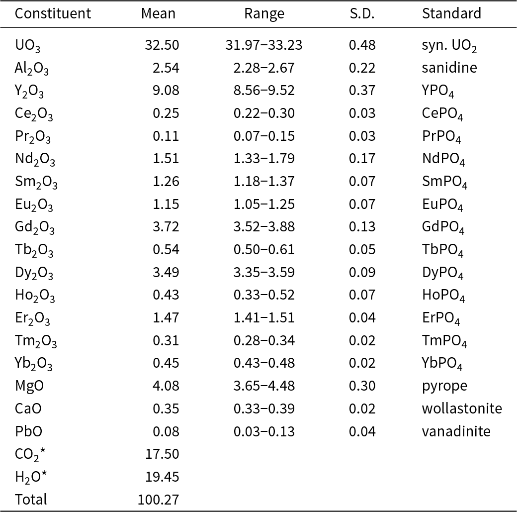
Electron probe microanalyses (EPMA; 7 points on the homogeneous aggregate of crystals) were performed at the Masaryk University in Brno (CZ) on a Cameca SX–100 electron microprobe operating in WDS mode. Analytical conditions were 15 kV accelerating voltage, 10 nA beam current and 10 μm beam diameter. These mild analytical conditions were used in order to minimise the electron beam-induced decomposition of the analysed areas. The raw X-ray intensities were corrected for matrix effects with a φρ(z) algorithm “X-PHI” (Merlet, Reference Merlet1994). Stoichiometric amounts of non-analysed elements (C, H and O) were included in the matrix correction procedure. Inter-REE coincidences were corrected by empirically determined correction factors. No other elements were detected, apart from locally elevated contents of Si (up to 0.10 wt. % SiO2). Because insufficient material is available to determine H2O amounts directly, it has been calculated based on the stoichiometry derived from the structure (U = 2 apfu, C = 7 apfu, H2O = 16 and OH calculated to keep the electroneutrality). Chemical analytical data for pendevilleite-(Y) are given in Table 1.
Chemical composition (wt.%) of pendevilleite-(Y)

Notes: S.D. – standard deviation; * derived from the structure
The empirical formula of pendevilleite-(Y) (calculated on the basis of 2 U apfu with CO32–, H2O derived from the structure and OH– to keep the electroneutrality) is Mg1.78[(Y1.42Gd0.36Dy0.33Nd0.16Er0.14Sm0.13Eu0.12Tb0.05Ho0.04Yb0.04Ce0.03Tm0.03Pr0.01)Σ2.86Ca0.11Pb0.01]Σ2.98Al0.88(UO2)2(CO3)7(OH)5.02(H2O)16. The ideal formula is Mg2Y3Al(UO2)2(CO3)7(OH)6(H2O)16, which requires MgO 4.76, Y2O3 20.01, Al2O3 3.01, UO3 33.80, CO2 18.20, and H2O 20.22, total 100 wt.%. Pendevilleite-(Y) is easily soluble in H2O at room temperature.
Raman spectroscopy of pendevilleite-(Y)
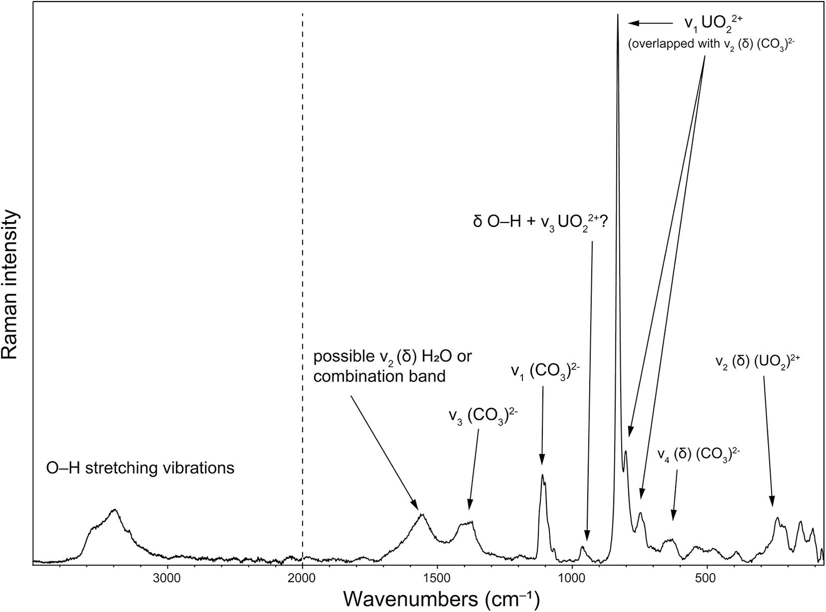
Raman spectroscopy of pendevilleite-(Y) (Fig. 3) was conducted on a Renishaw inVia micro-Raman spectrometer. Because significant fluorescence appeared using both 532 nm diode laser and a NIR (785 nm) laser, the spectrum was finally recorded using a 633 nm laser, which appeared to have the best signal-to-noise ratio (along with the lower thermal load and the consequent damages to the sample analysed). The final spectra were obtained with a laser power of ∼ 1mW through a 50× microscope objective with a numerical aperture N.A. = 0.5. The tentative assignments of the pendevilleite-(Y) spectrum were done, principally based on the papers by Čejka (Reference Čejka, Burns and Ewing1999), Koglin et al. (Reference Koglin, Schenk and Schwochau1979) and Anderson et al. (Reference Anderson, Chieh, Irish and Tong1980).
Raman spectrum of pendevilleite-(Y) recorded using a 633 nm diode laser.

A broad composite band, consisting of at least three overlapping bands, appearing from ca. 3600 to 3100 cm–1 is attributed to stretching O–H vibrations of the molecular H2O and OH groups. The shape of the overlapping bands in this region is characteristic of the hydrogen bonds in the structure of various strengths (bond lengths). Using the empirically derived equation of Libowitzky (Reference Libowitzky1999), the calculated O···O distances of the corresponding hydrogen bonds range between ∼3.0 and ∼2.7 Å. A weak band, located in the spectra of both minerals at around 1562 cm–1 is attributed to the ν2 (δ) (H2O) of the molecular water. Nevertheless, those vibration bands could also be assigned to the combination bands. Usually, the ν2 (δ) (H2O) tends to be located towards higher energies (1600 to 1650 cm–1). An asymmetric doublet (1415 and 1371 cm–1) is most probably connected with the activated split doubly-degenerate ν3 (CO3)2– antisymmetric stretching vibrations of the carbonate groups. There are three sharp (lower FWHM than others) overlapping vibrations at 1105, 1100 and 1065 cm–1 that should be connected with the ν1 (CO3)2– symmetric stretching vibrations. These bands are consistent with the presence of symmetrically non-equivalent carbonate units (Koglin et al., Reference Koglin, Schenk and Schwochau1979; Anderson et al., Reference Anderson, Chieh, Irish and Tong1980; Čejka, Reference Čejka, Burns and Ewing1999 and Reference Čejka2005, and references therein). We emphasise that there are seven independent C sites in the structure of pendevilleite-(Y) (see the structure description below). There is a weak component band at 963 cm–1 with a shoulder towards the lower energies; it can be ascribed to the ν1 (SiO4)4– symmetric stretching vibrations (probably of an admixture or small amount of Si in the crystals also documented by EPMA). A component band of the highest intensity, composed of overlapping bands at 831, 802 and 747 cm–1 (with pronounced shoulders), is attributed to the ν2 (δ) (CO3)2– bending vibrations and (in overlap) to the ν1 (UO2)2+ symmetric stretching vibrations. Using the empirical relation of Bartlett and Cooney (Reference Bartlett and Cooney1989), we can infer the corresponding U–O bond lengths from the wave numbers mentioned above (in the order given above): 1.78, 1.81 and 1.87. The structure refinement provided U–O bond lengths of 1.78–1.84 Å. Therefore, all observed overlapping bands in that region may contain the contribution of the ν1 (UO2)2+ symmetric stretching vibration. A doublet, 656 and 629 cm–1, is attributable to ν4 (δ) (CO3)2– bending vibrations. There are several bands, those at 538, 476 and 392 cm–1, which might be connected with (δ) (O–Si–O) or (δ) (Si–OH) and librations of H2O (or with the ν4 (δ) (SiO4) (see, e.g. Colmenero et al., Reference Colmenero, Plášil and Sejkora2019). Overlapping bands at 310, 239 and 215 cm–1 are attributed to a split doubly degenerate ν2 (δ) (UO2)2+ bending vibration. Bands of the lowest energies have been attributed to the lattice modes (Koglin et al., Reference Koglin, Schenk and Schwochau1979; Anderson et al., Reference Anderson, Chieh, Irish and Tong1980; Čejka, Reference Čejka, Burns and Ewing1999 and Reference Čejka2005).
Crystallography and 3D ED data collection
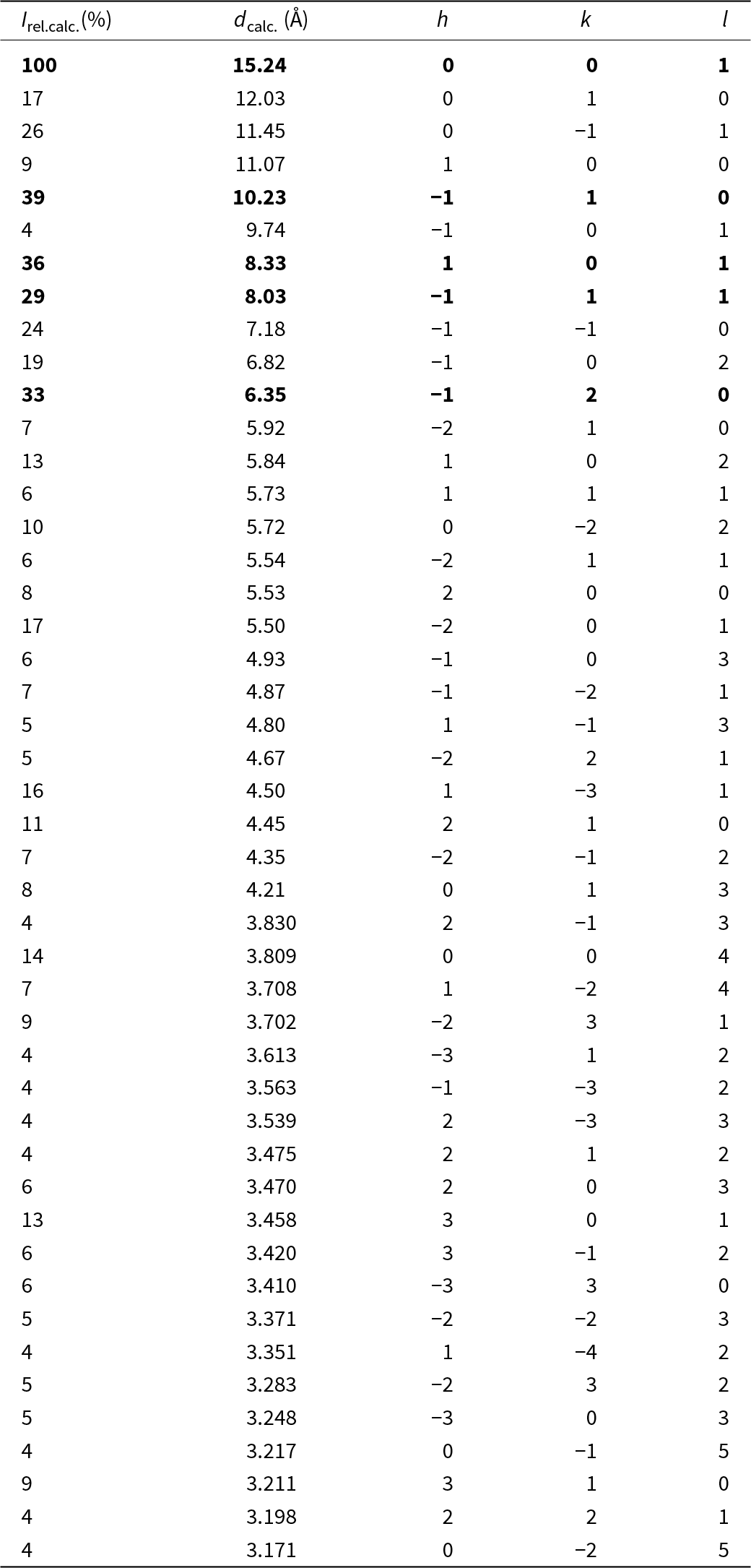
The material available and its nature prevented us from obtaining powder diffraction data for pendevilleite-(Y). Therefore, we only provide the pattern calculated from crystal structure data (Table 2).
Theoretical powder X-ray data (d in Å) for pendevilleite-(Y) based on the structure model obtained from the 3D ED data

Notes: Only calculated lines with I ≥ 4 are listed; the strongest lines are given in bold.
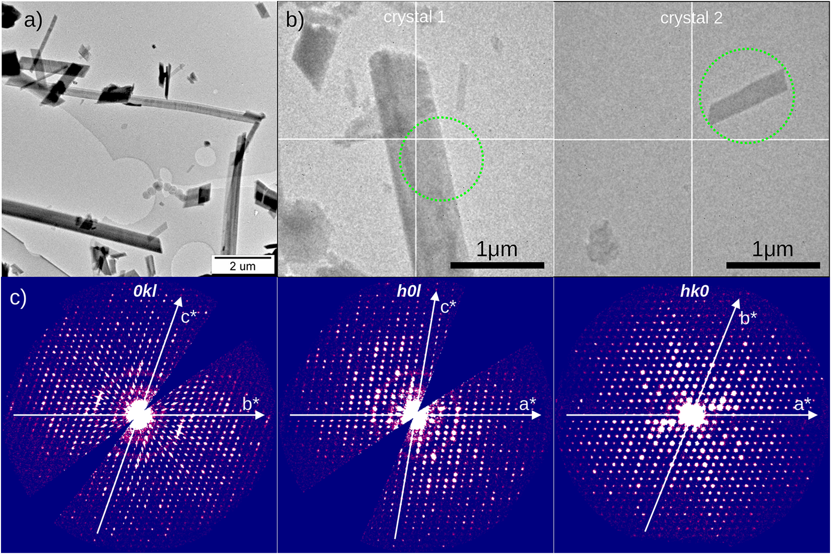
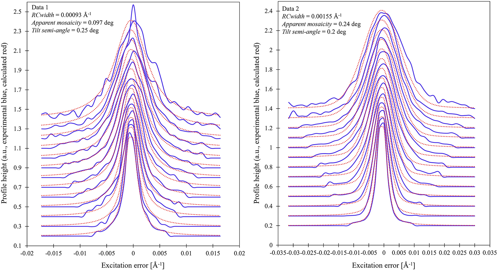
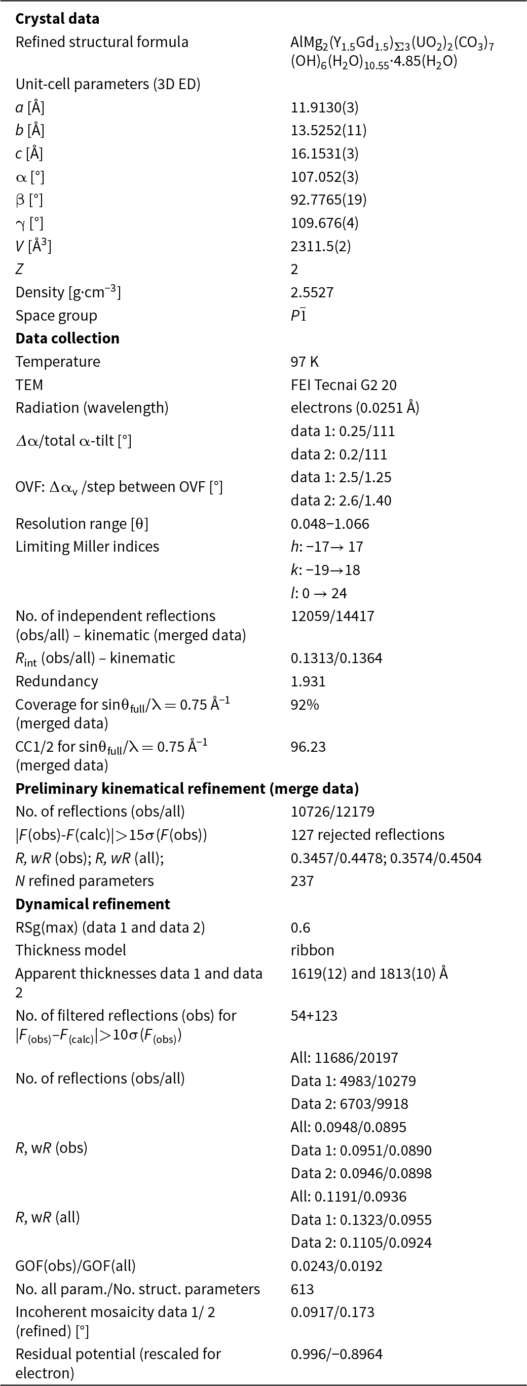
The single-crystal X-ray experiment on a Rigaku SuperNova diffractometer equipped with the microfocus Mo-source and Atlas S2 CCD detector failed due to the small size of the crystals, which diffracted poorly. Therefore, we employed 3-dimensional electron diffraction (3D ED) techniques using a transmission electron microscope (TEM) Gemmi and Lanza, Reference Gemmi and Lanza2019; Gemmi et al., Reference Gemmi, Mugnaioli, Gorelik, Kolb, Palatinus, Boullay, Hovmöller and Abrahams2019). The new mineral’s aggregate was gently crushed in a mortar with propanol and deposited on an Au-grid coated by a thin film of holey amorphous carbon. To prevent dehydration caused by the TEM high vacuum, the grid was plunged into liquid nitrogen and then transferred to the TEM using a Gatan cryo-transfer holder (Mugnaioli et al., Reference Mugnaioli, Lanza, Bortolozzi, Righi, Merlini, Cappello, Marini, Athanassiou and Gemmi2020; Steciuk et al., Reference Steciuk, Sejkora, Čejka, Plášil and Hloušek2021, Reference Steciuk, Majzlan, Rohlíček, Škoda, Sejkora and Plášil2024; Sejkora et al., Reference Sejkora, Steciuk, Marquez-Zavalia, Plášil and Dolníček2022). The 3D ED data were collected at 97 K with a continuous rotation mode in an FEI Tecnai G2 TEM (acceleration voltage of 200 kV, LaB6) equipped with a side-mounted hybrid single-electron detector ASI Cheetah M3, 512 x 512 pixels with high sensitivity and fast readout. For each selected crystal (Fig. 4), a series of non-orientated patterns are continuously collected by steps of 0.25° (data 1) and 0.25° (data 2), on all the accessible tilt ranges of the goniometer. The data collections are automated by the in-house software, including the tracking of the crystal (Plana-Ruiz et al., Reference Plana-Ruiz, Krysiak, Portillo, Alig, Estradé, Peiró and Kolb2020). In addition to the low-temperature experiment, low-illumination settings were used to further limit the beam-induced damage to the crystals. Continuous-rotation 3D ED data (cRED) reduction was performed using the computer program PETS2 (Palatinus et al., Reference Palatinus, Brázda, Jelínek, Hrdá, Steciuk and Klementová2019; Brázda et al., Reference Brázda, Klementová, Krysiak and Palatinus2022; Klar et al., Reference Klar, Krysiak, Xu, Steciuk, Cho, Zou and Palatinus2023). It includes the correction of geometrical and optical distortions to get accurate lattice parameters and a better integration of the reflections (Brázda et al., Reference Brázda, Klementová, Krysiak and Palatinus2022) (Supplementary Figs S1 and S2). The data quality of each crystal is represented by the rocking curve plots as well as the estimation of the crystal mosaicity, later used in the refinement (Fig. 5). The specific data processing for cRED data used in the structure solution and the refinement (with and without the dynamical approaches) is given extensively in detail by Klar et al. (Reference Klar, Krysiak, Xu, Steciuk, Cho, Zou and Palatinus2023). It includes introducing the overlapping virtual frames (OVFs) that aim to model experimental intensities from continuous rotation data by summing consecutive experimental diffraction patterns into a set of virtual frames. Each OVF is characterised by its angular range Δαv covered by the virtual frame and the angular step between two virtual frames (Table 3). The result of the data reduction is a hkl-type file obtained from merging the two data sets (Rint(obs/all) = 0.1313/0.1364). This file is used in the structure solution and the kinematical refinement. The two data sets are processed separately for the dynamical approach, where each OVF is considered independent (Palatinus et al., Reference Palatinus, Corrêa, Steciuk, Jacob, Roussel, Boullay, Klementová, Gemmi, Kopeček and Domeneghetti2015a, Reference Palatinus, Petříček and C.A2015b; Klar et al., Reference Klar, Krysiak, Xu, Steciuk, Cho, Zou and Palatinus2023). The structure was solved using Superflip (Palatinus and Chapuis, Reference Palatinus and Chapuis2007) implemented in Jana2020 (Petříček et al., Reference Petříček, Palatinus, Plášil and Dušek2023) and refined using DYNGO and Jana2020.
(a) General overview of pendevilleite-(Y) crystals morphology under TEM. (b) Crystal areas used in the 3D ED analysis (data 1 and data 2). (c) 0kl, h0l and hk0 sections of the reciprocal space of pendevilleite-(Y) reconstructed from the merged data set (data 1 + data 2).

Plots of the rocking-curve profiles (Camel plot) of the experimental 3D ED data 1 and data 2 at 95 K. The lowest blue curve is the average observed rocking curve in the range of 0.2 to 0.3 Å−1, and the next ones are obtained by steps of 0.1 Å−1. The red dotted curves are calculated from the Rocking curve width, the apparent mosaicity and the tilt semi-angle. I > 6*σ(I) reflections are involved in the Camel plot.

3D ED data collection and structure refinement details for pendevilleite-(Y)

Structure solution and refinement
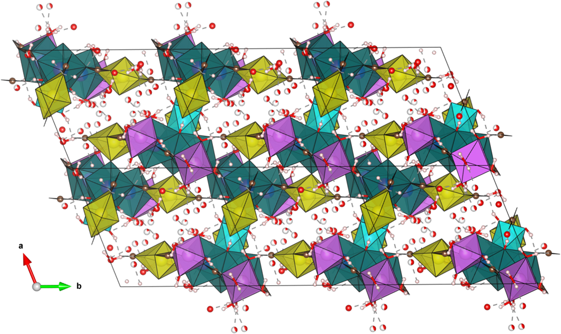
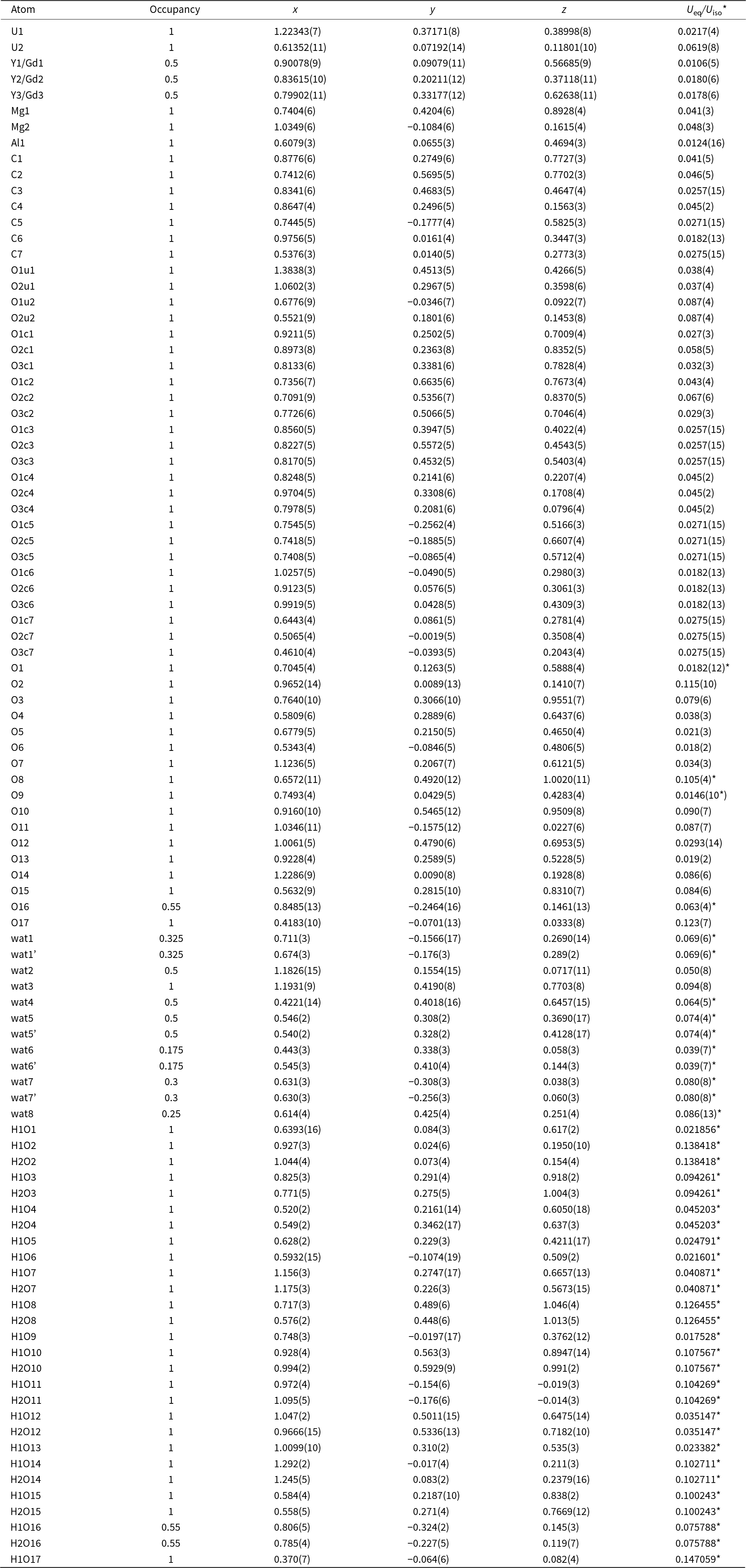
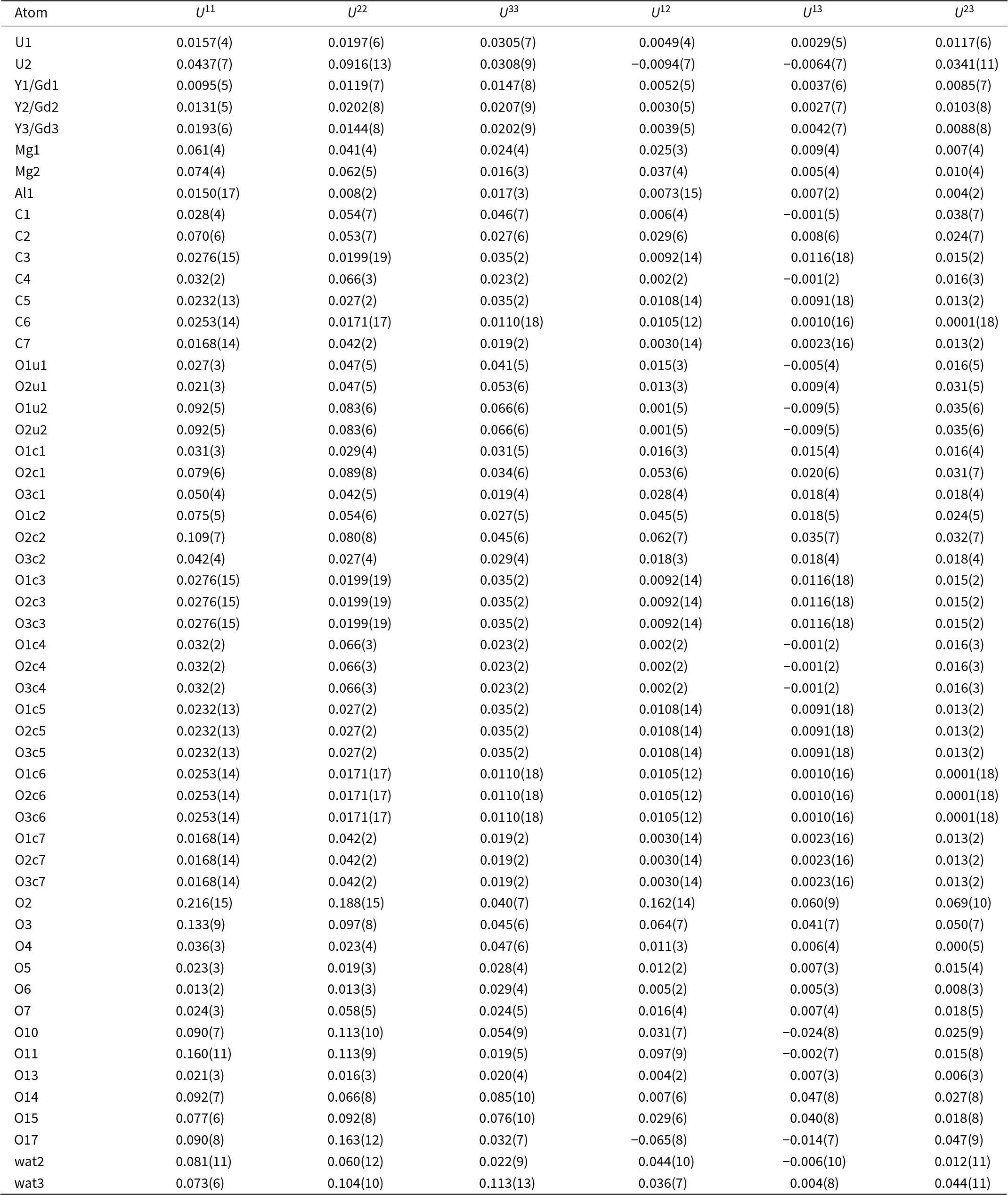
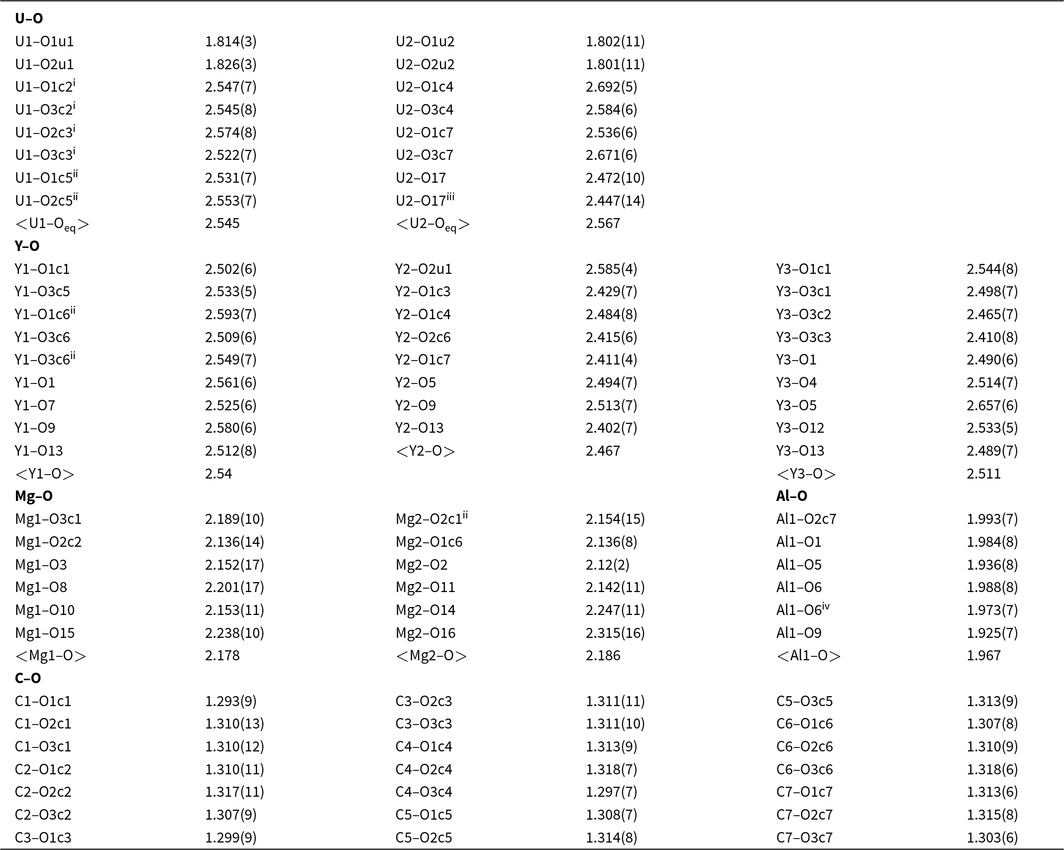
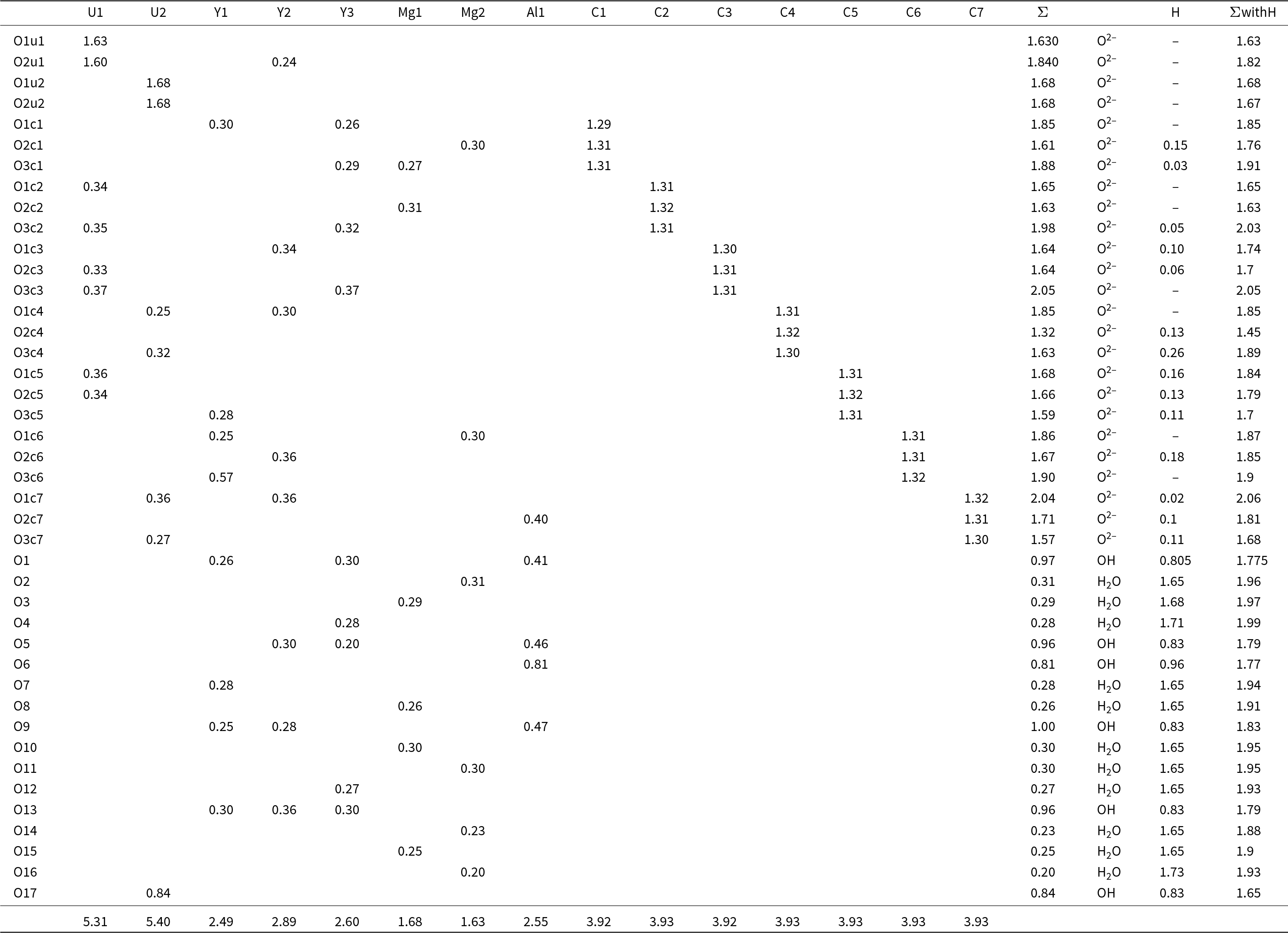
At 97 K, pendevilleite-Y is triclinic, with a = 11.9130(3) Å, b = 13.5252(11) Å, c = 16.1531(3) Å, α = 107.052(3)°, β = 92.7765(19)°, γ = 109.676(4)° and V = 2311.5(2) Å3. A coverage of 92% for sinθfull/λ = 0.75 Å–1 (Laue class –1) is reached by merging two data sets despite the strong preferential (001) orientation of the needle-like crystals on the grid. The initial model is obtained from the charge flipping algorithm in the triclinic unit cell, space group P  $\bar 1$, containing all the non-hydrogen atoms necessary for a primary interpretation. The cations present on each site were attributed according to the EPMA results and the coordination. The Y sites (labelled Y1, Y2 and Y3) are occupied by different proportions of Y3+, Ca2+ and the other Ln 3+, where the Ln was set to be represented by Gd3+ in the case of refinement from the electron data (Gd has the average Z from the Ln distribution detected by the WDS). According to the EPMA results, Gd is also usually the dominant among Ln. Even though U for Y/Ln substitution is possible, it is hard to model by the refinements in the presence of several other atoms. Indeed, for the refinement, all U is assumed to be in only the two sites U1 and U2. Soft restrictions were applied in the refinement on a few cation−oxygen distances and /or angles to ensure the geometry of the carbonate groups CO3, and the oxygen atoms of the linear uranyl groups [O=U=O]2+ (= 1.8 Å). The initial model was refined using the dynamical theory of diffraction, which considers multiple scattering (Palatinus et al., Reference Palatinus, Corrêa, Steciuk, Jacob, Roussel, Boullay, Klementová, Gemmi, Kopeček and Domeneghetti2015a, Reference Palatinus, Petříček and C.A2015b). The reflections of the 3D ED data involved in the refinement are filtered according to the main selection criteria RSg = 0.6. They are set to involve reflections that are properly covered by the experiment (Palatinus et al., Reference Palatinus, Corrêa, Steciuk, Jacob, Roussel, Boullay, Klementová, Gemmi, Kopeček and Domeneghetti2015a; Klar et al., Reference Klar, Krysiak, Xu, Steciuk, Cho, Zou and Palatinus2023). The refinement was carried out in several steps. First, the non-hydrogen atoms are refined with the occupancies of mixed sites set according to the EPMA results. From that refinement, hydrogen atoms were detected from the difference electrostatic potential map with isosurface levels above 2.5σ[ΔV(r)] together with the bond valence analysis: five hydroxyl groups (O1, O5, O6, O9, O13 and O17) and eleven bonded H2O molecules (O2, O3, O4, O7, O8, O10, O11 O12, O14, O15 and O16). The weakly-bonded H2O molecules (labelled wat1 to wat8) tend to be disordered in the structure even at 95 K. Therefore, the corresponding hydrogen sites are not visible from the difference electrostatic potential maps. All O–H distances are restricted to 1 Å and isotropic atomic displacement parameters (ADPs) for hydrogen are set as riding with an extension factor of 1.2. After introducing the refinement of isotropic incoherent mosaicity in the refinement (data1: 0.0917 deg; data2: 0.173 deg) R-values significantly dropped by 4 to 5%, and the final refinement leads to R(obs)/wR(obs) = 0.0948/0.0895 and R(all)/wR(all) = 0.1191/0.0936 for 613 parameter and Nobs/Nall = 11686/20197 (see Table 3). The residual potential map suggests that more very scarcely occupied water molecules are arranged in the channel running along the c axis. However, they were not added as they represent a very small contribution and the dynamical refinement of the low symmetry model with 100 atoms in the asymmetric unit cell, considering the mosaicity, took weeks to converge. Only atoms showing clear anisotropy in their displacement parameters were anisotropically refined to reduce the refinement time. The refined formula of pendevilleite-(Y) is AlMg2(Y1.5Gd1.5)Σ3.00(UO2)2(CO3)7(OH)6(H2O)10.55·4.85H2O, with Gd the average Ln based on the microprobe analysis. The details of the data collection and refinement are presented in Table 3, and the structure is displayed in Fig. 6. The atom coordinates and displacement parameters are given in Tables 4 and 5, and in the crystallographic information file (in supplementary material, see below), selected bond distances are shown in Table 6, and a bond valence analysis is shown in Table 7.
The structure of pendevilleite-(Y) projected down [001]. Uranyl polyhedra are yellow, Y/REE 3+ polyhedra green, Al-polyhedra blue, Mg-polyhedra purple and CO3 groups brown. Black solid lines outline the unit cells. Drawn using Vesta (Momma and Izumi, Reference Momma and Izumi2011).

Atom coordinates and displacement parameters (in Å2) for pendevilleite-(Y)

Atom anisotropic displacement parameters (Å2)

Selected bond distances (Å) for pendevilleite-(Y)

Symmetry codes: (i) –x+2, –y+1, –z+1; (ii) –x+2, –y, –z+1; (iii) –x+1, –y, –z; (iv) –x+1, –y, –z+1.
Bond-valence analysis for pendevilleite-(Y). Values are expressed in valence units.*

* Cation–O bond valence parameters are from Gagné and Hawthorne (Reference Gagné and Hawthorne2015). For simplicity, the Y/Gd sites have been labelled as Y. H atoms and non-bonded water are not included; mixed-site populations are considered in the calculations. Hydrogen-bond strengths are based on theoretical values given by Brown (Reference Brown2002).
Crystal structure of pendevilleite-(Y)
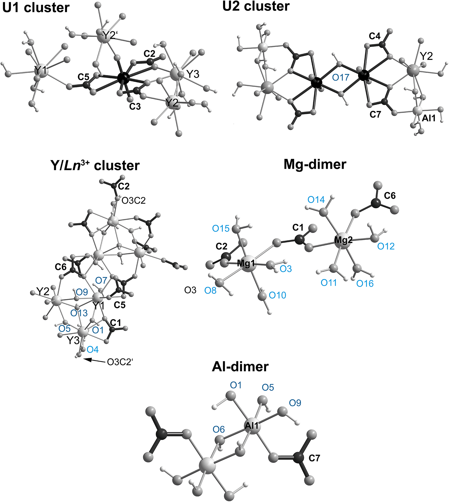
There are two U sites in the structure of pendevilleite-(Y). Both U1 and U2 sites are surrounded by eight O atoms forming squat UO8 hexagonal bipyramids. Nevertheless, the U1 site is chelated by the three CO32– groups to form the uranyl tricarbonate complex, [(UO2)(CO3)3]4– (Figs 6 and 7). The U2 site is chelated differently. It is linked only to two CO32– groups and the remaining two O atoms (O17 symmetrical equivalents) are shared with another U2-bipyramid. The O17 atom constitutes an OH group. Thus, the anionic complex comprising the U2 site is [(UO2)2(CO3)4(OH)2]6– (Fig. 7). Another fundamental building unit (FBU) of the structure of pendevilleite-(Y) is a large complex comprising six Y/Ln sites in coordination 9 and 8 (M1, M2 and M3 occupied by distinct proportions of Y3+, Ln 3+ and Ca2+). As mentioned earlier, the Ln has been modelled as Gd3+, which is usually the dominant Ln according to the EPMA results (and the average Z of the Ln corresponds closest to Gd). The summary composition of this polyoxometalate cluster is [(Y,Ln)6(OH)8(H2O)4(CO3)4]2+ (Fig. 7). There is one Al site in the structure. Two symmetrically related equivalents result in a dimer of composition [Al2O2(OH)8(CO3)22–]10–. Two of the (OH)– are located within the shared edge of the Al-octahedra; CO3 groups are linked monodentately (through the O2C7 atom). There are two sites in the structure occupied by magnesium. Both Mg1, and Mg2 are bonded to six ligands in octahedral coordination, forming a dimer of MgO2CO3(H2O)4 units via the C1O3 group. There are two other monodentately linked carbonate groups (C2O3 to Mg1 and C6O3 to Mg2) (Fig. 7). The FBUs are linked to form a sizeable heteropolyhedral framework structure (Fig. 6), dominated by the prominent Y, Ln-clusters, plane-parallel to (110), cross-linked by the U2 dimers, in the form of H-shaped structures. In the large channels of the framework (running parallel to c), there are at least height-independent partially occupied and disordered O sites of the H2O molecules. The chemical formula obtained from the structure refinement is M 1–M3(Y1.5Gd1.5)Σ3.00AlMg2(UO2)2(CO3)7(OH)6(H2O)10.55·4.85H2O (Z = 2).
Cation coordination in the pendevilleite-(Y) structure. For simplicity, the Y/Ln 3+ sites are labelled as Y1–Y3. The O atoms of the H2O groups are given in light blue, and those of the OH– groups in dark blue; symmetry equivalent atoms are labelled where necessary for clarity with an apostrophe.

Structural and chemical complexity of pendevilleite-(Y)
The structure of pendevilleite-(Y) is unique in the point of view of the structure architecture and different elements building it, so the complexity measures of its structure are of interest. The structural complexity was determined as the Shannon information content per atom (I G) and per unit cell (I G,total) following the approach of Krivovichev (Reference Krivovichev2012, Reference Krivovichev2013, Reference Krivovichev2014, Reference Krivovichev2016, Reference Krivovichev2017). The information-based structural complexity values were calculated using the software package TOPOS (Blatov et al., Reference Blatov, Shevchenko and Proserpio2014). The chemical complexity (Siidra et al., Reference Siidra, Zenko and Krivovichev2014) is estimated by considering the chemical formula as a message, where symbols correspond to different chemical elements. Calculated values for the structural complexity of pendevilleite-(Y) (including the contribution of the H-atoms, both determined and undetermined by the refinement; for the details see e.g. Gurzhiy et al., Reference Gurzhiy, Kalashnikova, Kuporev and Plášil2021) are 6.781 bits/atom and 1491.90 bits/cell. Based on this value, we can consider pendevilleite-(Y) as a very complex structure (Krivovichev, Reference Krivovichev2013). For comparison, we can refer to the complex sheet structure of lepersonnite-(Gd) (without the contribution of the H-atoms undetermined by the refinement; 2240.545 bits/cell), albrechtschraufite (1161.600 bits/cell) or grimselite (140.670 bits/cell). The chemical complexity, I chem, of pendevilleite-(Y) is 175.49 bits/formula. This is a particularly high value at a comparable magnitude of albrechtschraufite, MgCa4F2[UO2(CO3)3]2⋅17.29H2O (Mereiter, Reference Mereiter2013) (162.95 bits/formula), paddlewheelite, MgCa5Cu2[(UO2)4(CO3)12]⋅33H2O (Olds et al., Reference Olds, Plášil, Kampf, Dal Bo and Burns2018) (275.27 bits/formula), or the chemically-simple mineral grimselite, K3Na(UO2)(CO3)3(H2O) (Li and Burns, Reference Li and Burns2001; Plášil et al., Reference Plášil, Fejfarová, Skála, Škoda, Meisser, Hloušek, Císařová, Dušek, Veselovský, Čejka, Sejkora and Ondruš2012) (43.58 bits/cell). The furthermost chemically complex uranyl mineral is uranyl carbonate ewingite, Mg8Ca8(UO2)24(CO3)30O4(OH)12(H2O)138 (Olds et al., Reference Olds, Plášil, Kampf, Simonetti, Sadergaski, Chen and Burns2017a), with 1024.18 bits/formula.
Relation to other species
The heteropolyhedral frameworks are not unusual for U6+ structures, e.g. present in the mineral grimselite, K3Na[(UO2)(CO3)3](H2O) (Plášil et al., Reference Plášil, Fejfarová, Skála, Škoda, Meisser, Hloušek, Císařová, Dušek, Veselovský, Čejka, Sejkora and Ondruš2012), or leószilárdite, Na6Mg(UO2)2(CO3)6(H2O)6 (Olds et al., Reference Olds, Sadergaski, Plášil, Kampf, Burns, Steele, Marty, Carlson and Mills2017b) and others (Gurzhiy et al., Reference Gurzhiy, Kalashnikova, Kuporev and Plášil2021). However, the pendevilleite-(Y) structure is not closely related to any other known uranyl mineral. For instance, uranyl carbonate minerals andersonite, Na2Ca[(UO2)(CO3)3]·(5+1/3)H2O (Plášil and Čejka, Reference Plášil and Čejka2015; Gurzhiy et al., Reference Gurzhiy, Krzhizhanovskaya, Izatulina, Sigmon, Krivovichev and Burns2018) and grimselite, ideally K3Na[(UO2)(CO3)3](H2O) (Li and Burns, Reference Li and Burns2001; Plášil et al., Reference Plášil, Fejfarová, Skála, Škoda, Meisser, Hloušek, Císařová, Dušek, Veselovský, Čejka, Sejkora and Ondruš2012; Ghazisaeed et al., Reference Ghazisaeed, Majzlan, Plášil and Kiefer2018), have structures that are heteropolyhedral frameworks containing channels filled with H2O molecules, nevertheless, both are very distinctive from pendevilleite-(Y). Albrechtschraufite MgCa4F2[UO2(CO3)3]2⋅17–18H2O (Mereiter, Reference Mereiter2013) can be, to some extent, considered for a comparison. There are significant cavities in the crystal structures of both minerals, filled with molecular H2O, behaving to some extent as ‘zeolitic’ water. Albrechtschraufite possesses a structure with U:C ratio of 1:3, organised from FBUs with the overall formula MgCa3F2[UO2(CO3)3]⋅8H2O. It consists of a uranyl tricarbonate complex associated with a large cluster of Ca2+-polyhedra linked to Mg2+-octahedra. By assuming that in albrechtschraufite, Ca2+ plays the role of REEs, the ratio U:C:(Mg,Al):REEs goes from 2:7:3:3 in pendevilleite-(Y) to 2:6:2:8 (1:3:1:4) in albrechtschraufite. However, the fundamental feature of the albrechtschraufite structure is a paddle wheel (Mereiter, Reference Mereiter2013), which is not present in pendevilleite-(Y). Moreover, there is a unique feature in pendevilleite-(Y), which is a dimer of UO8 bipyramids of the composition U2O12(OH)2 (olation); this arrangement, involving bonding of two H atoms within a shared edge of uranyl bipyramids, has not been observed in minerals and inorganic compounds so far (only a dimeric unit bridged by the peroxo-group, Burns, Reference Burns2011; Qui and Burns, Reference Qui and Burns2013). In the Strunz system, pendevilleite-(Y) belongs in class 5.E, but has a distinctive overall U:C ratio.
Given the three octahedrally coordinated M sites present in pendevilleite-(Y) structure, we can expect with a high degree of certainty that there might be other new minerals. Among them, we can expect homovalent substitutions within Mg2+ octahedra, for instance, for Zn or Fe, or even for Cu2+ (that might involve some structural changes due to Jahn-Teller distorted polyhedra). To study these possible substitutions, it will be necessary to undertake very detailed EPMA studies of an extended set of specimens containing Ln-uranyl-carbonates from Kamoto-East and unfortunately, this material is currently scare..
Supplementary material
The supplementary material for this article can be found at https://doi.org/10.1180/mgm.2025.10095
Acknowledgements
The helpful comments of an anonymous reviewer, Vlad Gurhziy and a structure editor Peter Leverett, all helped improving the initial manuscript. This study was supported by the CzechNanoLab Research Infrastructure supported by MEYS CR (LM2023051) (GS and JP) and an OP VVV project (Geobarr CZ.02.1.01/0.0/0.0/16_026/0008459) (RS).
Competing interests
The authors declare none.