Introduction
Pulmonary arterial hypertension is a progressive disease characterised by pathological pulmonary vascular remodelling, leading to increased right ventricular afterload, right ventricular failure, and ultimately death.Reference Humbert, Kovacs and Hoeper1 Although contemporary medical therapies have contributed to improved survival and quality of life in both adult and paediatric populations, a substantial 5-year mortality rate—approaching 80%—persists among patients with severe or high-risk disease.Reference Haworth and Hislop2 Nonetheless, therapeutic options remain limited for paediatric patients with severe pulmonary arterial hypertension who are unresponsive to intensive medical therapy or experience rapidly progressive clinical deterioration secondary to right heart failure. Although lung transplantation is traditionally regarded as a palliative option, its accessibility is limited for many patients because of high costs, restricted availability of specialised centres, challenges in identifying suitable donors, elevated perioperative risks, and the potential for graft rejection.Reference Ivy, Rosenzweig and Abman3,Reference Hansmann, Koestenberger and Alastalo4
Over the years, innovative interventional approaches have been developed to alleviate right ventricular dysfunction in severe pulmonary arterial hypertension, offering viable alternatives to lung transplantation and enabling clinical improvement in high-risk paediatric patients.Reference Ivy, Rosenzweig and Abman3,Reference Hansmann, Koestenberger and Alastalo4 Based on the level of intervention, these approaches can be broadly categorised into: (1) pre-tricuspid procedures, which create a right-to-left shunt at the atrial level (atrial septostomy), and (2) post-tricuspid procedures, which establish a pulmonary-to-systemic anastomosis at the level of the great vessels (reverse Potts shunt) or create a ventricular-level shunt. These interventions aim to alleviate right ventricular pressure overload while preserving systemic perfusion, thereby optimising haemodynamics, improving symptoms and quality of life, and prolonging survival.Reference Humbert, Kovacs and Hoeper1,Reference Hansmann, Koestenberger and Alastalo4,Reference Doyle, McCrory, Channick and Conte5
The aim of this study is to evaluate the safety, efficacy, and clinical outcomes of 18 patient-specific interventional procedures performed in 12 paediatric patients with severe pulmonary arterial hypertension who were unresponsive to medical therapy or experienced rapid clinical deterioration secondary to right ventricular failure, thereby contributing valuable insights into individualised management strategies for this high-risk population.
Methods
Study design and population
This retrospective observational study was conducted between January 2019 and December 2024 in the Department of Pediatric Cardiology. We retrospectively reviewed the records of 12 children with severe pulmonary arterial hypertension who underwent interventional procedures. The diagnosis of pulmonary arterial hypertension was confirmed during cardiac catheterisation based on the following criteria: a mean pulmonary arterial pressure greater than 20 mmHg at rest, a pulmonary capillary wedge pressure less than 15 mmHg, and a pulmonary vascular resistance index greater than 3 Wood units·m2.Reference Humbert, Kovacs and Hoeper1
Indications for interventional procedures included paediatric patients with recurrent syncope, World Health Organization Functional Class III or IV symptoms, clinical signs of right ventricular failure, marked impairment in quality of life, or progressive haemodynamic deterioration.Reference Humbert, Kovacs and Hoeper1,Reference Hansmann, Koestenberger and Alastalo4 These criteria were applied both to patients who deteriorated despite the optimal combination of medical therapy and to those who required urgent intervention prior to the initiation of therapy because of acute decompensation. Failed medical therapy was defined as the persistence or progression of high-risk features despite at least 8–12 weeks of optimised dual oral therapy with an endothelin receptor antagonist and a phosphodiesterase type 5 inhibitor. Evidence of treatment failure included one or more of the following: recurrent syncope, persistent World Health Organization functional class III or IV symptoms, a ≥30% increase in N-terminal pro-brain natriuretic peptide levels, a ≥15% decline in 6-minute walk test distance, a ≥2 mm reduction in tricuspid annular plane systolic excursion, or resting or exertional arterial oxygen saturation ≤ 85%.
Failed medical therapy was defined as persistence or worsening of high-risk features despite at least 8–12 weeks of optimised dual oral therapy (endothelin receptor antagonist + phosphodiesterase type 5 inhibitor), evidenced by one or more of the following: recurrent syncope, persistence of World Health Organization Functional Class III/IV symptoms, ≥30% increase in N-terminal pro-brain natriuretic peptide, ≥15% decline in six-minute walk test distance, ≥2 mm reduction in tricuspid annular plane systolic excursion, or resting/exertional SaO2 ≤ 85%. Acute decompensation was defined as hospitalisation or ICU admission for right heart failure, or syncope accompanied by haemodynamic instability, including SaO2 ≤ 85%, rising serum lactate, or a progressive elevation in N-terminal pro-brain natriuretic peptide.
Patients were excluded if they had pulmonary hypertension secondary to left heart disease, were over 18 years of age, had significant comorbidities precluding intervention, or were receiving investigational treatments or participating in clinical trials involving therapies not approved for pulmonary arterial hypertension.
Data collection and Echocardiographic/Catheterisation findings
Clinical and demographic data—including the World Health Organization Functional Class, vital signs, current medical treatments (if applicable), six-minute walk test results, and laboratory values such as serum N-terminal pro-brain natriuretic peptide levels and electrocardiographic findings—were retrospectively obtained from the hospital’s electronic medical records. For patients who underwent multiple interventions, data were collected for both the pre-procedural evaluation and the most recent follow-up visit after the final procedure. Additionally, echocardiographic measurements—including right and left ventricular end-diastolic diameters, right ventricular systolic pressure, tricuspid regurgitation severity, and tricuspid annular plane systolic excursion—were recorded. Genetic testing for heritable pulmonary arterial hypertension (BMPR2, TBX4, EIF2AK4, and other panel genes) was performed in all patients
Written informed consent was obtained from the parents or legal guardians of all patients prior to cardiac catheterisation. All procedures were performed under general anaesthesia with an inspired oxygen fraction of 30%. In the patient at risk of malignant hyperthermia, intravenous propofol was used as the anaesthetic agent. Before the intervention, comprehensive right heart catheterisation was conducted to obtain detailed haemodynamic data, including pulmonary vascular resistance, systemic vascular resistance, mean pulmonary arterial pressure, systolic and diastolic pulmonary artery pressures, mean right atrial pressure, mean left atrial pressure, and the results of acute vasoreactivity testing. Acute vasoreactivity testing was performed using inhaled iloprost and supplemental oxygen, with a positive response defined as a reduction in mean pulmonary arterial pressure of at least 10 mmHg to an absolute value below 40 mmHg without any decrease in cardiac output.Reference Hansmann, Koestenberger and Alastalo4
Procedures
Interventional eligibility and procedure selection were determined by a pre-specified, multidisciplinary algorithm integrating: (i) clinical status (World Health Organization Functional Class, syncope, decompensation events), (ii) echocardiography, (iii) invasive haemodynamics (mean pulmonary arterial pressure, mean right atrial pressure, pulmonary vascular resistance index, cardiac index, systemic arterial saturation), and (iv) cardiac anatomy (presence/recanalizability of a ductus, number/size of atrial septal defects, ventricular septal defect. Ductal interventions, including recanalisation or stent-based enlargement of the ductus arteriosus, were prioritised whenever a ductus was present or could be recanalised. This strategy aimed to establish reverse Potts physiology, particularly in patients with supra- or near-systemic pulmonary artery pressures, preserved left ventricular function, and anticipated benefit from right ventricular unloading. When a significant ventricular septal defect was present, transcatheter ventricular septal defect closure was performed concurrently with ductal stenting in order to optimise haemodynamics.
In the absence of a suitable patent ductus arteriosus (PDA) or ventricular septal defect, atrial-level decompression was pursued. Balloon atrial septostomy was favoured in patients with a mean right atrial pressure ≤ 20 mmHg, resting SaO2 ≥ 85%, and no severe right ventricular failure or prohibitive left ventricular diastolic dysfunction. Relative contraindications included mean right atrial pressure > 20 mmHg, SaO2 < 85%, or advanced right ventricular failure, in which case atrial septostomy was either deferred, modified, or alternative decompressive approaches were considered.
The technique of atrial septostomy involved performing a transseptal atrial puncture using a Brockenbrough needle and a transseptal sheath with a dilator. After transseptal access was obtained, mean left atrial pressure was measured. Septostomy was initiated using a high-pressure 10 mm balloon, which is generally regarded in the literature as an effective target diameter for achieving atrial-level decompression. However, the final septostomy size was individualised according to patient size and haemodynamic response.Reference Boshoff, Butera, Gewillig, Butera, Chessa, Eicken and Thomson6 Subsequent graded dilations were performed, if necessary, based on echocardiographic assessment of defect size and systemic oxygen saturation. After each step, a 3-minute waiting period was observed to assess haemodynamic stability, during which mean left atrial pressure and arterial oxygen saturation were recorded. An atrial septal defect size of approximately 10 mm was targeted as optimal, and a post-procedural systemic oxygen saturation between 85% and 90% was considered desirable.Reference Humbert, Kovacs and Hoeper1,Reference Hansmann, Koestenberger and Alastalo4,Reference Doyle, McCrory, Channick and Conte5
In patients with multiple atrial septal defects or a large single atrial septal defect, the interventional plan was carefully designed to reduce right ventricular volume overload while avoiding a significant increase in right atrial pressure. This strategy was planned to reduce the volume burden associated with the left-to-right flow component in cases of bidirectional atrial-level shunting. Partial atrial septal defect closure was performed in patients with a Qp/Qs ratio ≥1.5. After closure or reduction in size of the most haemodynamically significant defect, the patency of the remaining interatrial communications was carefully assessed to ensure adequate atrial-level decompression. All procedures were performed under combined fluoroscopic and echocardiographic guidance.
Statistical methods
The findings of the study are expressed as frequencies and percentages. Normality was assessed using the Shapiro–Wilk test. Variables with a normal distribution are presented as mean ± standard deviation, while those without normal distribution are presented as median (minimum–maximum). Categorical variables were compared using the Chi-square test or the McNemar test for paired pre- and post-intervention comparisons. Numerical variables were compared using the independent samples t-test for normally distributed independent groups, the Mann–Whitney U test for non-normally distributed independent groups, the Paired Samples t-test for normally distributed paired data, or the Wilcoxon Signed-Rank test for non-normally distributed paired data. Pearson or Spearman correlation analyses were used to explore possible relationships between echocardiographic measurements and clinical variables, depending on the distribution and scale of the data. Data analysis was performed using SPSS 26.0 (IBM, USA), and a p-value of <0.05 was considered statistically significant.
Ethics committee approval
This study was conducted following the principles of the Declaration of Helsinki and was approved by the Turkish Ministry of Health and the institutional ethics committee for human research (Decision No: 1-24-493).
Results
Pre-procedural patient clinical, demographic, and haemodynamic characteristics
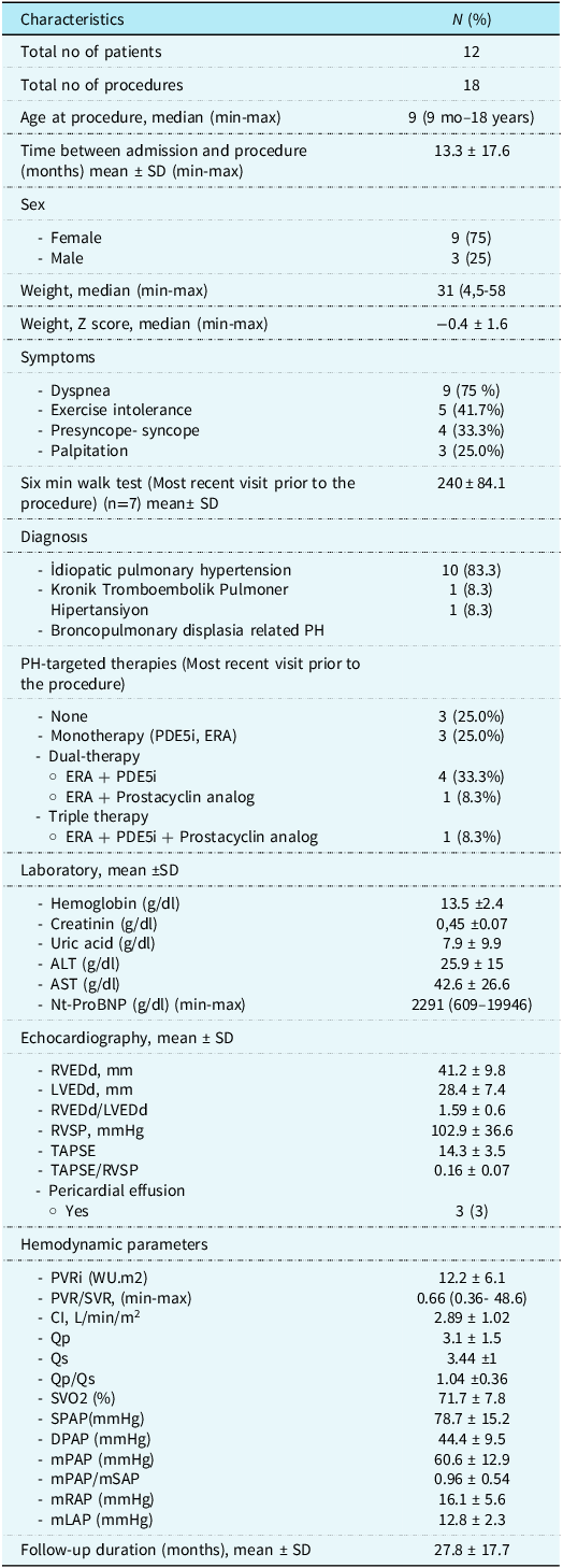
A total of 12 patients who underwent 18 interventional procedures were included. The clinical, demographic, echocardiographic, and haemodynamic characteristics of the patients before the initial procedure are presented in Table 1. Haemodynamic data in Table 1 represent baseline pre-interventional values obtained at diagnostic catheterisation immediately prior to the index procedure. The median age at the time of the first procedure was 9 years (9 months–18 years), and the mean time from hospital admission to the procedure was 13.3 ± 17.6 months. Of the patients, 9 (75%) were female, and 3 (25%) were male. The mean body weight was 31 (5,4–58) kg. Genetic testing was available in all 12 patients, and no pathogenic variants were identified.
Demographic, clinical, and haemodynamic characteristics of patients

ALT = Alanine Aminotransferase; AST = Aspartate Aminotransferase; CI = Cardiac Index; DPAP = Diastolic Pulmonary Arterial Pressure; ERA = Endothelin Receptor Antagonist; LVEDd = Left Ventricular End-Diastolic Diameter; mLAP = Mean Left Atrial Pressure; mPAP = Mean Pulmonary Arterial Pressure; mRAP = Mean Right Atrial Pressure; mSAP = Mean Systemic Arterial Pressure; NT-proBNP = N-terminal pro-Brain Natriuretic Peptide; PAH = Pulmonary Arterial Hypertension; PDE5-i = Phosphodiesterase Type 5 Inhibitor; PVRi = Pulmonary Vascular Resistance Index; Qp = Pulmonary Blood Flow; Qp/Qs = Pulmonary-to-Systemic Flow Ratio; Qs = Systemic Blood Flow; RV = Right Ventricle; RVEDd = Right Ventricular End-Diastolic Diameter; RVSP = Right Ventricular Systolic Pressure; sPAP = Systolic Pulmonary Arterial Pressure; SVO2 = Mixed Venous Oxygen Saturation; TAPSE = Tricuspid Annular Plane Systolic Excursion; WHO-FC = World Health Organization Functional Class; WU·m2 = Wood Units per Square Metre; Z score = Standard Deviation Score Based on Age-Specific Reference.
The most commonly reported symptom was dyspnoea, observed in 9 patients (75%). Exercise intolerance was noted in 5 patients (41.7%), presyncope or syncope in 4 patients (33.3%), and palpitations in 3 patients (25.0%). The mean six-minute walk test distance recorded at the most recent visit prior to the procedure (n = 7) was 240 ± 84.1 metres.
Idiopathic pulmonary arterial hypertension was diagnosed in 9 (81.8%) patients, while chronic thromboembolic pulmonary hypertension and bronchopulmonary dysplasia–associated pulmonary hypertension were each diagnosed in 1 (9.1%) patient. Electrocardiographic findings showed right ventricular hypertrophy with strain patterns in all patients.
Pulmonary vascular resistance index was 12.2 ± 6.1 WU·m2, and pulmonary vascular resistance/systemic vascular resistance was 0.66 (0.36–48.6). Mean pulmonary arterial pressure was 60.6 ± 12.9 mmHg, with a mean pulmonary arterial pressure/mean systemic arterial pressure ratio of 0.96 ± 0.54. Mean right atrial pressure was 16.1 ± 5.6 mmHg. None of the patients demonstrated a positive response to acute vasodilator testing.
Interventional procedures
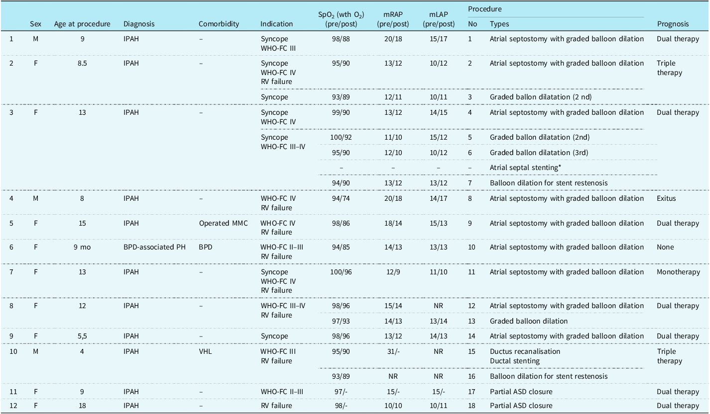
The patients’ demographics, clinical characteristics, procedural indications, interventional procedures, and prognosis are presented in Table 2.
Patient demographics, clinical indications, interventional procedures, and prognosis

ASD = Atrial Septal Defect; BPD = Bronchopulmonary Dysplasia; F = Female; IPAH = Idiopathic Pulmonary Arterial Hypertension; LAP = Left Atrial Pressure; M = Male; MMC = Myelomeningocele; NR = not recorded; RAP = Right Atrial Pressure; RV = Right Ventricle.
Patients 1, 5, 6, 7, and 9 underwent a single atrial septostomy with graded balloon dilatation, achieving sustained clinical improvement without the need for subsequent interventions during follow-up. In contrast, although initial clinical improvement was observed following the first procedure, repeat interventions were required in Patients 2, 3, and 8 due to restriction of the interatrial communication and subsequent symptom deterioration. For Patients 2 and 8, additional graded balloon dilatations were performed at 11 and 15 months, respectively, after the initial intervention. Both patients are currently maintained on dual targeted pulmonary arterial hypertension therapy consisting of a phosphodiesterase type 5 inhibitor and an endothelin receptor antagonist.
Patient 3 initially underwent atrial septostomy with graded balloon dilatation due to recurrent syncope episodes and World Health Organization Functional Class IV status, despite receiving dual targeted pulmonary arterial hypertension therapy with an endothelin receptor antagonist and a phosphodiesterase type 5 inhibitor. This resulted in marked clinical improvement. However, during regular monthly follow-up visits, recurrence of syncope and worsening symptoms were observed, attributed to restriction of the interatrial communication. Consequently, a second session of graded balloon dilatation was performed four months after the initial procedure, followed by a third session ten months later. After a one-year lapse in follow-up, the patient presented with clinical deterioration, having undergone atrial septal stenting at another medical institution, which subsequently required balloon dilatation due to in-stent restenosis. The patient is currently maintained on triple-targeted pulmonary arterial hypertension therapy consisting of an endothelin receptor antagonist, a phosphodiesterase type 5 inhibitor, and a prostacyclin analogue.
Patient 6 (bronchopulmonary dysplasia–pulmonary arterial hypertension, World Health Organization Group 3). This was a preterm infant (born at 31 weeks of gestation) with bronchopulmonary dysplasia–pulmonary arterial hypertension, receiving phosphodiesterase type 5 inhibitor therapy, and long-term home oxygen. Despite optimised bronchopulmonary dysplasia management—including lung-protective ventilation, diuretics, angiotensin-converting enzyme inhibitor, nutritional and aspiration precautions—the infant remained clinically unstable, with recurrent hospitalisations including multiple intensive care unit admissions, feeding difficulties, and poor weight gain. Echocardiography and angiography at 9 months confirmed the absence of a stentable ductus arteriosus. Given the persistent risk of right heart failure, balloon atrial septostomy was performed for decompression. Following the procedure, the patient demonstrated substantial improvement, including reduced hospitalisations, improved growth, and decreased oxygen dependency. Over subsequent years, all medications—including phosphodiesterase type 5 inhibitor, diuretics, and angiotensin-converting enzyme inhibitor—were gradually discontinued. By the age of 4.5 years, the child remained off all therapies, with no residual interatrial communication and no echocardiographic evidence of pulmonary hypertension at follow-up.
Patient 10 was a 4-year-old child with severe idiopathic pulmonary arterial hypertension, Von Hippel–Lindau disease, and PDA, referred for lung transplantation evaluation due to progressive clinical deterioration and recurrent ICU admissions despite receiving triple-targeted pulmonary arterial hypertension therapy (endothelin receptor antagonist, phosphodiesterase type 5 inhibitor, and a prostacyclin analogue). The patient had previously undergone two cardiac catheterisations: one for diagnostic evaluation and another to assess PDA suitability for closure using balloon occlusion testing. Transthoracic echocardiography demonstrated right heart chamber dilatation and severe tricuspid regurgitation, with no detectable PDA flow and no upper-to-lower extremity oxygen saturation difference. During catheterisation at our centre, the patient’s mean right atrial pressure was 31 mmHg, with a pulmonary vascular resistance index of 12.5 WU·m2 and a pulmonary-to-systemic blood flow ratio of 0.97. A comprehensive assessment of the PDA was undertaken to evaluate its feasibility for the creation of a reverse Potts shunt, as the markedly elevated mean right atrial pressure rendered atrial septostomy inappropriate. Angiographic imaging revealed no visible PDA. Multiple attempts with guidewires to cross the ductus were unsuccessful, consistent with prior functional closure following balloon occlusion testing. Subsequently, selective infusion of prostaglandin into the PDA ampulla via catheter elicited only minimal ductal flow. A second prostaglandin infusion was administered, after which a guidewire was successfully advanced through the residual PDA into the pulmonary artery and right ventricle. Sequential balloon dilatations were then performed, followed by implantation of a 6 × 22 mm Advanta™ V12 covered stent, effectively re-establishing ductal patency and creating an endogenous reverse Potts shunt for right ventricular decompression (Figure 1). Following the procedure, the pulmonary vascular resistance index decreased to 9.6 WU·m2. At the three-month follow-up, reduced stent flow and a diminished upper-to-lower extremity oxygen saturation gradient necessitated further gradual balloon dilatation. One year after the final intervention, the patient remains clinically stable on combination therapy, including an endothelin receptor antagonist, phosphodiesterase type 5 inhibitor, a prostacyclin analogue, furosemide, and dual antiplatelet therapy with aspirin and clopidogrel, with no hospitalisations required during follow-up.
(a), (b): patient 10– aortic angiography showing no flow through the ductus; (c): placement of a CTO wire into the pulmonary artery and right ventricle following ductal recanalisation; (d): dilatation of the ductus arteriosus with a non-compliant coronary balloon; (e), (f): deployment of a 6 mm × 22 mm Advanta™ V12 covered stent within the ductus.

Patient 11 was a 9-year-old female with a 5-year history of severe idiopathic pulmonary arterial hypertension, multiple bidirectional atrial septal defects with predominantly left-to-right shunting, and mitral valve prolapse. Despite dual therapy with a phosphodiesterase type 5 inhibitor and an endothelin receptor antagonist, she continued to experience fatigue and exercise intolerance, with oxygen saturation levels of 93–95% at rest and 84–85% during exertion. To reduce right ventricular volume overload while avoiding a significant increase in right atrial pressure, percutaneous closure of the haemodynamically most significant atrial septal defect was planned, allowing the smaller remaining defects to function as pop-off valves for right heart decompression. An 18 mm balloon-sizing catheter was advanced across the largest atrial septal defect under echocardiographic guidance. The stretched diameter measured 16.8 mm, and the defect was closed with an 18 mm Lifetech CeraFlex occluder. To ensure that the occluder disc did not inadvertently occlude the adjacent smaller atrial septal defects, a sizing balloon was simultaneously placed within one of the nearby defects during deployment; its stretched diameter measured 7.2 mm. Following closure of the most significant defect, echocardiography confirmed that the residual atrial septal defects, measuring 6.5 mm and 5 mm, remained patent—thus preserving right-to-left atrial decompression and supporting right ventricular unloading (Figure 2, Supplementary Video 1). At the 9-month follow-up, the patient showed significant improvement in exercise tolerance. N-terminal pro-brain natriuretic peptide levels showed a significant decline from 5404 to 355 pg/mL. Echocardiography revealed a stable device position, patent residual atrial septal defects, and a mild reduction in right ventricular size. The patient remains on endothelin receptor antagonist, phosphodiesterase type 5 inhibitor and aspirin.
Patient 11, transthoracic echocardiographic views, (a) Markedly dilated right heart chambers and a large atrial septal defect (ASD), b, c) Bidirectional shunting (left-to-right and right-to-left) across the wide ASD is demonstrated by colour Doppler imaging. (c, d) Post-closure echocardiography reveals decreased right heart dimensions with well-positioned device and moderate tricuspid regurgitation. (e) Two additional ASDs were identified adjacent to the primary defect.

Patient 12 was a 19-year-old female with a 3-year history of severe idiopathic pulmonary arterial hypertension and a large secundum atrial septal defect with bidirectional shunting, who presented with exertional dyspnoea and cyanosis. Resting oxygen saturation was 92%, dropping to 86% with exertion, and the six-minute walk test measured 400 metres. Despite ongoing therapy with endothelin receptor antagonist, phosphodiesterase type 5 inhibitor and a beta-blocker, the patient’s symptoms persisted. In addition to the pressure overload imposed by pulmonary arterial hypertension on the right ventricle, the volume load resulting from the bidirectional shunt across the atrial septal defect further exacerbated right ventricular haemodynamic stress. Therefore, partial atrial septal defect closure was planned to reduce right ventricular preload while preserving right-to-left atrial decompression as a protective mechanism. Balloon sizing revealed a stretched diameter of 31 mm, and a 32 mm fenestrated Lifetech CeraFlex™ atrial septal defect Occluder with a 7 mm central fenestration was selected. However, the 7 mm opening was deemed insufficient to allow adequate right atrial decompression. Therefore, additional fenestrations were manually created within the device during the procedure. The device was positioned across the atrial septal defect, but before its release, echocardiography did not clearly demonstrate flow through the manually created fenestrations. Therefore, balloon predilation was performed, and a 14 × 40 mm CP Stent™ was implanted across the central fenestration to ensure its long-term patency (Figure 3, Supplementary Video 2). At 2-month follow-up, the patient reported symptomatic improvement, accompanied by a reduction in N-terminal pro-brain natriuretic peptide levels, under continued dual pulmonary arterial hypertension therapy. The interatrial shunt remained patent, supporting controlled right heart decompression.
Patient 12, transthoracic echocardiographic views, (a) Markedly dilated right heart chambers and a large atrial septal defect (ASD), (b, c) Bidirectional shunting (left-to-right and right-to-left) across the wide ASD is demonstrated by colour Doppler imaging. (c, d) Post-closure echocardiography reveals decreased right heart dimensions with well-positioned device and moderate tricuspid regurgitation. (e) Two additional ASDs were identified adjacent to the primary defect.

Prognosis and follow-up findings
The procedure was uneventful in all cases, with clinical improvement observed post-intervention, and patients continued their anti-pulmonary arterial hypertension therapies. All patients were discharged after 2–3 days of in-hospital monitoring. However, Patient 4 had a pulmonary vascular resistance index of 21.3 WU·m2 and a mean right atrial pressure of 20 mmHg at cardiac catheterisation. Following atrial septostomy, the patient was discharged on dual pulmonary arterial hypertension-targeted therapy with an SpO2 of 80%. All follow-up, including the terminal admission, occurred at our centre. Three days after discharge, the patient returned to the emergency department with clinical deterioration and required intensive care. Clinical records and echocardiographic/haemodynamic assessments during this admission showed no evidence of procedure-related complications (e.g., pericardial effusion, tamponade, arrhythmia, or device-related obstruction). Despite aggressive medical management, the patient ultimately died. The death was primarily attributed to progressive right heart failure secondary to poor adherence to medical therapy.
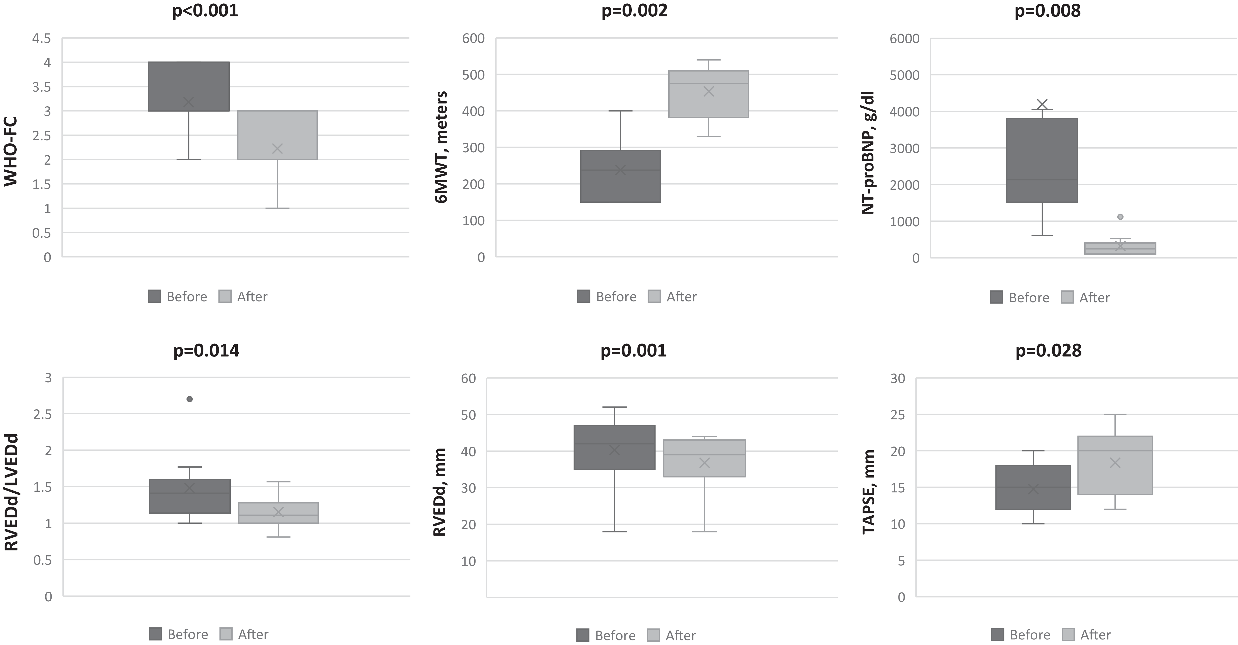
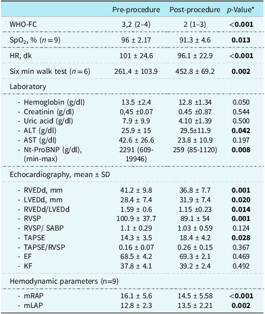
Comparison of pre- and post-procedural clinical, echocardiographic, and haemodynamic parameters is presented in Table 3. Heart rate significantly decreased (p < 0.01), and N-terminal pro-brain natriuretic peptide levels were markedly reduced (p = 0.008). The six-minute walk test distance increased significantly (p = 0.002), accompanied by an improvement in World Health Organization Functional Class (p < 0.001). Echocardiographic assessments demonstrated a significant reduction in the right ventricular end-diastolic diameters/left ventricular end-diastolic diameters ratio (p = 0.014) and an increase in tricuspid annular plane systolic excursion (p = 0.028) (Figure 4). Additionally, mean right atrial pressure decreased significantly after the intervention (p < 0.001). Importantly, post-procedural left atrial pressure remained below 18 mmHg in all patients.
Box-plot graphs illustrate the changes in clinical, functional, and haemodynamic parameters before and after the intervention. Statistical comparisons were performed using the Wilcoxon signed-rank test. WHO-FC = World Health Organization Functional Class; 6MWT = 6-minute walk test; NT-proBNP = N-terminal pro-Brain Natriuretic Peptide; RVEDd = right ventricular end-diastolic diameter; LVEDd = left ventricular end-diastolic diameter; TAPSE = tricuspid annular plane systolic excursion. To enhance the visual interpretability of NT-proBNP, the Y-axis was limited to the 0–6000 range. Outlier values above this range were excluded from the figure but included in the statistical analysis.

Comparison of clinical, laboratory, echocardiographic, and haemodynamic parameters before and after interventional procedures

Bold values indicate statistically significant (p < 0.05). * Wilcoxon signed-rank test.
ALT = Alanine aminotransferase; AST = Aspartate aminotransferase; EF = Ejection fraction; HR; Heart rate; KF = Fractional shortening; mLAP = mean Left atrial pressure; LVEDd = Left ventricular end-diastolic diameter; NT-proBNP = N-terminal pro-brain natriuretic peptide; mRAB = mean Right atrial pressure; RVEDd = Right ventricular end-diastolic diameter; RVSP = Right ventricular systolic pressure; SaBP = Systolic arterial blood pressure; SpO2 = Peripheral oxygen saturation; TAPSE = Tricuspid annular plane systolic excursion; WHO-FC = World Health Organization Functional Class.
Conversely, no statistically significant differences were observed in other echocardiographic parameters such as estimated right ventricular systolic pressure (p = 0.166) and EF (p = 0.469).
Discussion
Pulmonary arterial hypertension is a rare but progressively debilitating disease, particularly affecting children and young adults. Despite significant advancements in pharmacological therapies, many patients continue to experience persistent symptoms such as right ventricular dysfunction, reduced exercise capacity, and frequent hospitalisations.Reference Humbert, Kovacs and Hoeper1,Reference Ivy, Rosenzweig and Abman3 These clinical limitations highlight the need for individualised interventional strategies aimed at optimising haemodynamics and delaying disease progression.
In this study, we evaluated the effects of various interventional strategies implemented at our centre on clinical, haemodynamic, and echocardiographic parameters in paediatric patients with pulmonary arterial hypertension. Our findings indicate that, with appropriate patient selection, these procedures offer safe and effective therapeutic options that contribute to improved functional status and disease stabilisation. In addition to established interventions such as the Potts shunt and atrial septostomy, partial closure of pre-existing atrial septal defects stands out as a noteworthy approach, supporting haemodynamic improvement by regulating interatrial flow and balancing right ventricular load.
Atrial septostomy: balancing risks and benefits in severe PAH
Atrial septostomy is a palliative intervention designed to reduce right atrial pressure and increase cardiac output. Patients with pulmonary arterial hypertension and a patent foramen ovale have been observed to survive longer than those without,Reference Barst7,Reference Kothari, Yusuf, Juneja, Yadav and Naik8 suggesting that a controlled right-to-left shunt may decompress the right heart and augment left ventricular preload during acute rises in pulmonary vascular resistance.Reference Ivy, Rosenzweig and Abman3,Reference Micheletti, Hislop and Lammers9 In our cohort, atrial septostomy improved World Health Organization Functional Class, six-minute walk test distance, and N-terminal pro-brain natriuretic peptide levels, with resolution of syncope.
Graded balloon septostomy enables controlled, gradual creation of an atrial septal defect, incrementally increasing right-to-left shunting while preserving haemodynamic stability, and is preferred in many cases.Reference Kothari, Yusuf, Juneja, Yadav and Naik8,Reference Rothman, Sklansky and Lucas10 In our experience, it has been successful in most patients; however, spontaneous closure rates of 15–17% have been reported in adults,Reference Kothari, Yusuf, Juneja, Yadav and Naik8,Reference Micheletti, Hislop and Lammers9 and three of our patients required repeat interventions due to shunt restriction. Therefore, persistent clinical and echocardiographic follow-up is essential. In selected cases, interatrial stent implantation may provide a more durable solution; however, as observed in our patient, the stent may gradually narrow over time, necessitating repeated interventions.Reference Sandoval11 Furthermore, devices such as atrial flow regulators, specifically designed to maintain interatrial communication, may reduce the need for repeat procedures and support long-term haemodynamic stabilityReference Sivakumar, Rohitraj, Rajendran and Thivianathan12,Reference Butera, Piccinelli and Kolesnik13 However, in children, their use remains limited in many developing countries, including ours, due to restricted availability and high cost, and they are not approved for paediatric use in our country. Addressing these limitations is essential to facilitate the integration of these advanced technologies into personalised treatment strategies for complex pulmonary arterial hypertension cases.
Atrial septostomy can provide clinical benefits; however, its use is limited in patients with severe right ventricular failure, mean right atrial pressure ≥ 20 mmHg, or resting oxygen saturation ≤ 85%, as these conditions are associated with poor procedural tolerance and increased mortality riskReference Hansmann, Koestenberger and Alastalo4,Reference Sandoval11 Published studies report a 13% mortality rate during or shortly after the procedure, with a 30-day survival rate of 82%Reference Doyle, McCrory, Channick and Conte5,Reference Micheletti, Hislop and Lammers9 In our series, one patient—whose clinical, echocardiographic, and haemodynamic findings aligned with the high-risk profile defined by the Seventh World Symposium on Pulmonary HypertensionReference Ivy, Rosenzweig and Abman3—died shortly after the procedure, a situation attributed to nonadherence to prescribed medical therapy.
Moreover, the presence of a right-to-left shunt introduces potential risks, such as paradoxical embolism and mild systemic desaturation, which necessitate careful consideration during patient selection and vigilant post-procedural follow-up. Although a slight reduction in systemic arterial oxygen saturation may occur, the concomitant increase in cardiac output can lead to a net improvement in global tissue oxygen delivery.Reference Ivy, Rosenzweig and Abman3,Reference Micheletti, Hislop and Lammers9,Reference D’Alto, Romeo and Argiento14
To date, several questions regarding the benefits of atrial septostomy in pulmonary arterial hypertension remain unanswered. These include the optimal timing of the procedure, determination of the appropriate shunt size, long-term sustainability, and the risk of spontaneous shunt closure.
Reverse Potts shunt: clinical application and technical challenges
The reverse Potts shunt involves the creation of a right-to-left shunt between the aorta and the pulmonary artery to reduce pressure overload on the right ventricle in patients with systemic or suprasystemic pulmonary arterial hypertension. This approach has emerged as an effective palliative treatment option, particularly for patients with contraindications to atrial septostomy and those with advanced right ventricular dysfunction. An important advantage of the reverse Potts shunt over atrial septostomy is that it preserves coronary and cerebral oxygenationReference Ivy, Rosenzweig and Abman3,Reference Sandoval11,Reference Baruteau, Serraf and Lévy15 In our study, a patient with right heart failure and haemodynamic ineligibility for atrial septostomy successfully underwent transcatheter creation of a reverse Potts shunt by reopening a closed PDA. This intervention reduced pulmonary vascular resistance and subsequently decreased right ventricular workload, leading to marked clinical improvement and prevention of recurrent hospitalisations.
However, one of the major limitations of the reverse Potts shunt is the technical complexity associated with its implementation. If a PDA is present, patency can be re-established through transcatheter stent implantationReference Sandoval11,Reference Latus, Apitz and Moysich16 and appropriate stent selection remains a critical determinant of procedural success. Expandable stents may offer long-term patency advantages by reducing the risk of restenosis from endothelial proliferation, although balloon redilatation may still be required, as in our case. In the absence of a PDA, and when re-canalisation cannot be achieved with guidewire passage or intravenous prostaglandin infusion, alternative approaches are needed. While limited reports describe successful transcatheter creation of the shunt, surgical intervention remains the predominant method. Direct surgical anastomosis between the aorta and left pulmonary artery typically requires sternotomy and cardiopulmonary bypass, carrying additional risks such as perioperative bleeding and prolonged recovery.Reference Sandoval11,Reference Baruteau, Serraf and Lévy15
Recent data indicate that survival after a Potts shunt (5-year survival: 74.7%) is comparable to that following lung transplantation (5-year survival: 77.2%), supporting its role as a viable alternative in carefully selected high-risk paediatric pulmonary arterial hypertension patients.Reference Ivy, Rosenzweig and Abman3 Despite the growing global experience with the Potts shunt, several important questions remain unresolved, including optimal patient selection criteria, the role of concurrent atrial shunt closure, appropriate timing relative to lung transplantation, and strategies for managing patients who may require transplantation following shunt failure.
Partial atrial septal defect closure: a targeted and innovative strategy
In patients with pulmonary arterial hypertension, the management of an existing atrial septal defect presents a complex clinical dilemma. Isolated atrial septal defects in children are often incidental findings and are generally not considered the primary cause of pulmonary arterial hypertension.Reference Ivy, Rosenzweig and Abman3 Nonetheless, the presence of a patent foramen ovale or atrial septal defect has been linked to improved survival, suggesting that a controlled right-to-left interatrial shunt may confer protective effects by decompressing the right heart and attenuating the consequences of elevated pulmonary pressures.Reference Barst7,Reference Micheletti, Hislop and Lammers9 Recognising this potential benefit, the Seventh World Symposium on Pulmonary Hypertension recommended an individualised, stepwise strategy, incorporating deep patient phenotyping to guide the use of pulmonary arterial hypertension-targeted therapy alongside surgical or percutaneous complete or partial shunt closure.Reference Ivy, Rosenzweig and Abman3
The rationale for partial atrial septal defect closure in pulmonary arterial hypertension is to mitigate the combined haemodynamic burden of pressure and volume overload. A large, unrestrictive atrial septal defect with predominant left-to-right shunting leads to chronic right ventricular volume overload, progressive dilation, and dysfunction.Reference Humbert, Kovacs and Hoeper1,Reference Kovacs, Bartolome and Denton17 Conversely, an unregulated right-to-left shunt can cause profound systemic desaturation, impairing exercise capacity and predisposing to end-organ injury.Reference Humbert, Kovacs and Hoeper1,Reference D’Alto, Romeo and Argiento14,Reference Kovacs, Bartolome and Denton17 Partial closure offers a haemodynamic compromise, reducing right ventricular volume overload while preserving a protective right-to-left shunt during pressure surges. This controlled decompression strategy allows the right heart to offload pressure when needed, while limiting the detrimental effects of excessive left-to-right shunting. Importantly, in patients with severe pulmonary arterial hypertension and left-to-right shunting, partial closure may counterbalance the volume overload that can result from right ventricular afterload reduction with pulmonary arterial hypertension-targeted therapy. By optimising shunt size, this approach helps prevent systemic hypoxaemia, maintain right heart decompression, and support functional capacity, potentially slowing right ventricular failure progression. While adult studies have shown favourable outcomes with fenestrated device closure, this strategy has not been specifically evaluated in children.Reference Bruch, Winkelmann and Sonntag18,Reference Abdelkarim, Levi, Tran, Ghobrial and Aboulhosn19 In our series, Patients 11 and 12 who underwent partial atrial septal defect closure demonstrated marked symptomatic improvement, increased exercise capacity, reduced right ventricular dilation, and decreased N-terminal pro-brain natriuretic peptide levels during follow-up.
Partial atrial septal defect closure can be achieved using custom-made fenestrated occluder devices, which allow for controlled interatrial shunting after the procedure. However, as demonstrated in our case, the fenestration size may be suboptimal, necessitating additional interventions such as stent implantation to maintain adequate right-to-left shunting. Manual creation of a fenestration is another option, but the resulting orifice is often too small to sustain sufficient interatrial flow and tends to close over time.Reference Abdelkarim, Levi, Tran, Ghobrial and Aboulhosn19 Since sustained interatrial communication is essential for preserving haemodynamic stability in this patient population, we believe that manual fenestration is unlikely to represent a reliable long-term solution.
In patients with multiple atrial septal defects, selectively closing the most haemodynamically significant defect while leaving smaller defects patent can be an effective and physiologically sound strategy. This approach alleviates right ventricular volume overload while preserving the capacity for pressure decompression, thereby maintaining haemodynamic balance. Device selection is crucial, as the occluder discs must not inadvertently cover adjacent defects, which could diminish the intended decompressive effect. In complex atrial septal anatomies, surgical downsizing of the defect offers an alternative, allowing precise control over shunt size and location. Nevertheless, surgical intervention carries inherent risks and should be pursued judiciously.
However, interventional device closure also entails specific procedural risks, such as device embolisation, residual shunting that may necessitate re-intervention, and difficulties in accurately determining the optimal fenestration size. Further multicenter studies are needed to define appropriate patient selection criteria, determine the ideal timing for partial atrial septal defect closure, and identify the most effective device types and fenestration dimensions.
Haemodynamic and clinical improvements
In our study, interventional procedures were associated with significant haemodynamic and clinical improvements. Notable enhancements in heart rate, N-terminal pro-brain natriuretic peptide levels, six-minute walk test distance, and World Health Organization Functional Class align with previous reports demonstrating the capacity of such interventions to alleviate symptoms and improve functional capacity.Reference Ivy, Rosenzweig and Abman3,Reference Doyle, McCrory, Channick and Conte5,Reference Sandoval11,Reference Chiu, Zuckerman and Turner20 Among patients undergoing atrial septostomy, the observed reduction in mean right atrial pressure indicated effective atrial decompression. Moreover, increases in tricuspid annular plane systolic excursion and the tricuspid annular plane systolic excursion/right ventricular systolic pressure ratio reflected improved right ventricular systolic performance and enhanced ventriculo-arterial coupling. The decrease in the right ventricular end-diastolic diameters/left ventricular end-diastolic diameters ratio further suggested favourable reverse remodelling of the right ventricle. Collectively, these findings highlight the beneficial effects of individualised interventional strategies in achieving both symptomatic relief and structural and functional cardiac improvement in appropriately selected patients.
Timing of interventional procedures in paediatric PAH
Optimal timing of interventional strategies—such as atrial septostomy, partial atrial septal defect closure, and reverse Potts shunt—is a critical determinant of outcomes in paediatric pulmonary arterial hypertension. Delayed intervention can lead to irreversible right ventricular failure, whereas prematurely performed procedures may precipitate haemodynamic instability. Consequently, individualised decision-making, guided by clinical trajectory and invasive haemodynamic assessment, remains essential.
Evidence from previous studies supports the concept that timely decompressive interventions, when implemented before the onset of overt right ventricular dysfunction, can improve transplant-free survival. However, current guidelines—largely extrapolated from adult populations—provide limited clarity on the optimal timing and indications for these procedures in children. For example, although syncope and World Health Organization functional class III/IV status are widely recognised triggers for atrial septostomy in adults, no such consensus exists regarding the timing of other interventional strategies in paediatric pulmonary arterial hypertension.Reference Humbert, Kovacs and Hoeper1,Reference Hansmann, Koestenberger and Alastalo4
In high-risk paediatric patients, conventional pharmacological treatment algorithms may fail to achieve adequate or timely clinical improvement. Given the rapid disease progression and limited right ventricular reserve in such cases, interventional procedures can evolve from adjunctive measures into urgent, potentially life-saving, and even first-line therapeutic options. From this perspective, physiology-driven, patient-specific interventions—such as atrial septostomy, partial atrial septal defect closure, and reverse Potts shunt—should not be viewed as deviations from standard treatment pathways, but rather as integral components of advanced management strategies.
In our series, the only patient who died after atrial septostomy had a mean right atrial pressure of 20 mmHg, placing him in the high-risk category.Reference Ivy, Rosenzweig and Abman3 This observation illustrates the limitations of performing septostomy at such an advanced stage and, despite ongoing debate in the literature, supports our opinion that atrial septostomy should be considered earlier in paediatric patients, before the development of severe haemodynamic compromise. Moreover, the frequently cited threshold of mean right atrial pressure > 20 mmHg as an indicator of high risk is largely derived from adult data; in children, even lower right atrial pressure values may carry significant risk. In addition, because procedures in children are typically performed under general anaesthesia, the measured mean right atrial pressure may be lower than in the awake state, potentially underestimating the true haemodynamic burden.
Ultimately, both the timing and technical approach should be determined by an experienced, multidisciplinary team, with careful integration of patient-specific haemodynamic and anatomical considerations, risk–benefit evaluation, procedural feasibility, and the potential sequencing of interventions to optimise safety, efficacy, and long-term outcomes.
Limitations
This study has several limitations that should be acknowledged. First, the retrospective, single-centre design inherently introduces selection bias and limits the generalisability of the findings. The relatively small sample size, although reflecting the rarity and complexity of paediatric pulmonary arterial hypertension requiring individualised interventional strategies, reduces the statistical power of subgroup analyses and limits the ability to perform multivariate modelling. Additionally, the heterogeneity of underlying aetiologies (e.g., Idiopathic pulmonary arterial hypertension, bronchopulmonary dysplasia-associated pulmonary arterial hypertension, pulmonary arterial hypertension with congenital heart defects) and variations in the timing and type of interventions (partial atrial septal defect closure, atrial septostomy, or reverse Potts shunt) may confound interpretation of outcomes.
Conclusion
In conclusion, the management of severe pulmonary arterial hypertension in paediatric patients through interventional procedures requires careful consideration of timing, patient selection, and procedural strategy. Our findings demonstrate that such interventions can lead to significant improvements in both echocardiographic parameters and clinical status in children with severe pulmonary arterial hypertension and right heart failure. A unique contribution of our study is the demonstration that partial atrial septal defect closure in paediatric pulmonary arterial hypertension can alleviate right ventricular volume overload while preserving protective decompression, thereby supporting haemodynamic stability and enhancing clinical outcomes. Considering and implementing the intervention early in the disease course—before progression to a high-risk state—may play a pivotal role in reducing mortality. Prospective multicentre and large-scale studies are warranted to validate these findings and guide future clinical practice.
Supplementary material
The supplementary material for this article can be found at https://doi.org/10.1017/S104795112611141X
Acknowledgements
None.
Author contributions
IE and EGT contributed to idea, concept, design, and writing. IE and DB contributed to supervision and critical review. EGT contributed to the literature search, material and data collection, analysis, and interpretation. All authors reviewed the manuscript.
Financial support
This research received no specific grant from any funding agency.
Competing interests
The authors declare no conflict of interest.