Introduction
Testosterone (T) concentrations decline in older men, and lower T levels have been associated with increased incidence of adverse clinical conditions, including decreased sexual function and cardiovascular, metabolic and central nervous system diseases.Reference Yeap1 Our knowledge on these issues is mainly based on association studies unable to demonstrate cause-and-effect relationships, and only a limited number of prospective studies have been performed. In addition to age, T production is influenced by several lifestyle factors and co-morbidities. Late-onset hypogonadism (LOH), instead of andropause or male menopause, is the currently recommended term for the development of hypogonadism with ageing.Reference Jones2 In this review, we first summarize briefly the physiology of T production in ageing men, and then discuss the association of T with mortality and common diseases affecting elderly men.
Basic physiology of testosterone
T, the most important male sex hormone, is synthesized by the interstitial Leydig cells of the testis under regulation of the gonadotrophin luteinizing hormone (LH). T, or its metabolite 5α-dihydrotesterone (5α-DHT), are essential for all masculine functions of the body, including sexual differentiation, development of secondary sex characteristics, spermatogenesis, the masculine features of the muscle–bone apparatus, male-pattern hair distribution and male sexual behaviour.Reference Nieschlag and Behre3 A single regulatory step, the binding of T or 5α-DHT to the androgen receptor, is able to evoke this variety of structural and functional responses.
Testicular androgen synthesis starts from cholesterol, originating from multiple sources: (a) de novo synthesis from acetyl coenzyme A, (b) stored cholesteryl esters, (c) exogenous lipoprotein-supplied cholesterol and (d) plasma membrane-derived cholesterol following hormonal stimulation.Reference Azhar, Reaven, Payne and Hardy4 The next critical and LH-regulated step is the rapid transfer of cholesterol from the outer to the inner mitochondrial membrane, mediated by two carrier proteins, the steroidogenic acute regulatory protein (StAR)Reference Clark and Stocco5 and the peripheral-type benzodiazepine receptor (PBR).Reference Papadopoulos, Liu and Culty6 The first and rate-limiting step of steroid biosynthesis at the mitochondrial inner membrane is the conversion of cholesterol to pregnenolone, catalysed by the cytochrome P450 cholesterol side chain cleavage enzyme (CYP11A1) and auxiliary electron transferring proteins. For the following steps, pregnenolone is translocated to the smooth endoplasmic reticulum, to be converted via 17-hydroxypregnenolone, dehydroepiandrosterone (DHEA) and androstenedione to T.
The daily T production is 6–7 mg and about 95% of it originates from the testes, the rest through peripheral conversion of adrenal androgenic precursors to T. The testis also produces small amounts of 5α-DHT, but most of this hormone is formed in peripheral androgen target tissues. Likewise, a small proportion of androstenedione and T are aromatized to oestrogens and they seem to have important physiological functions both within the testis and extragonadally,Reference Payne and Hales7 in particular in bone formation and the hypothalamic feedback regulation of gonadotrophins.
The secretion of steroid hormones is assumed to be a passive process due to their lipid solubility and ease of transit through cell membranes. Accordingly, there is no intratesticular storage of bioactive steroid hormones, except for their sulphate conjugates, and the regulation of circulating steroid hormone concentrations occurs mainly at the level of their biosynthesis. There is diurnal variation in T secretion due to night-time accentuation of LH secretion.Reference Spratt, O'Dea, Schoenfeld, Butler, Rao and Crowley8 Secretion pulses of serum T after the LH secretion peaks (average interval of 2 h) are inconsistent, apparently due to sluggish response of human testicular steroidogenesis to acute gonadotrophin stimulation,Reference Martikainen, Huhtaniemi and Vihko9 and to the buffering effect of steroid hormone binding to plasma transport proteins.Reference Bhasin, Kronenberg, Melmed, Polonski, Wilson and Larsen10
In circulation, the majority of T is bound to sex-hormone-binding globulin (SHBG, 44%), and albumin (54%), and only about 2% appears in unbound free form. Only free T is biologically active, i.e. able to penetrate to androgen target cells. The avid binding of T to SHBG prevents this, but because the binding of T to albumin is loose, this fraction together with free T, that is about 50% of total plasma T, is considered to form the so-called bioavailable fraction of androgens. Plasma SHBG level is under endocrine regulation,Reference Bhasin, Kronenberg, Melmed, Polonski, Wilson and Larsen10 increasing clearly by oestrogen and ageing, decreasing somewhat by androgens, and clearly by obesity. If the hypothalamic–pituitary–testicular axis functions normally, changes in SHBG levels do not affect the balance of bioactive androgens, due to fast compensatory changes in the feedback regulation.
Like all steroid hormones, androgens initiate their effects at the cellular level by interacting with high-affinity nuclear receptors.Reference Bhasin, Kronenberg, Melmed, Polonski, Wilson and Larsen10, Reference Heemers and Tindall11 The androgen receptor (AR or NR3C4) is a member of the nuclear receptor superfamily of ligand-activated transcription factors, along with those of the other steroid hormones. Unliganded AR occurs predominately in the cytoplasm where it is complexed with several proteins, including various heat shock proteins. AR binds to specific androgen response elements (AREs) in the promoter and enhancer regions of androgen-responsive genes. Highest levels of AR are found in androgen target tissues, such as the accessory male sex organs. The same receptor protein binds both T and 5α-DHT, the latter with up to 10-fold higher affinity. This may function as an amplifying mechanism of androgen action in many target organs, for example the prostate and hair follicle.
The main androgenic functions include the regulation of gonadotrophin secretion, regulation of initiation and maintenance of spermatogenesis, male-type differentiation of the sexual organs during embryogenesis, stimulation of sexual differentiation during puberty and control of male sexual behaviour and potency. An alternative way of grouping the androgen effects is to define them as androgenic, anabolic and psychological. The anabolic androgen actions on muscle are predominantly due to T action, whereas its effects on bone, at least partly, require aromatization of T to oestrogens. LH is the main regulatory hormone of T production, and these two hormones form a classical feedback regulatory loop of the hypothalamic–pituitary–gonadal axis.
Measurement of testosterone production
Testicular androgen production is usually monitored by measuring serum total T levels using immunological techniques (radioimmunoassay, enzyme immunoassay). The levels in men are normally high enough to be reliably measured with these techniques, but if the levels are low, as in children, women and hypogonadal men, the more specific and sensitive mass spectrometric techniques are recommended.Reference Rosner, Auchus, Azziz, Sluss and Raff12 The analyses can be supplemented by SHBG measurements, which allow the calculation of free T.Reference Vermeulen, Verdonck and Kaufman13 Direct free T measurements are not considered reliable, and the reliable method of equilibrium dialysis is too expensive for routine clinical use.Reference Rosner, Auchus, Azziz, Sluss and Raff12 Total T measurements are usually sufficient in the assessment of testicular T production in ageing men, but free T may provide additional information in cases suggestive of borderline hypogonadism.Reference Wu, Tajar and Pye14
Changes in testosterone production and action during ageing
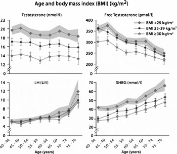
T production decreases with ageing.Reference Kaufman and Vermeulen15, Reference Perheentupa and Huhtaniemi16 According to a large recent study (European Male Ageing Study (EMAS), n = 3220)Reference Wu, Tajar and Pye14 serum level of total T decreased between 40–49 and 70–79 years of age on average from the level of 17.6 to 15.9 nmol/l (0.5% per year) and that of free T from 350 to 230 pmol/l (1.1% per year, Figure 1). The higher magnitude of the latter change is explained by simultaneous increase in the level of SHBG, whereby the proportion of free T declines. A concomitant increase of circulating LH levels with age reveals that the decrease of testicular function is primary, as the feedback regulatory link attempts to compensate it by increase of LH. Longitudinal studies detect an additional secular decline in T levels, which may be as high as the per year cross-sectional decline.Reference Travison, Araujo, Hall and McKinlay17 Selection bias of cross-sectional studies, increasing adiposity, smoking cessation and accumulation of endocrine disrupting compounds in the environment have been suggested as causes of this trend. Age-related decreases of similar magnitude have been reported in total T (0.8% per year) and in free and albumin-bound T (2% per year) also in other studies.Reference Kaufman and Vermeulen15, Reference Feldman, Longcope and Derby18 If we consider as the criterion of low T a level below the lower limit of the reference range, i.e. <10 nmol/l, then the frequency of biochemical hypogonadism increases from 8.5% in 40- to 49-year-old men to 13.5% in 70- to 79-year-old men.Reference Wu, Tajar and Pye14 If calculated free T is used as the criterion, the frequency of hypogonadism increases in the oldest age group threefold. However, these figures do not equate with genuine LOH, because the majority of ageing men with low T remain asymptomatic.
Relationship between age, body mass index (BMI) and hormones in middle-aged to ageing men. The data are from the European Male Ageing Study (n = 3220). The cohort was stratified according to BMI into three groups: normal weight (BMI < 25 kg/m2), overweight (BMI 25–29 kg/m2), and obese (BMI ≥ 30 kg/m2). Mean (95% CI in shaded area and vertical lines) total T, free T and SHBG were significantly lower at all ages in the overweight and obese compared with non-obese men. The total T and SHBG age trends in the three BMI categories were similar (indicating no interaction between BMI and age); the free T age trend in the obese group was less steep than in the other two groups (indicating an interaction between BMI and age). From ref. 14, with permission.

The most important factor in the decline of T in adult men is overweight (in particular increased waist/hip ratio), and thereafter various co-morbidities.Reference Wu, Tajar and Pye14 In the EMAS study, when the body mass index (BMI) increased from <25 kg/mReference Jones2 to >30 kg/mReference Jones2, total T decreased by 4 nmol/l (about 25%). Being mildly overweight (BMI >25 kg/mReference Jones2) decreases serum T more than the increase of age from 40 to 80 years; the trend is similar with free T (Figure 1). Low T is a risk factor of imminent metabolic syndrome and type 2 diabetes.Reference Laaksonen, Niskanen and Punnonen19 Age-related decline of T is primary testicular (associated with increased LH levels), whereas the overweight-associated decline is secondary hypothalamic (no compensatory increase in LH).Reference Kaufman and Vermeulen15 This is important because lowering of circulating cholesterol may reduce the substrate for androgen production. Some,Reference Dobs, Schrott and Davidson20 but not all,Reference Hyyppä, Kronholm, Virtanen, Leino and Jula21, Reference Hall, Page, Travison, Montgomery, Link and McKinlay22 studies have shown mild decrease in T levels during cholesterol-lowering treatment with statins.
Androgens and mortality
An independent inverse association between serum T concentration and mortality has been demonstrated in several longitudinal studies.Reference Lehtonen, Huupponen and Tuomilehto23–Reference Shores, Moceri, Gruenewald, Brodkin, Matsumoto and Kivlahan28 In cause-specific analysis, increased rate of mortality existed in cardiovascularReference Laughlin, Barrett-Connor and Bergstrom24, Reference Khaw, Dowsett and Folkerd25 and respiratoryReference Laughlin, Barrett-Connor and Bergstrom24 diseases, and in some,Reference Khaw, Dowsett and Folkerd25 but not in all,Reference Laughlin, Barrett-Connor and Bergstrom24 studies also in cancer deaths. However, in the population-based Caerphilly study no association between total T and all-cause mortality was found.Reference Smith, Ben-Shlomo, Beswick, Yarnell, Lightman and Elwood29 Similarly, based on the Massachusetts Male Aging Study, Araujo et al.Reference Araujo, Kupelian, Page, Handelsman, Bremner and McKinley30 concluded that endogenous sex hormone levels have only relatively weak associations with mortality. They found that, in multivariate-adjusted models, higher free T was associated with mortality from ischaemic heart disease and inversely with mortality from diseases of the respiratory system, whereas total T was unrelated to mortality. Total T may not be the only anabolic marker of poor prognosis, because concomitant low baseline concentrations of bioavailable T, IGF-1 and DHEA sulphate increased the hazard ratios for mortality compared with low value in only one of them.Reference Maggio, Lauretani and Ceda26
Androgens and atherosclerosis
Male gender is one of the risk factors for coronary artery disease (CAD). Androgens, or the lack of oestrogens, have been regarded as the proximate cause of this risk factor.Reference Liu, Death and Handelsman31, Reference Wu and von Eckardstein32 There is substantial evidence that levels of the sex hormones T, oestradiol (E2) and DHEA are associated with cardiovascular disease (CVD) risk.Reference Liu, Death and Handelsman31–Reference Mendelsohn and Karas35 Androgens have important influence on cardiovascular function, and androgen receptors are expressed in vascular tissues, including the endothelium, smooth muscle and myocardium.Reference Wu and von Eckardstein32
Among 39 studies on the relationship between circulating T and CAD, sixteen demonstrated lower levels of T in CAD patients compared with healthy controls, 16 showed no difference in T, but none suggested association of high levels of T with CAD.Reference Wu and von Eckardstein32 The largest study including 2512 menReference Yarnell, Beswick, Sweetnam and Riad-Fahmy36 showed no difference in basal T between men with and without ischaemic heart disease during a 5-year follow-up. Similarly, in two large long-term prospective cohort studies, no correlation between baseline T levels and subsequent development of CAD was found in middle-aged and elderly men.Reference Barrett-Connor and Khaw37, Reference Harman, Metter, Tobin, Pearson and Blackman38
Low serum T levels have been associated with faster progression of subclinical atherosclerosis,Reference Hak, Witteman, de Jong, Geerlings, Hofman and Pols39, Reference Muller, van den Beld, Bots, Grobbee, Lamberts and Van Der Schouw40 and an independent inverse association between T and occurrence of aortic atherosclerosis,Reference Hak, Witteman, de Jong, Geerlings, Hofman and Pols39 and T and carotid intima-media thicknessReference Mäkinen, Järvisalo and Pöllänen41 have been demonstrated. Low levels of total and free T were detected in male subjects with angiographically measured coronary atherosclerosisReference Phillips, Pinkernell and Jing42 but no association between peripheral arterial disease and T was found in another study.Reference Price, Lee and Foekes43 This discrepancy may be attributable to the fact that peripheral arterial disease encompasses subjects with less severe atherosclerosis than those with severe aortic atherosclerosis.Reference Hak, Witteman, de Jong, Geerlings, Hofman and Pols39 Likewise, no association between endogenous T and CAD or cardiovascular disease events was found in the Honolulu Heart Program,Reference Phillips, Yano and Stemmermann44 and in some other studies.Reference Yarnell, Beswick, Sweetnam and Riad-Fahmy36, Reference Barrett-Connor and Khaw37, Reference Phillips, Yano and Stemmermann44–Reference Ärnlöv, Pencina and Amin47 Recently, low T levels have been associated with the occurrence of stroke or transient ischaemic attacksReference Yeap, Hyde and Almeida48 and with arterial stiffness.Reference Hougaku, Fleg and Najjar49 Interestingly, E2 has been associated in an inverse manner with cardiovascular disease risk, especially in older men.Reference Ärnlöv, Pencina and Amin47
In summary, the cross-sectional data suggest an association between low T and CAD, but a significant and independent association has not been confirmed in large prospective studies. Extreme androgen deficiency may be associated with increased cardiovascular morbidity but physiological levels may be cardioprotective.
T therapy and coronary heart disease
In hypogonadal men, replacement therapy with T may have beneficial effects on bone density,Reference Snyder, Peachey and Hannoush50 muscle mass,Reference Snyder, Peachey and Berlin51, Reference Sih, Morley, Kaiser, Perry, Patrick and Ross52 body compositionReference Snyder, Peachey and Berlin51–Reference Snyder, Peachey and Berlin53 and cognition,Reference Cherrier, Asthana and Plymate54, Reference Moffat, Zonderman, Metter, Blackman, Harman and Resnick55 but considerable controversy still remains regarding the indications for T supplementation in ageing men.Reference Rhoden and Morgentaler56
T replacement therapy may improve exercise tolerance in chronic stable angina,Reference English, Steeds, Jones, Diver and Channer57 and infusion of T into coronary arteries seems to improve coronary artery blood flow in CAD patients.Reference Webb, Adamson, de Zeigler and Collins58–Reference Rosano, Leonardo and Pagnotta60 The sum of ECG ST-segment depressions immediately after the exercise test decreases after T therapy.Reference Jaffe61 Studies on T replacement therapy have not revealed increased incidence of cardiovascular disease or events such as myocardial infarction, stroke or anginaReference Hajjar, Kaiser and Morley62 or other major adverse effects.Reference Nair, Rizza and O'Brien63 Thus therapeutic doses of exogenous T may have beneficial effects on cardiovascular risk factors, but the data do not justify uncontrolled use of T in the prevention of cardiovascular disease,Reference Wu and von Eckardstein32 and definitive assessment of the long-term effects of T replacement therapy on cardiovascular health requires more prospective, placebo-controlled studies.Reference Rhoden and Morgentaler56
Metabolic effects of androgens
Metabolic syndrome, initially named as syndrome X,Reference Reaven64 is a cluster of central obesity (measured as increased waist circumference), raised triglycerides, reduced HDL-cholesterol, raised blood pressure and increased fasting plasma glucose.Reference Alberti, Zimmet and Shaw65 The syndrome helps to identify subjects at high risk of CVD and type 2 diabetes mellitus.
The prevalence of both metabolic syndrome and hypoandrogenaemia increases with advanced age. Therefore, it is not surprising that they may occur simultaneously, and the existence of a hypoandrogenic-metabolic syndrome in men has been suggested.Reference Gould, Kirby and Amoroso66 The prevalence of obesity is high among men with isolated hypogonadotropic hypogonadism referred to hospital for treatment of obesity,Reference Hofstra, Loves, van Wageningen, Ruinemans-Koerts, Jansse and de Boer67 and over a wide age range men with type 2 diabetes mellitus have a lower free T concentration than non-diabetic men.Reference Fukui, Soh, Tanaka, Kitagawa, Hasegawa, Yoshikawa, Miki and Nakamura68
The relationship between T and insulin sensitivity has been demonstrated both in cross-sectional and longitudinal studies. In elderly men, there is positive correlation between T and insulin sensitivity, and hypogonadal men are twice as insulin resistant as eugonadal men.Reference Pitteloud, Mootha and Dwyer69 Withdrawal of T treatment increases insulin resistance in middle-aged men with idiopathic hypogonadotropic hypogonadism without any changes in body composition,Reference Yialamas, Dwyer, Hanley, Lee, Pitteloud and Hayes70 suggesting that insulin sensitivity can be modulated by T also in an adipose-tissue-independent manner.
In the prospective Massachusetts Male Aging Study, lower levels of total T and SHBG and the existence of clinical androgen deficiency at baseline each predicted later development of metabolic syndrome, especially in initially non-obese men.Reference Kupelian, Page, Araujo, Travison, Bremner and McKinlay71 Similarly, during an 11-year follow-up, men with total T, free T and SHBG concentrations at the lower fourth had a clearly increased risk of developing metabolic syndrome and diabetes.Reference Laaksonen, Niskanen and Punnonen19 On the other hand, metabolic syndrome itself is a risk factor for later development of hypogonadism.Reference Laaksonen, Niskanen, Punnonen, Nyyssönen, Tuomainen, Valkonen and Salonen72
Studies on effects of exogenous androgens or androgen deprivation on metabolism are scarce, and have usually been small, short and often performed in prostate carcinoma patients. Studies on androgen deprivation in patients with prostate cancer indicate increased risk of developing dyslipidaemia, insulin resistance, hyperglycaemia and metabolic syndrome.Reference Hakimian, Blute, Kashanian, Chan, Silver and Shabsigh73 In androgen deficiency, T treatment reduces the accumulation of visceral fat, prevents the loss of muscle and fat-free body mass but does not affect total body and abdominal subcutaneous fat masses.Reference Allan, Strauss, Burger, Forbes and McLachlan74 In type 2 diabetic hypogonadal men, T treatment improves insulin sensitivity and reduces total cholesterol and visceral adiposity.Reference Kapoor, Goodwin, Channer and Jones75
The intimate connections between T and metabolic syndrome suggest a common underlying cause. One possibility is the hypogonadal-obesityReference Cohen76 or hypogonadal-cytokine-obesityReference Kapoor and Jones77 cycle where, at low T, an increased triglyceride uptake to visceral adipocytes takes place, resulting in increased adipose mass. This favours catabolism of T to E2, and the increase in visceral adiposity further worsens hypoandrogenaemia and the metabolic syndrome.
Low serum T concentration is associated in skeletal muscle with markers of impaired mitochondrial oxidation, also known to be associated with metabolic syndrome, suggesting a mechanism by which T could influence metabolic syndrome.Reference Pitteloud, Mootha and Dwyer69 Insulin sensitivity seems to correlate with testicular capacity to produce T, rather than with the pituitary gonadotrophin reserve.Reference Pitteloud, Hardin and Dwyer78
In summary, obesity is a central component of metabolic syndrome and an important factor contributing to hypoandrogenaemia. In these situations, weight reduction remains the cornerstone of treatment.
Androgens and physical function, frailty and falls
Muscle strength is a key factor in maintaining independence in older people, while decreased muscle strength is a risk factor for falls, frailty and disability.Reference Bhasin and Tenover79, Reference Morley, Baumgartner, Roubenoff, Mayer and Nair80 Muscle strength and skeletal muscle mass decrease with ageing but are only weakly associated with circulating T concentrations.Reference Baumgartner, Waters, Gallagher, Morley and Garry81, Reference Roy, Blackman, Harman, Tobin, Schrager and Metter82 Walking speed and the ability to rise from a chair and to climb stairs both correlate, although moderately, with muscle strength.Reference Knutzen, Brilla, Caine, Chalmers, Gunter and Schot83 Low T correlates with poorer performance of activities of daily living,Reference Breuer, Trungold and Martucci84 and severe illness and low body mass index, components of frailty in older men, are associated with low T.Reference Morley, Kaiser, Sih, Hajjar and Perry85 In institutionalized men, low T levels are associated with greater mortalityReference Shores, Moceri, Gruenewald, Brodkin, Matsumoto and Kivlahan28, Reference Dong, Hawker, McWilliam, Bangah, Burger and Handelsman86 and decreased physical function.Reference Shores, Moceri, Gruenewald, Brodkin, Matsumoto and Kivlahan28 Observational epidemiological studies have shown an association not only between T and muscle mass and strength, but also between T and physical performance and fall risk.Reference Schaap, Pluijm and Smit87, Reference Orwoll, Lambert and Marshall88
Of frailty components, grip strength and physical activity, but not exhaustion, slow walking or weight loss, are associated with total T, whereas SHBG correlates with weight loss, exhaustion and physical activity.Reference Mohr, Bhasin, Kupelian, Araujo, O'Donnell and McKinlay89 In clinical trials in healthy older men and in men with chronic illnesses, T supplementation improves muscle strengthReference Urban, Bodenburg and Gilkison90–Reference Bhasin, Woodhouse and Casaburi92 and self-reported physical function.Reference Snyder, Peachey and Hannoush93
Lean and fat mass of the body are strongly associated with grip strength and physical function, indicating the possibility that T influences physical function via an indirect association with body composition.Reference Araujo, Travison and Bhasin94 In a recent study, handgrip strength in ageing men correlated with serum bioavailable, but not with total, T.Reference Chu, Tam and Kung95
Some androgen trials in older men have reported improvements in muscle strengthReference Bhasin, Woodhouse and Casaburi92, Reference Bhasin, Storer and Berman96, Reference Storer, Magliano and Woodhouse97 and performance-based measures of physical function,Reference Brill, Weltman and Gentili98, Reference Page, Amory and Bowman99 but other trials have failed to confirm these improvements.Reference Snyder, Peachey and Hannoush93, Reference Clague, Wu and Horan100 T administration is associated with dose-dependent increases in skeletal muscle mass, leg strength and power, but may not improve muscle fatiguability, walking speed and stair climbing power.Reference Storer, Woodhouse and Magliano101 A recent meta-analysis of major randomized, controlled trials of androgen treatment concluded that T replacement therapy has beneficial effects on muscle strength.Reference Ottenbacher, Ottenbacher, Ottenbacher, Acha and Ostir102
Androgens and bone
In men trabecular bone loss starts early, but cortical loss later in life, together with decreasing bioavailable oestrogen and T levels.Reference Ebeling103 In older men the skeletal effects of T may mainly be mediated by oestrogens produced by aromatization from T.Reference Clarke and Khosla104, Reference Vandenput and Ohlsson105 When the association of sex steroids with bone mineral density (BMD) was studied in 20- to 90-year-old men, subjects with low free E2 or T levels were more likely to have low BMD, whereas low SHBG level was protective for osteopenia.Reference Paller, Shiels and Rohrmann106 The association of BMD and free E2 was strongest among men over 70 years of age, whereas free T and BMD showed no association in this age group. Similarly, in adult males both total and free E2 were associated with proximal femur strength, whereas total or free T were not.Reference Travison, Araujo and Beck107
Decreased BMD strongly predicts later occurrence of fractures. Two large prospective studies have suggested the importance of oestrogens rather than T in the prevention of fractures in older men. Subjects with lowest bioavailable E2 or highest SHBG levels had a greater risk for non-vertebral fractures, whereas bioavailable T was not independently associated with the risk.Reference LeBlanc, Nielson and Marshall108 It was highest in men who were at the same time in the lowest quartiles of bioavailable T and E2 and in the highest quartile of SHBG. In another study, total and free E2 and T were all inversely, and SHBG directly, associated with fracture risk, but in the multivariate analysis only free E2 and SHBG had an independent association with the risk.Reference Mellström, Vandenput and Mallmin109
The effect of androgen supplementation on fracture prevention has not been investigated in elderly males, but some small-scale studies using BMD as a surrogate end-point have been published. In older men and women, increases in hip and spine BMD after DHEA sulphate therapy were associated with increased E2 levels but not with total or free T concentrations, suggesting that the beneficial effects were not caused directly by DHEA sulphate.Reference Jankowsky, Gozansky, Kittelson, Van Pelt, Schwartz and Kohrt110 In another study, a 2-year treatment with DHEA, vitamin D and calcium increased spine BMD in elderly women but not in elderly men.Reference Weiss, Shah, Fontana, Lambert, Holloszy and Villareal111 Administering intramuscular T enanthate treatment for 12 months to men with decreased T levels increased lumbar but not femoral neck BMD.Reference Basurto, Zarate, Gomez, Vargas, Saucedo and Galvan112 Similarly, a meta-analysis of trials including 365 men concluded that intramuscular (but not transdermal) T therapy moderately increases lumbar BMD, whereas the results in femoral neck were inconclusive.Reference Tracz, Sideras and Bolona113
Thus in some body areas, T therapy may beneficially affect BMD, possibly through a secondary oestrogen action. In elderly men, lifestyle changes, prevention of falls, supplementation of calcium and vitamin D and in some situations bisphosphonates remain the key components in fracture prevention.
Androgens and cognitive function
The brain possesses receptors for all steroid hormones, and androgen receptors are expressed in multiple brain areas.Reference Simerly, Chang, Muramatsu and Swanson114 The primary androgen is T, which can be metabolized to the more potent androgen 5α-DHT. It is also converted to E2 by aromatase, an enzyme found in brain and other non-reproductive tissues. Because of the decrease of serum concentrations of T with age, it has been hypothesized that the age-related decline in sex hormone levels in men may be associated with the decline in cognitive function.Reference Lamberts, Van Den Beld and Van Der Lely115 Conspicuously, T levels in older healthy men are lower among those who possess the E4 allele of apolipoprotein E, a potent contributor to Alzheimer's disease (AD) risk.Reference Hogervorst, Lehmann, Warden, McBroom and Smith116
Several population-based studies dealing with the relationship of endogenous sex hormones and cognitive function, such as memory, executive functions and processing speed in elderly men have been published.Reference Moffat, Zonderman, Metter, Blackman, Harman and Resnick55, Reference Barrett-Connor, Goodman-Gruen and Patay117–Reference Muller, Aleman, Grobbee, de Haan and van der Schouw120 Men with higher levels of bioavailable T had better scores on the Blessed Information-Memory-Concentration (BIMC) test, although the associations were relatively weak. In these elderly men, high T predicted better performance on several tests of cognitive function. Linear and non-linear associations were found, suggesting that an optimal level of sex hormones may exist for some cognitive functions.Reference Barrett-Connor, Goodman-Gruen and Patay117
In ageing men, the subsequent development of AD was inversely associated with free T index (FTI), and calculated free T concentrations were lower in men who developed AD.Reference Moffat, Zonderman and Metter119 The suppression of T for the management of prostate cancer resulted in a twofold increase in plasma β-amyloid concentrations in elderly men,Reference Gandy, Almeida and Fonte121 and T was found to be protective for human primary neurons in culture independent of oestrogen action.Reference Hammond, Le, Goodyer, Gelfand, Trifiro and LeBlanc122 These data, combined with previous studies in men without dementia, suggest the possibility that T may inhibit cognitive decline and reduce the incidence, or delay the age of onset, of AD in men.Reference Cherrier, Asthana and Plymate54, Reference Moffat, Zonderman, Metter, Blackman, Harman and Resnick55, Reference Barrett-Connor, Goodman-Gruen and Patay117, Reference Moffat and Hampson123 A linear association of T with cognitive function seems to be true especially in the oldest age category, and it could be hypothesized that, due to the increasing vulnerability of ageing men, decreased T levels in them lead to an imbalance of cognitive functioning, while in younger men the balance is preserved by other factors.Reference Muller, Aleman, Grobbee, de Haan and van der Schouw120
T supplementation has not been found to affect cognitionReference Sih, Morley, Kaiser, Perry, Patrick and Ross52, Reference Cherrier, Asthana and Plymate54, Reference Emmelot-Vonk, Verhaar and Nakhai Pour124–Reference Haren, Wittert, Chapman, Coates and Morley126 even in studies where an increase in lean body mass or other metabolic effects were recorded.Reference Emmelot-Vonk, Verhaar and Nakhai Pour124 Studies with T supplementation in men with cognitive impairment of AD disease have yielded mixed findings.Reference Cherrier, Matsumoto and Amory127–Reference Lu, Masterman and Mulnard129
In summary, the association of T and certain cognitive functions in men is curvilinear, which suggests the presence of an optimal sex hormone level. T supplementation has not been shown to affect cognitive function.
T and sexual dysfunction
Low T results in decreased libido.Reference Camacho and Reyes-Ortiz130, Reference Travison, Morley, Araujo, O'Donnel and McKinlay131 Erectile dysfunction (ED) and lower urinary tract symptoms affect quality of life of older men.Reference Rosen132 The prevalence of ED rapidly increases with age, especially after 60 years.Reference Shiri, Koskimäki and Hakama133, Reference Halden, McLachlan and Pitts134 As demonstrated recently in the European Male Ageing Study, subjects with lowest levels of total, free and bioavailable T also receive lowest scores in the overall sexual functioning questionnaire.Reference O'Connor, Corona and Forti135 The studies evaluating the effect of T treatment on ED and libido have been small and of short duration. A meta-analysis based on 656 evaluable subjects receiving either T or placebo for 1–36 months concluded that T moderately improves erectile function, the number of successful intercourses, sexual thoughts and motivation, and overall sexual satisfaction.Reference Isidori, Giannetta and Gianfrilli136 The effects were seen in subjects with a low baseline T concentration but were much weaker or absent in eugonadal men. The recent international guidelines on the treatment of LOH list men with ED and/or diminished libido and T deficiency as candidates for T therapy.Reference Wang, Nieschlag and Swerdloff137 In borderline cases, a short therapeutic trial may be justified, but in any case, the absence of therapeutic effect calls for reassessment of the causal mechanisms behind the symptoms and cessation of T therapy. Sexual dysfunction is often associated with chronic conditions, such as cardiovascular disease, obesity and diabetes.Reference Camacho and Reyes-Ortiz130 T and phosphoesterase-5 inhibitors may have synergistic effects in LOH.Reference Wang, Nieschlag and Swerdloff137 Amongst others, prostate and breast cancer are contraindications for T therapy, and all men above 45 years of age should be screened for them and polycythaemia before, and at regular intervals during, T therapy.Reference Jones2, Reference Wang, Nieschlag and Swerdloff137
Conclusions and future directions
Decreasing T production is associated with male ageing, but it leads in only a minority of men to symptomatic hypogonadism, i.e. LOH, when the commonest complaints are related to sexual function. Rather than ageing per se, associated overweight, and other co-morbidities, are the major causes for the decline in T. Prevention of ageing-associated weight gain is apparently the most effective way to avoid LOH. T replacement therapy can be used in selected patients, but it should be limited to those with a clearly defined LOH diagnosis. Follow-up of prostatic, haematological and cardiovascular risk factors is important during the treatment. Long-term risks of androgen therapy are not yet known. An interesting future concept is whether T, in addition to replacement therapy, could also be used as anabolic pharmacotherapy of frailty in elderly people.
Conflicts of interest
The authors declared no conflicts of interest.