Implications
Capacitation is one of the most important aspects of semen assessment. Biophysical changes associated with viability have been reported in bovine spermatozoa, but studies regarding the effect of capacitation on sperm volumetric characteristics are lacking. The results of this study show that using volumetrics as a biophysical marker could improve the evaluation of capacitated and non-capacitated spermatozoa. This is the first report regarding the use of morphometry as a technique for sperm assessment during capacitation. The present observations suggest an application of computerized technologies on sperm capacitation assessment, which could be of interest for improving animal reproduction performance.
Introduction
In mammals, sperm emerging from the male reproductive tract require a preparation period before they are able to fertilize the oocyte. The period during which sperm acquire the ability to fertilize oocytes, which is known as sperm capacitation, includes complex mechanisms that are poorly understood (Bedfort, Reference Bedford1983; Pons-Rejraji et al., Reference Pons-Rejraji, Bailey and Leclerc2009; Breininger et al., Reference Breininger, Cetica and Beconi2010; Pérez Aguirreburualde et al., Reference Pérez Aguirreburualde, Fernández and Córdoba2012). During capacitation, sperm undergo a series of ultrastructural and functional modifications that are collectively required for binding to the zona pellucida and subsequently for induction of an exocytotic event called the acrosome reaction (Harrison, Reference Harrison1996; Pons-Rejraji et al., Reference Pons-Rejraji, Bailey and Leclerc2009). This physiological process enables the release of acrosomal enzymes to penetrate the egg after the membrane fusion between both gametes (de Lamirande et al., Reference de Lamirande, Leclerc and Gagnon1997; Breininger et al., Reference Breininger, Cetica and Beconi2010).
The mechanism of capacitation is not well understood and involves changes in sperm intracellular ion concentrations (including calcium (Ca2+) influx, cytosolic rise of pH and an increase in cAMP concentrations; Baldi et al., Reference Baldi, Luconi, Bonaccorsi, Krausz and Forti1996). These changes include changes in plasma membrane fluidity by the removal of adsorbed components from the sperm surface, such as lipids and cholesterol, with the resulting decrease in the cholesterol/phospholipid (C/P) molar ratio, an increase in metabolism (mitochondrial activity) and motility (hyperactivation) (Gadella and Van Gestel, Reference Gadella and Van Gestel2004; Witte and Schäfer-Somi, Reference Witte and Schäfer-Somi2007). It appears that capacitation can be achieved in vitro using defined media containing certain components such as salt solutions with appropriate concentrations of electrolytes (Ca2+) and bicarbonate (HCO3−), which facilitate changes in lipid architecture of the sperm plasma membrane due to phospholipid scrambling), metabolic energy sources (glucose) and serum albumin (as the primary protein source and cholesterol acceptor; Garcia-Herreros et al., Reference Garcia Herreros, Aparicio, Nunez, Garcia-Marin, Gil and Pena Vega2005; Gadella et al., Reference Gadella, Tsai, Boerke and Brewis2008). However, studies in cattle have shown that in vitro capacitation can be accomplished in media containing heparin, which binds to the bovine spermatozoon through a ligand–receptor interaction, promoting capacitation (Parrish et al., Reference Parrish, Susko-Parrish and First1985; Pons-Rejraji et al., Reference Pons-Rejraji, Bailey and Leclerc2009; Pérez Aguirreburualde et al., Reference Pérez Aguirreburualde, Fernández and Córdoba2012). To achieve this physiological process in bovine sperm by heparin requires heparin binding to sperm and the culture media must contain HCO3−, Ca2+ and have a pH of 7.0 or greater for at least 4 h during in vitro incubation conditions (Bergqvist et al., Reference Bergqvist, Ballester, Johannisson, Lundeheim and Rodríguez-Martínez2007). Thus, heparin is considered a physiological capacitation inducer in bovine sperm and for instance a strong inducer of the acrosome reaction (Hung and Suarez, Reference Hung and Suarez2012).
Understanding the specific role of biophysical factors is important for fertility investigations and for improving the success of assisted reproductive technologies. Because of that, different analysis methods have been developed, as the morphometric sperm head analysis (ASMA) includes a computerized analysis system that provides a series of accurate, objective and repeatable parameters that have been reported for several different species such as caprine, ovine, porcine, rabbit, equine, non-human primates and bovine species (Davis et al., Reference Davis, Gravance and Casey1993; Gravance et al., Reference Gravance, Lewis and Casey1995; Gravance and Davis, Reference Gravance and Davis1995; Garcia-Herreros et al., Reference Garcia-Herreros, Aparicio, Baron, Garcia-Marin and Gil2006; Marti et al., Reference Marti, Aparicio and Garcia-Herreros2011a; Valle et al., Reference Valle, Nayudu, Leal and Garcia-Herreros2012; Garcia-Herreros et al., 2014a). Development of the ASMA system enables the evaluation of new sperm characteristics that until now have not been possible, such as sperm nuclear shape assessment using Fourier functions and harmonic amplitudes (Ostermeier et al., Reference Ostermeier, Sargeant, Yandell and Parrish2002). Thus, some physiological studies have been carried out using the ASMA, giving relevant information about breed differences, sexual maturity effects, environmental and seasonal effects, fertilization capacity or detection of subfertile individuals (Marti et al., Reference Marti, Aparicio and Garcia-Herreros2011b; Marti et al., Reference Marti, Aparicio, Leal and Garcia-Herreros2012; Valle et al., Reference Valle, Nayudu, Leal and Garcia-Herreros2012), as well as about methodological studies such as the effect of sperm cryopreservation process, sperm subpopulation characteristics, sperm concentration and staining influence (Garcia-Herreros et al., Reference Garcia-Herreros, Baron, Aparicio, Santos, Garcia-Marin and Gil2008; Valle et al., Reference Valle, Arakaki, Carvalho, Muniz, Leal and Garcia-Herreros2013; García-Herreros and Leal, Reference García-Herreros and Leal2014a). It appears that these variations in sperm morphometry among species, breeds, age or other natural characteristics are a consequence of the physiological or biochemical properties of their membranes (Gadella et al., Reference Gadella, Flesch, van Golde and Colenbrander1999). Previous studies have demonstrated that sperm head morphometry can be used as a potential diagnostic tool for detecting biophysical changes associated with sperm viability in bovine spermatozoa (García-Herreros and Leal, Reference García-Herreros and Leal2014b). Therefore, these sperm head structural changes during sperm in vitro capacitation processes make the bovine species a potentially useful model for studying the biophysical changes during heparin-induced capacitation (Parrish et al., Reference Parrish, Susko-Parrish, Winer and First1988). This is the first report concerning the role of sperm morphometry as an objective diagnostic tool for detecting volumetric changes during capacitation process in bovine spermatozoa.
Thus, the objectives of this research were as follows: (i) to study changes in sperm morphometry in order to determine the role of this biophysical factor as a potential marker of bovine sperm capacitation or acrosome reaction under different in vitro capacitating and non-capacitating incubation conditions and (ii) to investigate the ability of sperm head morphometry analysis as an objective tool for the detection of acrosome-reacted sperm in bovine species under in vitro capacitating conditions.
Material and methods
Media
All reagents used in this study, unless otherwise stated, were of analytical grade and were purchased from Sigma-Aldrich Chemical Company (Sao Paulo, Brazil). All media were prepared using the method described by Garcia-Herreros et al. (Reference Garcia Herreros, Aparicio, Nunez, Garcia-Marin, Gil and Pena Vega2005), adapted to bovine species. Tyrode’s complete medium with heparin (TCMH) – the sperm capacitating medium – consisted of 114 mM NaCl, 3.2 mM KCl, 0.5 mM MgSO4, 0.4 mM NaH2PO4, 5.5 mM glucose, 1 mM sodium pyruvate, 22 mM sodium lactate, 2 mM CaCl2, 25 mM NaHCO3, 20 mM HEPES, 6 mg/ml BSA, 10 µg/ml heparine and phenol red, and it was equilibrated with 5% CO2 Tyrode’s basal medium (TBM) – the sperm non-capacitating medium – a variant of TCMH medium, which was prepared by omitting heparin, CaCl2 and NaHCO3−. Finally, to study the effect of the absence of heparin on the sperm capacitation process but in the presence of other capacitation-inducer components such as CaCl2, NaHCO3 and BSA, Tyrode’s complete medium heparin-free (TCM; a variant of TCMH capacitating medium) was prepared by just omitting heparin. All media were prepared on the day of use and maintained at an osmolarity of 300 mOsm/kg at pH 7.45 and 38°C.
Ejaculate collection and sperm washing
This study was conducted using commercial artificial insemination (AI) doses (CRV Lagoa S.L., Sao Paulo, Brazil) from five healthy Nellore bulls of proven fertility (data provided from the AI centre related to offspring after AI). All bulls were maintained under uniform nutritional and environmental conditions to minimize external factor differences and effects on semen quality. Five ejaculates from each bull, which is a total of 25 ejaculates, were collected and diluted to 80×106 sperm/ml, packaged in 0.25 ml straws (range 15 to 25×106 sperm per dose) and cryopreserved on a regular basis in a commercial extender (CRV Lagoa, S.L.). After thawing, diluted semen from each of the five bulls was pooled to avoid individual effects during experiments, centrifuged once (10 min, 800×g) and washed twice with TBM. Samples of 1.5 ml containing ∼1×10⁸ sperm/ml were incubated in a water bath at 38ºC in TBM, TCM or TCMH medium for 6 h, respectively. All the experiments were repeated five times (n=5).
Semen processing and sperm analysis
Sperm capacitation status and viability analysis
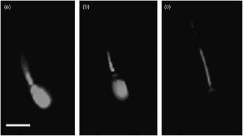
Chlortetracycline (CTC) staining was used to determine the capacitation status of the sperm after 0 (after 10 min), 3 and 6 h of incubation in TBM, TCM or TCMH medium, respectively, as described by Garcia-Herreros et al. (Reference Garcia Herreros, Aparicio, Nunez, Garcia-Marin, Gil and Pena Vega2005), adapted to bovine species. After being washed and incubated in a plastic test tube, 784 µl of spermatozoal suspension containing 30×106 sperm/ml were stained using 16 µl of ethidium homodimer-1 staining solution (11.67 mM). Subsequently, the suspension was centrifuged at 1400×g for 5 min through 4 ml of 3% polyvinyl pyrrolidone in phosphate bufffered saline (PBS). The supernatant was removed by aspiration and the pellet was resuspended in 45 µl of WS–PVA solution (140 mM NaCl, 20 mM HEPES and 0.1% polyvinyl alcohol) and mixed thoroughly with 45 µl of CTC staining solution (750 mM CTC, 5 mM dl-cysteine, 130 mM NaCl and 20 mM Tris–HCl at pH 7.8 prepared immediately before use). The next step included the addition of 8 µl of 12.5% (w/v) paraformaldehyde in 0.5 M Tris–HCl (pH 7.4). Finally, 5 µl of the fixed spermatozoal suspension was placed on a glass slide and mixed well with an equal amount of antifade solution (1,4-diazobicycol[2.2.2]octane, DABCO dissolved in glycerol : PBS [9 : 1]). The droplet was then covered with a 22×22 mm coverslip and the slide firmly but gently pressed under two folds of a tissue paper to absorb any excess fluid. Colourless nail varnish was used to seal the edges. Slides were prepared in duplicate and were stored in the dark at 4°C for further analysis (always within 24 h). Evaluations were made using an epifluorescence microscope (Nikon Eclipse 80i, Tokyo, Japan). Sperm viability was confirmed using an excitation wavelength of 530 to 560 nm and an emission wavelength of 580 nm. The CTC staining patterns were evaluated under blue–violet illumination (excitation 330 to 380 nm, emission 420 nm). Only intact sperm (ethidium homodimer-1 negative, live sperm) were assessed for CTC staining patterns. Two-hundred viable sperm were classified into three patterns as follows (Garcia Herreros et al., Reference Garcia Herreros, Aparicio, Nunez, Garcia-Marin, Gil and Pena Vega2005): F pattern (intact): fluorescence detected over the entire region of the sperm head; B pattern (capacitated): fluorescence detected in the sperm head, except in the postacrosomal region; and, finally, AR pattern (acrosome-reacted): weak fluorescence observed over the sperm head plus a bright band in the equatorial segment (Figure 1).
Different patterns of chlortetracycline (CTC) staining during capacitation process in bovine spermatozoa. From left to right: (a) ‘F’ pattern corresponds to non-capacitated and acrosome-intact spermatozoa; (b) ‘B’ pattern corresponds to capacitated and acrosome-intact spermatozoa and finally (c) ‘AR’ pattern corresponds to acrosome-reacted spermatozoa. Scale bar indicates 7 μm.

Sperm motility analysis
Aliquots of sperm suspension were withdrawn to analyse subjective motility after 0 (10 min), 3 and 6 h of incubation in TBM, TCM or TCMH medium, respectively. Glass slides were pre-warmed to 38°C, and 7 µl of the sperm suspensions were placed and covered with 22×22 coverslips. Samples were diluted with the corresponding incubation medium to give a final concentration of about 50×106 sperm/ml. Analyses were then immediately carried out with a phase–contrast Nikon Eclipse E200 microscope (Nikon).
Computerized sperm morphometric analysis
Slides were prepared after 0 (10 min), 3 and 6 h of incubation in TBM, TCM or TCMH medium, respectively, by placing the sperm sample on the clear end of a frosted microscope slide to give a final concentration of about 100×106 sperm/ml and dragging the drop across the slide to create a thin feathered smear (two smears per ejaculate). Duplicated sperm smears from each sample were stained as described by Marti et al. (2012). Finally, samples were air-dried, mounted and permanently sealed with Eukitt® mounting medium (Fluka BioChemika, Buchs SG, Switzerland; 22×50 coverslips). The prepared slides were used for computerized morphometric analysis using a commercially available system (Motic Corporation Ltd, Hong Kong, China), equipped with a Nikon Eclipse E200 (Nikon) microscope with a 100× oil immersion bright-field objective lens. The video signal was acquired by a MotiCam 2000 digital camera (CMOS ½″; Motic Corporation Ltd), mounted over the microscope and connected to a Pentium P8400 4GB processor, as described (Marti et al., Reference Marti, Aparicio and Garcia-Herreros2011a). The configuration of the computer system included the interface Motic Images Plus 2.0 ml (Motic China Group Ltd) imaging analysis software. Digitized images were made up of 1 920 000 pixels (picture elements) and 256 grey levels. At least 500 sperm per sample were randomly captured in two slides per sample in the manual acquisition mode of the program. Data were compiled and stored for further analysis. Sperm heads were displayed on the monitor at equivalent brightness, and all cells that did not overlap with debris or other cells were considered for analysis. The search, capture and morphometric analysis in all slides were carried out by the same person. Each sperm head was measured for four primary dimensional parameters (area (A, µm2), perimeter (P, µm), length (L, µm) and width (W, µm)) and three head shape-derived parameters (Ellipticity represented by (L/W), Elongation represented by (L−W)/(L+W), and Rugosity represented by (4лA/P2)). These morphometric descriptors were chosen to provide maximal statistical information with minimal number of parameters. Measurements of each sample were saved in an Excel© (Microsoft Corporation, Redmond, WA, USA) compatible database by the software for further analysis.
Statistical analyses
All analyses were performed using the statistical package SPSS version 15.0 for Windows software (SPSS Inc., Chicago, IL, USA). Data matrix from all the sperm analysed by the computer-assisted analysis represented more than 8300 observations, each one defined by the seven morphometric descriptors specified above. The mean and standard error of the mean were calculated for descriptive statistics. Kolgomorov, Smirnov and Levene tests were used to check the normality and homogeneity of data variance distribution, respectively. When the data did not adjust to a normal Gaussian distribution, non-parametric tests were used and a log transformation was performed before running a multivariate ANOVA to study the effect of incubation media or incubation period on each morphometric dimension and shape values. Differences between incubation time and capacitation status in each incubation medium were evaluated by the Kruskall–Wallis test. Descriptive statistics were used to establish the medium, minimum and maximum values and the percentiles 10% to 90% for every morphometric variable. Two processes of sperm classification were performed. The first one consisted of the definition of head size to define small, normal and large sperm. The threshold for each class was established on the basis of the area values, considering the 25th percentile as small and the 75th percentile as large sperm. The second one was the determination of sperm head shape as normal, elliptic, rough and elongated, using the percentiles 25th and 75th to define sperm with normal values of each shape parameter. After defining these classes, a number of sperm heads belonging to each group were analysed. The same procedure was used, according to the amount of intact (viability %) or motile (motility %) to classify the sperm measured. To characterize the differences between non-capacitatied/capacitated, non-capacitated/acrosome-reacted or capacitated/acrosome-reacted bovine sperm, allowing for all the morphometric variables, discriminant analysis was carried out. To study the frequency of distribution of sperm within each size and shape category, a χ 2-test was used. The Spearman non-parametric test was used to study the correlations among sperm head morphometric parameters and the capacitation status of bull sperm as assessed by CTC staining. Significance was set at P<0.05.
Results
Bovine sperm quality parameters under capacitating and non-capacitating incubation conditions
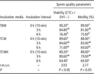
Sperm quality parameters under capacitating and non-capacitating incubation conditions and the different incubation intervals are represented in Table 1. All sperm samples analysed had lower percentage values for all variables assessed (membrane integrity and motility parameters) under capacitating and non-capacitating incubation conditions from 0- (after 10 min) to 6-h incubation periods. After 3 h of incubation, there were no statistical differences regarding sperm motility or membrane integrity percentages comparing TBM, TCM and TCMH incubation conditions (P>0.05; Table 1). However, after 6 h of incubation, a decreasing tendency was observed in all samples studied regarding sperm motility and viability parameters (P<0.05).
Effect of incubation period on sperm quality parameters under capacitating and non-capacitating incubation conditions in bovine spermatozoa (0 to 6 h)

TBM=Tyrode’s basal medium; TCM=Tyrode’s complete medium heparin-free (TCM); TCMH=Tyrode’s complete medium with heparin; CTC=chlortetracycline staining; and EH1=ethidium homodimer-1.
All sperm sample pools were obtained from five clinically healthy bulls that have been used for semen collection in the insemination centre. Values are means. r.m.s.e.: root mean square error (residual).
P represents the significance of the model. Different superscripts (a to c) in a column indicate significant differences among incubation period and media (P<0.05). Results are derived from five identical experiments.
Sperm capacitation status under capacitating and non-capacitating incubation conditions in bovine spermatozoa
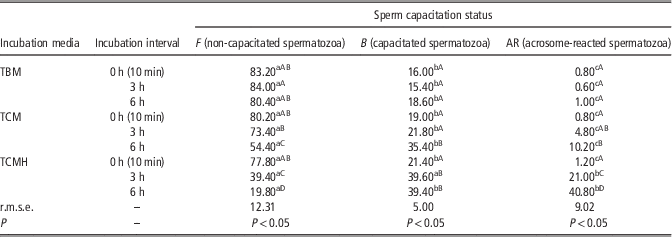
Sperm capacitation patterns (percentages) under capacitating and non-capacitating incubation conditions are represented in Table 2. Sperm incubated under TCMH conditions showed a significant increase in the percentage of capacitated (B pattern) and AR pattern sperm after the 3-h incubation period (P<0.01). This tendency increased drastically after the 6-h incubation period under the same TCMH conditions (P<0.01). Sperm incubated under TBM conditions showed no significant differences in the percentage of B or AR patterns over time (P>0.05).
Effect of incubation period on sperm capacitation status under capacitating and non-capacitating incubation conditions in bovine spermatozoa (0 to 6 h).

TBM=Tyrode’s basal medium; TCM=Tyrode’s complete medium heparin-free; and TCMH=Tyrode’s complete medium with heparin.
Pooled sperm samples from five bulls were incubated under TBM, TCM and TCMH up to 6 h at 38°C. Values are means. r.m.s.e.: root mean square error (residual).
P represents the significance of the model. Within column comparisons (A to D) refer to F (uncapacitated), B (capacitated) or AR (acrosome-reacted) patterns as determined by CTC staining (P<0.05). Within row comparisons (a to c) refer to incubation media (TBM, TCM and TCMH) and incubation period (0, 3 and 6 h) (P<0.01).
Sperm head morphometric analysis under capacitating and non-capacitating incubation conditions in bovine spermatozoa
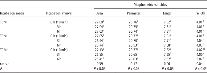
Table 3 shows the effect of time interval and incubation medium on area, perimeter, length and width descriptors. Interestingly, both time interval and incubation media resulted in significant changes in sperm dimensional parameters depending on the interval and media used (P<0.01). After 3 h under TCMH conditions, there were significant differences related to all sperm dimensional characteristics compared with sperm incubated under TBM conditions or under TCM conditions (P<0.01). After 3 h, sperm incubated under TBM or TCM conditions showed similar and no significant differences regarding morphometric characteristics (P>0.05). Under TBM incubation conditions, there were no time-dependant changes in all ASMA sperm morphometric descriptors. Area, perimeter, width and length were maintained over time (P>0.05). Even under TCM incubation conditions, sperm dimensional characteristics were maintained after 3 h of incubation (P>0.05). Only under TCMH incubation conditions (Supplementary Figure S1), a significant reduction in area, perimeter, width and length was observed after 3 and 6 h of incubation (P<0.01).
Effect incubation period on sperm head morphometric dimensional parameters under capacitating and non-capacitating incubation conditions in bovine spermatozoa (0 to 6 h)

TBM=Tyrode’s basal medium; TCM=Tyrode’s complete medium heparin-free; and TCMH=Tyrode’s complete medium with heparin.
The results were obtained from 25 ejaculates (five ejaculates from each bull). Values are means. r.m.s.e.: root mean square error (residual).
P represents the significance of the model. The total number of spermatozoa analysed was 2653, 2881 and 2798 (TBM, TCM and TCMH, respectively). Different superscripts (a to e) in a column indicate significant differences among incubation period and media (P<0.01).
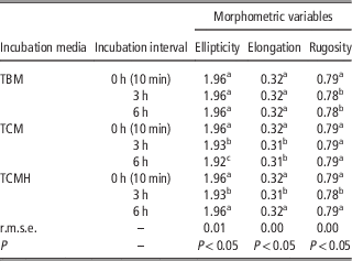
Table 4 and Supplementary Figure S2 show the effect of time interval and incubation medium on ellipticity, elongation and rugosity sperm shape parameters. No significant differences were observed in sperm incubated under TBM conditions over time with respect to all shape descriptors (P>0.05). However, after 3 and 6 h, there were significant differences related to all the shape parameters when samples were incubated under TCM and TCMH compared with TBM conditions, showing a significant decrease of ellipticity, elongation and rugosity (P<0.01).
Effect incubation period on sperm head morphometric shape parameters under capacitating and non-capacitating incubation conditions in bovine spermatozoa (0 to 6 h)

TBM=Tyrode’s basal medium; TCM=Tyrode’s complete medium heparin-free; and TCMH=Tyrode’s complete medium with heparin.
The results were obtained from 25 ejaculates (five ejaculates from each bull). Values are means. r.m.s.e.: root mean square error (residual).
P represents the significance of the model. The total number of spermatozoa analysed was 2653, 2881 and 2798 (TBM, TCM and TCMH, respectively). Different superscripts (a to c) in a column indicate significant differences among incubation period and media (P<0.01).
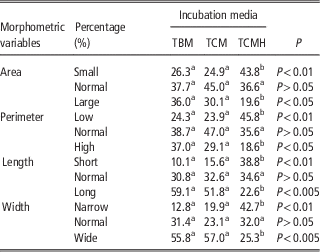
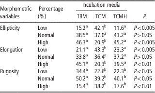
Tables 5 and 6 show the frequency of distribution (%) of sperm dimensional and shape parameters, respectively. Significant differences were observed in the distribution of sperm incubated under TBM, TCM and TCMH incubation conditions over time with respect to all dimensional and shape descriptors (P<0.01).
Frequency of distribution (percentage) of sperm dimensional parameters showing different characteristics of area, perimeter, length and width under capacitating and non-capacitating incubation conditions in bovine spermatozoa (6-h incubation period)

TBM=Tyrode’s basal medium; TCM=Tyrode’s complete medium heparin-free; TCMH=Tyrode’s complete medium with heparin.
χ 2-test.
P represents the significance of the model. Different superscripts (a to b) in a row indicate significant differences among incubation media (P<0.05).
Frequency of distribution (percentage) of sperm shape parameters showing low, normal and high degree of ellipticity, elongation and rugosity under capacitating and non-capacitating incubation conditions in bovine spermatozoa (6-h incubation period)

TBM=Tyrode’s basal medium; TCM=Tyrode’s complete medium heparin-free; and TCMH=Tyrode’s complete medium with heparin (TCMH).
χ 2-test.
P represents the significance of the model. Different superscripts (a to b) in a row indicate significant differences among incubation media (P<0.05).
Correlation level of different capacitation status and the sperm morphometric dimension and shape parameters in bovine spermatozoa
Relationships among sperm capacitation status and different morphometric variables were studied to determine the degree of correlation among them after 6-h incubation under different capacitating and non-capacitating conditions. The statistical correlation study revealed strong positive or negative correlations depending on the sperm morphometric descriptor analysed (mainly area parameter) and the percentages of sperm showing non-capacitated (F pattern), capacitated (B pattern) and AR pattern status (P<0.01). Under TCM incubation conditions, significant correlations were found between area and the percentage of sperm showing F pattern (r=0.792; P<0.01), between area and the percentage of B pattern (r=0.681; P<0.01) and between area and AR pattern (r=−0.804; P<0.01). Moreover, under TCMH incubation conditions, a significant correlation was observed between the percentage of sperm showing F pattern and the parameter area (r=0.855; P<0.01). However, significant correlations were observed between area and the percentage of sperm showing B pattern (r=0.662; P<0.01) and between the parameter area and the percentage of sperm showing AR pattern (r=−0.875; P<0.01).
Discussion
To the best of the authors’ knowledge, this is the first description of bovine sperm head morphometrics as an objective diagnostic tool for detecting volumetric changes under capacitating and non-capacitating incubation conditions. Clear relationships were found among selected morphometric dimensional and shape parameters and the percentage of acrosome-reacted sperm. However, this relationship was not found between sperm morphometric parameters and the percentage of capacitated sperm. The changes observed with respect to acrosome-reacted sperm were a decrease in all sperm head dimensional parameters (area, perimeter, width and length) and in all sperm head shape parameters (ellipticity, elongation and rugosity) under TCMH incubation conditions. Area dimensional parameter showed a strong negative statistically significant correlation with the percentage of acrosome-reacted sperm, as assessed by the CTC assay. Acrosome reaction in bovine sperm has been related to a significant glycoprotein re-distributions on the sperm head (Wolfe et al., Reference Wolfe, James, Mackie, Ladha and Jones1998). The staining pattern of the AR sperm assessed by the CTC assay showed that the morphometric patterns were distinct from those morphometric characteristics of uncapacitated or capacitated sperm where there were no changes in sperm head morphometry under TCMH incubation conditions. This may suggest that the protein re-distribution together with the possible loss of acrosomal membrane structure during late events of AR under capacitating conditions, required for sperm to be fully competent to interact with the oocyte and complete fertilization, are totally different compared with capacitated sperm (Gabriele et al., Reference Gabriele, D'Andrea, Cordeschi, Properzi, Giammatteo, De Stefano, Romano, Francavilla and Francavilla1998; Bucci et al., 2012). It is possible that protein distribution and the loss of acrosomal membrane structure after 3-h incubation that showed a substantial, but not significant, increase of AR staining pattern was not enough to detect dimensional or shape changes during the capacitation process, in contrast with the dimensional and shape characteristics that were detected after 6-h incubation. This suggests that some of the changes in staining patterns, particularly those in which a region of staining is lost in the AR population, could be the result of the removal of surface-adhered material during late-capacitation, as suggested by previous researchers (Dravland and Joshi, Reference Dravland and Joshi1981). Moreover, we have to take into account that in many cell types, changes in cholesterol content of the plasma membrane have been correlated with changes in protein–protein interactions among plasma membrane-associated proteins (Tannert et al., Reference Tannert, Töpfer-Petersen, Herrmann, Müller and Müller2007).
Several studies have described an active participation of the sperm plasma membrane in the process of capacitation, mainly through loss of cholesterol (Ehrenwald et al., Reference Ehrenwald, Parks and Foote1988; Cross Reference Cross1998). The cholesterol efflux produces an increase in membrane fluidity and a decrease in the cholesterol/phospholipid ratio, which directly or indirectly impacts protein function, such as that of surface ion channels and enzymes (Davis, Reference Davis1981; Gadella et al., Reference Gadella, Tsai, Boerke and Brewis2008). In the present study, under capacitating conditions, the cholesterol/phospholipid re-distributions could undergo changes quite rapidily due to the observed significant shift in AR staining pattern after just 6-h incubation with the corresponding changes in sperm head morphometry. This fact together with the loss of acrosomal membrane structure during late events of AR could be responsible for the increase in membrane permeability and the final loss of cell volume in the bovine sperm due to changes in ion fluxes across the membrane, mainly HCO3−, Na, K and Ca2+, which have been implicated during sperm capacitation (Baldi et al., Reference Baldi, Luconi, Bonaccorsi, Krausz and Forti1996; Bucci et al., 2012). In addition, the re-distribution of protein components in the sperm head plasma membrane shown in several species is consistent with the hypothesis of the biophysical change that occurs during capacitation process, as shown in the present study (Gadella and Van Gestel, Reference Gadella and Van Gestel2004). This was particularly evident when bovine sperm were challenged with 6-h incubation at 38°C under capacitating conditions with heparin. It is interesting to note that the uncapacitated ‘F’ and capacitated ‘B’ sperm populations were homogeneous with respect to the distribution of CTC-binding sites. However, such homogeneity among freshly isolated F and B sperm populations was not found in the AR population in the present study.
The functional significance of this heterogeneity in the distribution of CTC-binding sites in AR sperm population is not clear, but it may indicate that not all capacitation and acrosome reaction-associated changes are strictly dependent on the inclusion of HCO3−, BSA and Ca2+ in the capacitating medium, but at least in bovine sperm it depends on heparin binding as well (Parrish et al., Reference Parrish, Susko-Parrish, Winer and First1988). Thus, heparin in bovine sperm is crucial for the final biophysical mechanism of fusion between the plasma membrane and the outer acrosomal membrane, and as a consequence the loss of cell volume at the final stage of acrosome reaction (Vadnais et al., Reference Vadnais, Galantino-Homer and Althouse2007). In bovine-capacitated sperm, there were no significant changes in cell volume compared with non-capacitated sperm even after exposure to BSA, which is commonly used to mediate sterol efflux (Ehrenwald et al., Reference Ehrenwald, Parks and Foote1988). However, the presence of heparin did induce a loss of cell volume and an increase in the AR pattern in bovine sperm, suggesting that this association exerts effects on bovine sperm membranes (Therien et al., Reference Thérien, Moreau and Manjunath1999). The reduction of cell volume emerged only when heparin, Ca2+ and HCO3− were present in the medium, and revealed that bovine sperm require these stimuli to show the full extent of membrane changes and as a result undergo acrosome reaction.
This finding provides a refinement to existing studies of capacitation and acrosome reaction. Yet, in addition to these points of basic science, our findings also suggest a clinical application for sperm morphometry as a biophysical marker for detecting acrosome reaction-associated membrane changes in bovine sperm. Changes in dimensional and shape characteristics in response to specific stimuli for the acrosome reaction could provide a diagnostic tool for predicting a male’s reproductive status based on the proportion of sperm that are capable of responding to such stimuli. This information could be used in AI centres to make broad classifications regarding a male’s fertility or could be used to help guide specialists when choosing between techniques such as intracytoplasmic sperm injection or in vitro fertilization (Garcia-Herreros et al., Reference Garcia-Herreros, Carter, Villagómez, Macaulay, Rath, King and Lonergan2010). Furthermore, because this potential assay is based on membrane responses, it might also be useful when evaluating or comparing media or conditions used to handle sperm in vitro or in sperm cryopreservation processes.
Conclusions
In conclusion, in vitro capacitating incubation conditions affected dimensional but not shape characteristics in bovine spermatozoa. In addition, acrosome reaction but not capacitation was related to significant decreases in the sperm head morphometrical parameters under in vitro capacitating incubation conditions in bovine spermatozoa. Finally, the substantial information derived from this study provided new and relevant knowledge, which might have a practical use for future studies using sperm head morphometry as a potential biophysical marker for detecting volumetric changes during capacitation process in bovine spermatozoa.
Acknowledgements
The authors thank Agustín Sánchez-Domínguez for excellent technical assistance. Dr García-Herreros was funded by Spanish Government (MAE, 2010-IA) and Ecuadorian Government(SENESCYT-Prometeo Project-2014/2015).
Conflicts of Interest
None of the authors of this paper has a financial or personal relationship with other people or organizations that could inappropriately influence or bias the content of the paper.
Supplementary material
To view supplementary materials for this article, please visit http://dx.doi.org/10.1017/S1751731115000129