Introduction
Mesoporous materials have attracted the attention of many researchers because of their many properties, including a high degree of pore order on the micrometer scale, narrow pore-size distribution,[Reference Adhikari, Mishra and Nayak1] high surface area,[Reference Moshe and Markovich2] excellent thermal stability,[Reference Corma3] and hydrothermal stability.[Reference Kortesuo, Ahola and Kangas4] Therefore, they have been applied in many fields, such as chemistry, optoelectronics, electromagnetism, materials science, and environmental science.[Reference Kumara, Stephenson and Murray5–Reference Chowdhuri, Singh and Ghosh7] Mesoporous materials have also been used as nanocarriers in the biomedical field owing to their super-porous structure and adsorption properties.[Reference Han, Wang and Bai8–Reference Perez-Esteve, Ruiz-Rico and De-La Torre10] It is of technical interest to produce a shell-like capsule in which release is controlled by permeation through the shell material, but the eventual collapse of the capsule makes it difficult to ensure the mechanical stability of the coating as a whole.[Reference Kumara, Stephenson and Murray5] Environmentally friendly carbon and silica materials appear to be ideal mesoporous sources because of their low-cost and unique mechanical, thermal, and chemical properties.[Reference Minati, Speranza and Bernagozzi11–Reference Li, Li and Pan13] For example, Valdés-Solís et al. fabricated hollow mesoporous carbon capsules in nanosized catalysts.[Reference Valdés, Valle and Sevilla14] Adhikari et al. synthesized mesoporous silica and hollow mesoporous silica spheres applied for chemotherapeutic drug delivery.[Reference Adhikari, Mishra and Nayak1] However, the preparation process required calcination,[Reference Chang, Sha, Guo, Jiao, Wang and Yang15] harsh conditions, and high energy consumption.[Reference Amanpreet, Manchanda and Kruk16] Chen et al. synthesized mesoporous polydopamine (MPDA) nanoparticles with the triblock copolymer pluronic F127 (EO106PO70EO106) and trimethylbenzene (TMB) as the organic templates.[Reference Chen, Xing and Wang17] Polydopamine (PDA) has better biologic safety and biodegradability than carbon and silica materials as dopamine itself is a biomimetic material. Furthermore, because the reactive groups include both amino and catechol groups of PDA, it can be used as an ideal secondary reaction platform, which is also very important for its application in drug carriers.
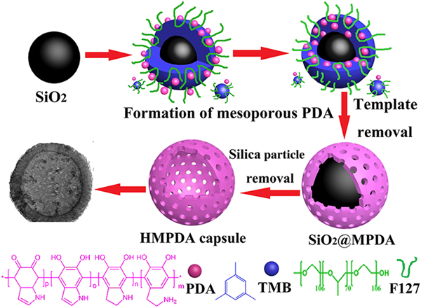
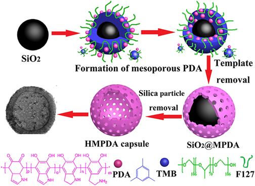
Herein, we report the synthesis of a novel type of core–shell-structure SiO2@MPDA submicrometer particle by an organic–organic self-assembly process that is driven by π–π interactions of TMB and PDA. To remove the core of silicon particles, 2% HF solution was used to remove the silica templates; hollow mesoporous polydopamine (HMPDA) microcapsules were then obtained. The synthesis process of the HMPDA microcapsules is illustrated in Scheme 1. The obtained HMPDA microcapsules have excellent adsorption properties for methylene blue (MB) and doxorubicin (DOX), an anticancer drug.
Synthetic preparation routes of HMPDA microcapsules.

Material and methods
Materials
MB (82%) was purchased from Sigma-Aldrich. Other chemicals were supplied by Aladdin Industrial, Inc. (Shanghai, China).
Preparation of SiO2@MPDA core–shell composites
SiO2 particles measuring approximately 300 nm in diameter were prepared by the Stober–Fink–Bohn method.[Reference Tao, Ma and Chen18,Reference Jiang, Bertone and Hwang19] The SiO2@MPDA particles were synthesized by a one-pot process. First, 0.36 g of F127 and 625 µL of 13,5-TMB were added to a mixture of 65 mL of H2O and 60 mL of ethanol. After 30 min of stirring, a clarified solution was obtained. Then, 90 mg of Tris and 60 mg of dopamine were dissolved in 4 mL of H2O, and the mixture was then added to the above solution. Finally, 20 mg of prepared SiO2 nanoparticles were added to the solution and mechanically stirred for 24 h at room temperature. A dark brown suspension was obtained after stirring. Then, 50 mL of acetone was added to the suspension and the solid powders were separated by centrifugation (11,000 rpm, 10 min). To remove F127 and TMB, the separated powders were dispersed in 80 mL of ethanol and 40 mL of acetone with sonication for 30 min, and then separated by centrifugation (11,000 rpm, 10 min) (this cycle was repeated three times). SiO2@MPDA core–shell composites were thus obtained.
Preparation of HMPDA microcapsules
A 2% HF solution was used to dissolve and remove the silicon particles, and HMPDA microcapsules were obtained after centrifugation and washing with distilled water.
Adsorption experiments for MB
To compare the absorption of SiO2@MPDA particles and HMPDA microcapsules with MB, the same mass of samples (2 mg) was added into the same concentration of MB solution (0.05 mg/mL). The adsorption capacities could be obtained using ultraviolet–visible-light (UV-Vis) measurements by removing the supernatant at certain time intervals.
In vitro drug release
Preparation of simulated body fluid (SBF) solution proceeded as follows: 0.8 g of NaCl, 0.4 g of KCl, 0.14 g of CaCl2, and 1.0 g of Dextran 40,000 were dissolved in 1000 mL of H2O, adjusted to pH = 7.4 by dilute HCl.
A total of 5 mg of HMPDA microcapsules were dispersed in 20 mL of DOX water solution (0.05 mg/mL) and stirred for 24 h in dark conditions. Then, 5 mg of DOX-loaded HMPDA composites were dispersed in 2 mL of SBF solution in dialysis bags (14,000 kDa) immersed in 100 mL of SBF at 37 °C and stirred for 72 h. A total of 3 mL of SBF solution was removed by centrifugation to detect the released amount of DOX at certain time intervals. Meanwhile, 3 mL of fresh SBF was added to the original system.
Results and discussion
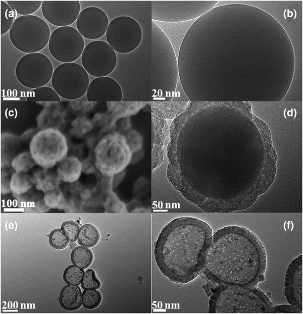
Transmission-electron-microscopy images of SiO2 nanoparticles are shown in Figs. 1(a) and 1(b). The nanoparticles had a regular spherical shape and were approximately 300 nm in diameter. Figure 1(c) shows the field-emission scanning-electron-microscopy (FESEM) image of the SiO2@MPDA composites in which a porous structure can be clearly observed on the surface of the particles; the pore diameter was approximately 10 nm. The high-resolution-transmission-electron-microscopy images in Fig. 1(d) show the core–shell-structure SiO2@MPDA microparticles. The core–shell structures can be clearly observed, and the diameter of the core was approximately 300 nm, which was consistent with the size of silica nanoparticles. The shell is a porous film with a thickness of approximately 40–50 nm. The mechanism for generating mesoporous PDA structures is the effect of π–π stacking interactions between PDA structures and the π-electron-rich TMB molecules. Ethanol and Tris were employed as the co-solvent and catalyst, respectively, for the polymerization of PDA. The primary PDA nanoparticles in the MPDA synthesis have a high affinity toward π-electron-rich TMB for emulsion-templated self-assembly.[Reference Chen, Xing and Wang17] PDA with a mesoporous structure can be obtained by extracting and dissolving the template. The TEM images of HMPDA microcapsules are shown in Figs. 1(e) and 1(f), in which the hollow and mesoporous structures on the wall can be clearly observed. The diameter of the hollow microcapsules was approximately 300 nm, which was less than that of the SiO2@MPDA microparticles, because the microcapsules will shrink after drying.
TEM images (a,b) of SiO2 particles; FESEM images of MPDA particles (c); TEM images of SiO2@MPDA (d); and MPDA microcapsules (e,f).

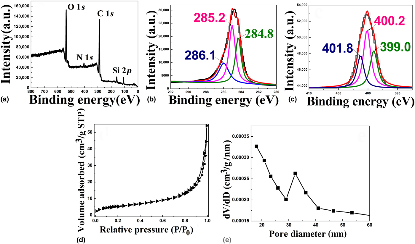
X-ray photoelectron spectroscopy (XPS) was used to study the surface chemical composition of the SiO2@MPDA core–shell composites, as shown in Fig. 2. Figure 2(a) shows the XPS survey spectrum with a stronger carbon (C 1s), oxygen (O 1s), as well as the weaker nitrogen (N 1s) peak, which come from PDA. The weak peak of Si 2p comes from the “core” SiO2 nanoparticles. The C 1s spectra of the MPDA film in Fig. 2(b) was deconvoluted using three Gaussian components at approximately 284.8, 285.2, and 286.1 eV, respectively. The peak near 284.8 eV conforms to benzene carbon atoms. The peak around 285.2 eV corresponds to C-O carbon atoms. The peak near 286.1 eV is assigned to CN carbon atoms. These carbon atoms come from the dopamine molecules. The N 1s spectra in Fig. 2(e) were fitted with three components: C = N-R (399.0 eV), R1-NH-R2 (400.2 eV), and R-NH2 (401.8 eV).[Reference Dalsin, Hu, Lee and Messersmith20–Reference Xu, Liu and Wang23] Therefore, the existence of MPDA molecules on the surface of the particles has been convincingly proved by the XPS spectrum of SiO2@MPDA nanoparticles.
XPS survey (a) including high-resolution C 1s (b) and N 1s (c) spectra of SiO2@MPDA composites; nitrogen adsorption isotherm (d) and pore-size distribution (e) of SiO2@MPDA composites.

The mesoporous structure of SiO2@MPDA nanoparticles was investigated by the nitrogen adsorption–desorption method in Figs. 2(d) and 2(e). Upon reference to the IUPAC nomenclature, the plot of SiO2@MPDA nanoparticles is found to belong to a type-IV isotherm, which has a typical mesoporous structure.[Reference Hu, Sun and Huang24] The specific surface area and total pore volumes of SiO2@MPDA composites were 22.57 m2/g and 0.084 cm3/g, respectively. Owing to the folding structure of HMPDA microcapsules after drying, the adsorption properties of HMPDA microcapsules cannot be determined by the nitrogen adsorption–desorption method. The adsorption of HMPDA microcapsules was studied by comparing the amount of dye adsorption of HMPDA microcapsules and SiO2@MPDA composites.
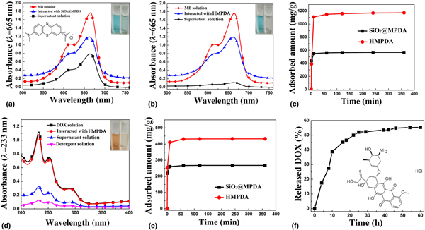
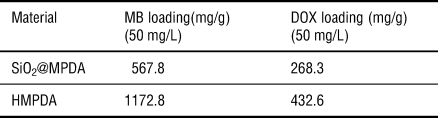
The adsorption behaviors of the HMPDA microcapsules and SiO2@MPDA core–shell nanoparticles toward MB at different time intervals were researched. Figures 3(a) and 3(b) show the UV-Vis absorption spectra of the MB in a mixture containing SiO2@MPDA core–shell nanoparticles and HMPDA microcapsules. The adsorption capacities of SiO2@MPDA particles and HMPDA microcapsules toward MB were determined to be 567.8 and 1172.8 mg/mg [as shown in Fig. 3(c)], respectively. Therefore, by comparison, it can be found that the adsorption capacity of HMPDA microcapsules to dyes was much larger than that of SiO2@MPDA particles, so the pore volume of the former should also be much larger than that of the latter. The adsorption properties of HMPDA on drugs were also studied. DOX, an anti-cancer drug,[Reference Liu, Guo and Odusote25] was used as a model drug to load into HMPDA capsules. Figure 3(d) shows that HMPDA microcapsules can also adsorb DOX. The average loading capacity of HMPDA microcapsules toward DOX could reach 432.6 mg/g; meanwhile, the average loading capacity of SiO2@MPDA particles toward DOX was approximately 268.3 mg/g, as shown in Fig. 3(e).
UV-Vis absorption spectra of MB solution in mixture containing SiO2@MPDA nanoparticles (a) and HMPDA microcapsules (b); adsorption capacities of SiO2@MPDA particles and HMPDA microcapsules toward MB(c); UV-vis absorption spectra of supernatant DOX solution at different time intervals (d); adsorption capacities of SiO2@MPDA particles and HMPDA microcapsules toward DOX(e); release profiles of DOX from HMPDA microcapsules in PBS (pH = 7.4) (f).

The DOX-loaded HMPDA microcapsules were then dispersed in Na2HPO4-NaH2PO4-buffered saline solution (pH 7.4) to determine the drug's sustained-release properties. Figure 3(f) shows the release curve of the maximum absorbance of DOX at different times. The fast release of DOX was, at first, 25 min, and the release amount was approximately 50.5% due to the surface mesoporous structure of PDA. The slower release appeared later, and the release maximum reached 55.7%. Therefore, DOX could be released slowly in HMPDA microcapsules.
The adsorption capacity q e can be calculated by Eq. (1), while the desorption efficiency R (%) can be calculated by Eq. (2):
 $$q_e = V(\hbox{Cinitial drug-Cdrug in supernatant})/m \, \hbox{drug carrier},$$
 $$R = (\hbox{Cinitial drug-Cdrug in supernatant})/ \hbox{Cinitial drug} \times 100 \%. $$
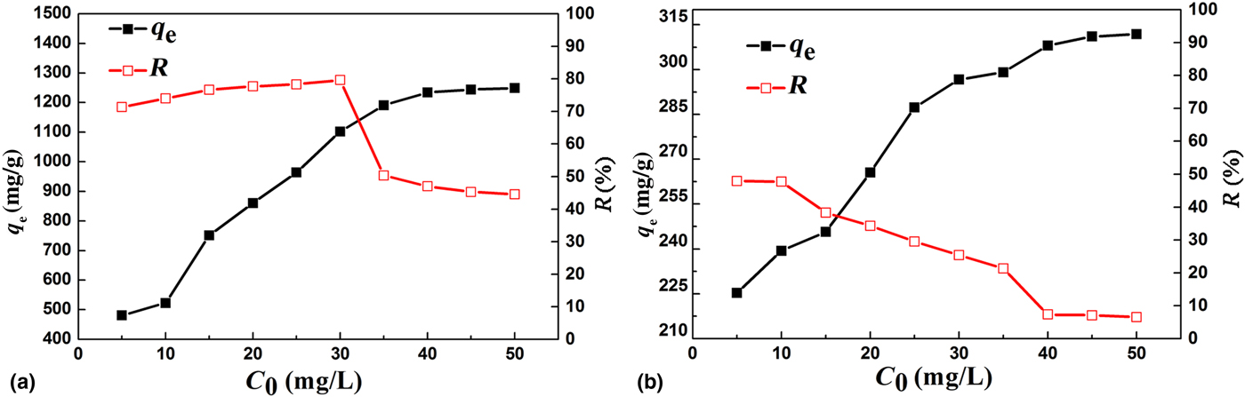
The effects of initial concentration C 0 on the q e and R of MB and DOX are shown in Fig. 4, from which it can be seen that both the adsorption amount of MB and DOX almost increased linearly with increasing C 0 at the first stage, and then increased gradually until remaining constant. The adsorption amount nearly reached saturation at C 0 = 35 mg/L for MB and 40 mg/L for DOX. The driving force for adsorption became larger with increasing C 0, which resulted in the increase of R, and when the active sites were nearly exhausted and finally reached the saturation state, R decreased with further increase of C 0.
Effect of initial concentration on q e and R of MB (a) and DOX (b) on SiO2@MPDA particles.

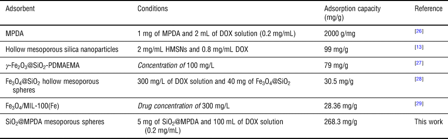
In addition, compared with the reported drug adsorption carriers, HMPDA microcapsules have a larger adsorption capacity for drugs. A comparison of the results in the present study with the reported systems[Reference Li, Li and Pan13,Reference Xing, Zhang and Chen26–Reference Kawashita, Tanaka and Ueno29] for DOX removal is summarized in Table I. As can be seen from the table, the SiO2@MPDA mesoporous spheres prepared in this paper have a relatively large adsorption capacity of DOX. Meanwhile, compared with SiO2@MPDA spheres, HMPDA microcapsules have a larger loading capacity on MB and DOX, as shown in Table II, indicating that HMPDA microcapsules can be used as an efficient nano-drug carrier.
Comparison of results of the present study for adsorption capacity of DOX with those of other reported systems.

Comparison of adsorption capacity in the same concentration of SiO2@MPDA spheres with HMPDA spheres.

Conclusions
In summary, we developed a simple and effective method to synthesize HMPDA microcapsules. Because of its unique cavity structure and mesoporous wall, HMPDA has a high adsorption capacity to dyes and drug molecules, as well as a good sustained-release effect. In addition, its good biologic safety is also helpful to its application in drug carriers.
Acknowledgments
We thank LetPub (http://www.letpub.com) for its linguistic assistance during the preparation of this manuscript.
Funding
We acknowledge financial support from the Natural Science Fund Project of Gansu Province, China (18JR3RA109) and the Foundation of a Hundred Youth Talents Training Program of Lanzhou Jiaotong University, China.