Purpose
Computed tomography (CT)-guided brachytherapy is the standard treatment planning method adapted for accurate delineation of target volumes.Reference Blechacz and Gores1–Reference Weber, Jarnagin and Klimstra3 Thus, it provides safe dose escalation without harming the adjacent normal structures.Reference Shimada, Kishi and Hata4, Reference Kamphues, Seehofer and Eisele5 CT improves the overall 5-year survival rates by about 3·5%, with the greatest impact on small volume treatments.Reference Ricke, Mohnike and Pech6–Reference Ishii, Furuse and Nagase10 Advanced planning systems provide an option for applicator modelling that helps in reducing catheter reconstruction time. However, they offer a material library only for specific gynaecological applicators.12 Auto reconstruction is possible when there is a difference between CT density of applicators and surrounding tissue.Reference Milickovic, Baltas, Giannouli, Lahanas, Uzunoglu and Zamboglu13 CT density is almost similar for the two metal stents used in the biliary duct system. In other words, both intra-hepatic and extra-hepatic (used for intraluminal brachytherapy (ILBT)) metal stents introduce artefacts in the CT scans. The planner’s expertise plays a major role in reconstructing the correct passage of the ILBT guide tube that recoils and overlaps with the intra-hepatic drain stent. Digitally reconstructed radiograph (DRR) is a useful tool in brachytherapy planning when there is no other image guidance avialable.Reference Milickovic, Baltas, Giannouli, Lahanas and Zamboglou14 In this study, we used DRR to reconstruct the ILBT catheter. We contoured the catheter’s passage in each transverse CT section by verifying with DRR view. Thus, it helps in quick reconstruction, reducing the overall planning time. It leads to accurate catheter reconstruction, whereas any displacement could lead to erratic dose calculation for a high-dose-rate brachytherapy source.
Technique
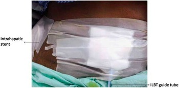
A 64-year-old woman with a known case of Non Hodgkin’s Lymphoma (Stage I, small cell type) was treated with external beam radiotherapy on January 2004. Recently, she presented with complaints of recurrent vomiting, stomach ache and symptomatic obstructive jaundice. CT investigation revealed an ill-defined lesion at the porta hepatis encasing the right hepatic duct and the common hepatic duct as well as revealed a moderate dilatation of left hepatic duct. Positron emission tomography suggested relapse lymphoma. Ultrasound-guided biopsy confirmed hilar cholangiocarcinoma (CCA) as the second malignancy. To remove the block in common biliary duct, an intra-hepatic stent was inserted at the right side. In addition, percutaneous transhepatic biliary drainage (as extra hepatic drain) was performed for the palliation of obstructive jaundice caused by CCA (Figure 1). ILBT is considered to reduce the risk of recurrent stent occlusion of the self-expanding metal stent.
Patient image showing stent insertions.

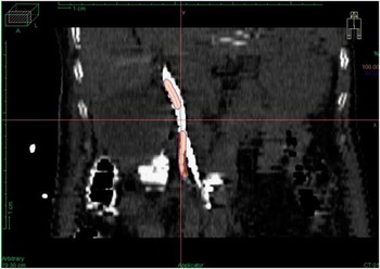
The ILBT dummy wire was inserted into the guide tube (LumenCath; Nucletron, an Elekta company, Stockholm, Sweden), which was 1,500-mm long. CT simulation was performed, and the images were exported to Oncentra MasterPlan (Nucletron, an Elekta company) treatment planning system (TPS). Metal stents causing artefacts in the CT slices produced a distorted view of the overlapped dummy tube with the right hepatic stent at the level of T11. Therefore, in addition to distortion due to metallic stents, the passage of the ILBT tube is more complex compared with oesophageal/endobronchial applications. Thus, catheter tracking became difficult. Figure 2 shows the misplaced catheter with an average displacement of 7·2 mm from the original.
Incorrect reconstruction of catheter.

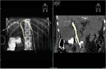
TPS generates an automatic DRR view (Bone window) at the patient’s midplane. The ILBT guide tube was medially placed and the intra-hepatic drain tube was laterally away with respect to the 11th thoracic vertebra (T11) as per the DRR. Therefore, we decided to contour the guide tube passage using Bone window of DRR (Figure 3).
Corrected reconstruction using digitally reconstructed radiograph.

Results and Discussions
Catheter reconstruction becomes easier in the complex bile duct passage with this method. It can be used for quick verification of catheter reconstruction. Two lengths (3 and 3·4 cm) of irradiation separated by a distance of 2 cm were marked, starting at 5 mm from the tip of the guide tube. A dose of 6·5 Gy was prescribed at 2·5 mm from the central axis of the catheter. Total dose prescribed was 32·5 Gy in five fractions (low dose rate equivalent: 53·2 Gy).
ILBT guide tube contour reduces the overall planning time. Planning could be verified easily with this method when computed tomography was used as the only image guidance. Source tracking can be improved with exact placement of the catheter. Although demonstrated for ILBT planning, this method could be used for planning soft tissue sarcoma implants with flexible plastic tubes that could result in overlapped view in adult thigh/knee.
Acknowledgement
I sincerely acknowlege the effort of Dr. InduShekar and Dr. Kranthi Kumar for this HDR procedure.