Gaucher disease is characterised by glucocerebroside accumulation in lysosomes of macrophages due to β-glucosidase enzyme deficiency and notable clinical heterogeneity. It is inherited in an autosomal-recessive manner. The glucocerebrosidase gene-1 that causes the disease is located on chromosome 1q21. Glucocerebroside, the defective gene product, is stored in the lysosomes of the reticuloendothelial system. According to the presence and severity of neurological symptoms, three different clinical forms of Gaucher disease are described: type I, non-neuronopathic (95%); type II, acute neuronopathic; and type III, chronic neuronopathic. Type IIIA is a subgroup characterised by Gaucher disease with myoclonus and dementia, type IIIB by early recognised gaze limitation and aggressive systemic involvement, and type IIIC by cardiovascular calcification.Reference Grabowski, Petsko and Kolodyn 1 In type IIIC, cardiovascular calcification may be accompanied by oculomotor apraxia. In our case, the patient presented with frequent recent-onset syncope, although he showed gaze limitations at an early age. We present this case because of the atypical clinical findings and the rare nature of type IIIC Gaucher disease with severe cardiovascular manifestations during teenage years.
Case report
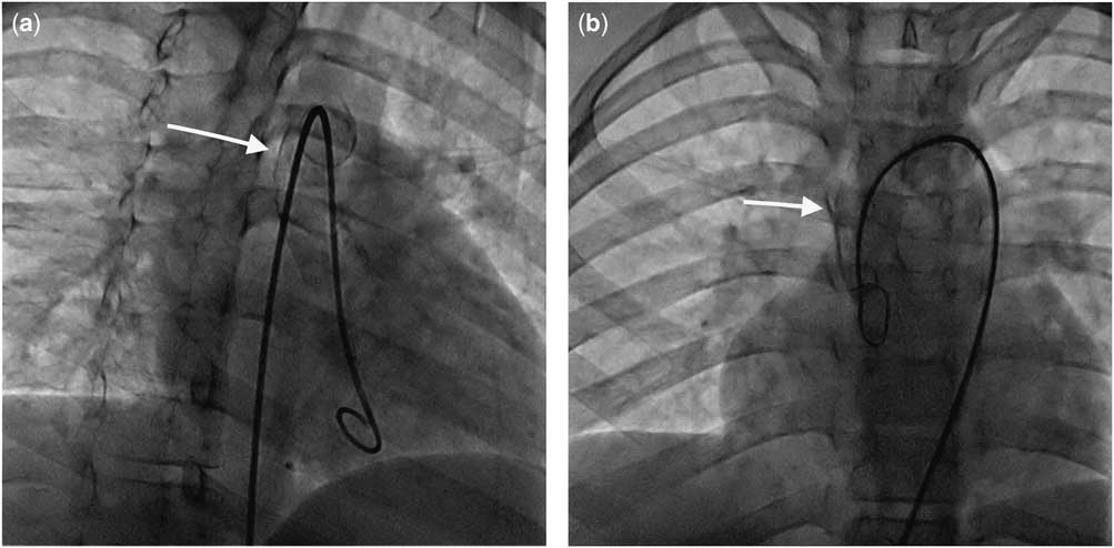
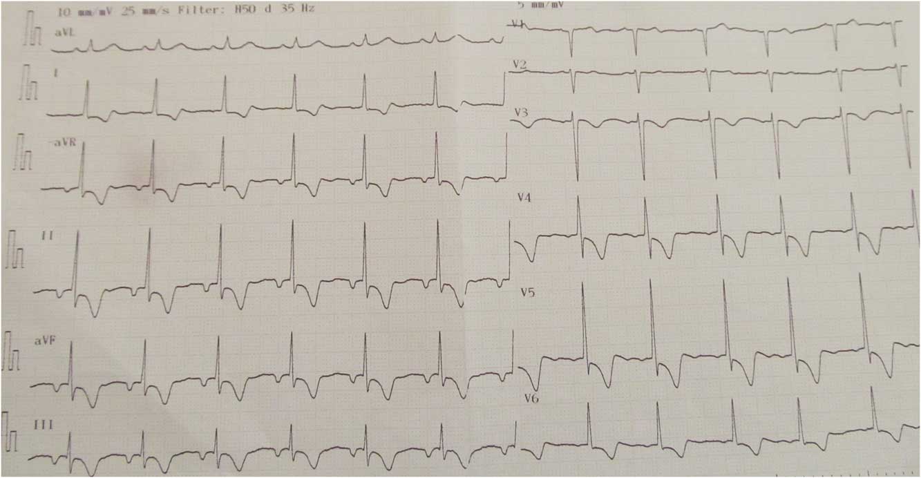
A male patient aged 15 years and 8 months was admitted with a 3-month history of syncope during physical activity. His parents were first cousins. He had two healthy sisters and two healthy brothers, with no other known disease in the family. Physical examination revealed bilateral gaze limitation in all directions, ptosis, a 3/6 systolic murmur, an 8-cm splenomegaly, pectus excavatum, and mild scoliosis. His complete blood count, thyroid function, liver and renal functions, and lipid profile were normal. Electrocardiography revealed left ventricular hypertrophy with lateral lead strain findings. Aortic and mitral valve thickening with fibrosis was detected on transthoracic echocardiography. Aortic valve stenosis (maximum gradient of 86 mmHg, mean gradient of 45 mmHg), second-degree mitral regurgitation, and second-degree aortic regurgitation were detected (Supplementary video 1). Left heart cardiac catheterisation and angiography demonstrated a calcified ascending aorta – porcelain aorta – up to the transverse arch, with diffuse calcified plaques in the left subclavian artery, left common carotid artery, and left main coronary artery (Fig 1, Supplementary video 2). Valvar aortic stenosis with significant stenosis was confirmed. Splenomegaly and cholelithiasis were found using abdominal ultrasonography. His Z-score was −3.2 g/cm2 for bone densitometry (with a normal range from −1 to +1). Storage cells were observed during bone marrow aspiration. We suspected a metabolic disease with a differential diagnosis of familial hypercholesterolaemia, Niemann–Pick disease, and Gaucher disease; Gaucher disease was the most likely diagnosis because of the presence of splenomegaly, aortic calcification, and oculomotor apraxia. The patient was diagnosed with Gaucher disease type IIIC when we found a leucocyte β-glucosidase level of 1.6 (6.5–18.9) nmol MU/mg protein along with the clinical findings. Treatment with imiglucerase (60 U/kg/2 weeks) was initiated. Gene mutation analysis revealed a homozygous mutation of c.1342G>C (p.Asp448His) (D409H) in the glucocerebrosidase gene-1. Aortic valve replacement was planned, but his parents declined the operation because of the high mortality risk quoted. After 6 months since the initiation of enzyme-replacement therapy and cardiac medical treatment with β-blocker therapy and angiotensin-converting enzyme inhibitor, the patient presented with syncope to the emergency department, with symptoms and signs suggestive of myocardial infarction (Fig 2). His cardiac enzyme levels were extremely high [troponin T: 0.5 ng/ml (0.000–0.025) and creatinine kinase-MB: 78 ng/ml (0.00–6.22)]. Unfortunately, the patient died despite resuscitation efforts with the administration of intravenous fluid, dopamine, dobutamine, and adrenalin. The family declined a postmortem examination.
( a and b ) Aortic calcification of the ascending aorta and aortic arch is shown without contrast during cardiac catheterisation. Right anterior oblique and anteroposterior views (arrows).

Electrocardiography showing extended anterolateral ischaemic T-valve changes with negative T waves from V3 to V6.

Discussion
Constrictive pericarditis in Gaucher disease type IIIC, widespread infiltration of the myocardium with Gaucher cells, and calcification of the ascending aorta, aortic valve, and mitral valve have been reported in the literature.Reference Chabás, Cormand and Grinberg 2 In addition to these calcified locations, we also detected widespread calcification of the left carotid–subclavian artery and left main coronary artery in our case, and we explained the reasons for a more severe clinical course of the condition in our patient compared with previously reported cases.
Congenital oculomotor apraxia may occur as a limitation to voluntary and involuntary eye movements.Reference Cogan, Chu, Reingold and Barranger 3 Such patients make rapid head movements to compensate for their gaze limitation. Acquired oculomotor apraxia has been described in Gaucher disease and may be a unique neurological finding.Reference Chabás, Cormand and Grinberg 2 , Reference Bohlega, Kambouris, Shahid, Al Homsi and Al Sous 4 In our case, extraocular muscle involvement and oculomotor apraxia were present. Uninvestigated gaze limitation, which began in early childhood, resulted in delayed diagnosis and the failure to appreciate worsening cardiovascular disease.
Manifestations of bone disease in Gaucher disease patients include infarction, avascular necrosis, lytic lesions, osteosclerosis, and osteoporosis with associated pain. Enzyme-replacement therapy and substrate-inhibition treatment may be useful under such conditions.Reference Grabowski, Petsko and Kolodyn 1 In our case, mild osteoporosis accompanied by bone pain was present; however, no lytic lesions were observed. Bone pain resolved soon after the initiation of enzyme-replacement therapy.
The spectrum of Gaucher disease is quite variable, ranging from asymptomatic disease to very severe disease. Phenotypic expression and severity may be different even in identical twins. It has a unique phenotype associated with homozygosity for the D409H mutation commonly found in the Mediterranean population.Reference Aabha 5 Reported patients with Gaucher disease with the single allele D409H heterozygous mutation have neither cardiac disease nor ophthalmoplegia; however, patients with the D409H homozygous mutation have severe cardiac involvement.Reference Horowitz and Zimran 6 Furthermore, it has been suggested that there may be a homozygous mutation specifically linked to cardiac involvement.Reference Horowitz and Zimran 6 Our case was homozygous for the D409H mutation, and it is assumed that the severity of his clinical findings was due to the homozygosity of this mutation, as previously suggested.
Therefore, it is confirmed that multisystemic involvement occurs in Gaucher disease type IIIC, and that cardiovascular calcification and oculomotor apraxia have a more severe course with homozygous D409H mutations. Our case illustrates that a late diagnosis leads to disease progression and increased morbidity and mortality. In conclusion, despite the fact that enzyme-replacement therapy can reduce glucocerebroside storage, relieving some symptoms, such as bone pain, cannot resolve existing calcifications; therefore, early diagnosis would significantly reduce morbidity and mortality in such patients.
Acknowledgements
None.
Financial Support
The authors declare that this study has received no financial support.
Conflicts of Interest
No conflicts of interest were declared by the authors.
Supplementary material
To view supplementary material for this article, please visit https://doi.org/10.1017/S1047951117000579