INTRODUCTION
Due to the increasing control of vectorial and transfusional transmission of Chagas' disease, vertical transmission has become more relevant for public health, as it is a continuous source of newly infected infants born to T. cruzi-infected mothers.
The risk of congenital transmission seems to vary according to different epidemiological factors, such as the parasite strain, the parasitaemia levels of the mother, the existence of previous lesions in the placenta and the geographical area (WHO, 2002).
Although it has been proposed that intrauterine infection with T. cruzi might induce spontaneous abortion (Hernandez-Matheson et al. Reference Hernandez-Matheson, Frankowski and Held1983), little is known about the effect of T. cruzi infection on pregnancy outcome. In our laboratory we have previously demonstrated that infection of female mice with the myotropic K98 T. cruzi clone results in a high rate of fetal resorption associated with inflammatory foci and parasite persistence at the genital tract (Solana et al. Reference Solana, Celentano, Tekiel, Jones and González Cappa2002). However, the mechanisms involved in tissue damage during the chronic stage of the disease are still under debate. The low number of parasites close to the lesions suggests that host cell destruction could be mediated by self-reactive B and T clones triggered by the (i) persistence of local inflammatory responses, (ii) intense polyclonal lymphocyte activation at the acute phase of infection, or (iii) cross-reactivity between parasites and organ-specific self antigens (Savino et al. Reference Savino, Villa-Verde, Mendes-da-Cruz, Silva-Monteiro, Perez, Aoki, Bottasso, Guiñazú, Silva-Barbosa and Gea2007). Despite these facts, the presence of parasites seems to be crucial in the development of chronic Chagas' disease pathology (Zhang and Tarleton, Reference Zhang and Tarleton1999). In agreement with this, the diminution of parasite load during the acute phase by parasiticide therapy has been reported to reduce the severity of myocarditis and myositis at the chronic phase of the disease (Marinho et al. Reference Marinho, D'Imperio Lima, Grisotto and Alvarez1999). However, no data regarding modulation of tissue damage during pregnancy have been reported.
At present, treatment of Chagas' disease is usually based on the administration of the nitroderivative N-benzyl-2-nitro-1-imidazole acetamide (benznidazole, Bz). This compound has been found to be effective in patients with an acute infection. In contrast, the beneficial effect of treatment during the chronic phase is still a controversial issue. Nevertheless, there are reports recommending parasiticide treatment of the whole population during the asymptomatic indeterminate phase in order to prevent the onset of cardiomyopathy (Viotti et al. Reference Viotti, Vigliano, Lococo, Bertocchi, Petti, Alvarez, Postan and Armenti2006). It must be taken into account that, besides the trypanocide effect of Bz, the therapy is able to synergize with the host immune response to induce an immune-resistant phenotype. In this regard, studies in experimental models have shown that the administration of Bz increases parasite destruction by enhancing macrophage phagocytosis and inducing higher levels of IFN-γ and nitric oxide by splenocytes from Bz-treated mice (Murta et al. Reference Murta, Ropert, Alves, Gazzinelli and Romanha1999). When T. cruzi-infected children are treated with Bz during the early-indeterminate disease, a shift of the innate immunity towards a type-1 immune profile is observed as NK and CD8+ cells increase their IFN-γ production in response to parasite antigens (Sathler-Avelar et al. Reference Sathler-Avelar, Vitelli-Avelar, Massara, Borges, Lana, Teixeira-Carvalho, Dias, Elói-Santos and Martins-Filho2006).
The aim of this work was to investigate whether the reduction of parasite load before mating has a beneficial effect on pregnancy outcome in mice infected with a myotropic T. cruzi clone that induces fertility failures.
MATERIALS AND METHODS
Mice and parasites
Female C3H/HeN(H-2k) mice (6–8 weeks old) were bred and housed at the animal facilities of the Departamento de Microbiología, Parasitología e Inmunología, Facultad de Medicina; Universidad de Buenos Aires. All procedures requiring animals were performed in agreement with institutional guidelines. T. cruzi parasites of the K98 clone (derived from the CA-I strain; González Cappa et al. Reference González Cappa, Chiale, Del Prado, Katzin, Martín, Isola, Abramo Obrego and Segura1980) were kept by passages in CF1 mice every 6 weeks.
Experimental groups
Mice were separated into the following experimental groups: uninfected untreated mice (N); uninfected Bz-treated mice (N-Bz); T. cruzi-infected untreated mice; and T. cruzi-infected Bz-treated mice.
Experimental design
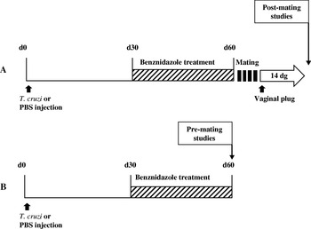
The rationale of the experimental work is outlined in Fig. 1. Briefly, T. cruzi-infected and control mice were subjected to Bz treatment before mating and pregnancy outcome was studied by evaluating fetal resorption and placental injury on day 14 of pregnancy (post-mating studies) (Fig. 1A). To determine the immune status at the time of mating, another group of mice that received parasiticide treatment was killed at the end of the therapy (pre-mating studies) (Fig. 1B). Uninfected untreated and T. cruzi-infected untreated mice were also subjected to post- and pre-mating studies. Additional groups of T. cruzi-infected (Bz-treated and untreated) mice were followed for 80 days in order to quantify parasitaemia.
Experimental design. Mice were infected with Trypanosoma cruzi (K98 clone) or injected with phosphate-buffered saline (PBS) at day 0. At day 30 post-infection (p.i.), they were treated daily with 120 mg/kg/day of benznidazole (Bz) by oral gavage up to day 60 p.i. (cross-hatched line). During the same period, a control group received vegetal oil by the same route. On day 60 p.i., a group of uninfected untreated mice; uninfected Bz-treated mice; T. cruzi-infected untreated mice and T. cruzi-infected Bz-treated mice was subjected to pre-mating studies that included lymphocyte phenotyping, measurement of IFN-γ production and analysis of IgG isotypes against live trypomastigotes by flow-cytometry. Another group of uninfected untreated mice; uninfected Bz-treated mice; T. cruzi-infected untreated mice and T. cruzi-infected Bz-treated mice was mated at the end of Bz therapy. The day of vaginal plug appearance was considered day 0 of gestation (d.g.). After 14 d.g. (open arrow), mice were killed to perform post-mating studies: quantification of fetal resorptions and histopathological studies on fetoplacental units.

Infection and chemotherapy treatment
Mice were infected by the intradermal (in the hind foot-pad) inoculation of 20 μl of blood containing 5×10⁴ trypomastigotes (tryp) of T. cruzi. Control mice received an equal volume of phosphate-buffered saline (PBS) by the same route. Thirty days later, infected and uninfected mice were daily treated with N-benzyl-2-nitro-1-imidazolacetamide (benznidazole, Bz, Roche) for 30 days at a daily dose of 120 mg/kg of body weight. Drug pills were crushed in a mortar and resuspended in corn oil. Each mouse received 0·20 ml of this suspension by oral gavage. This method was performed using an 18-gauge stainless animal feeding needle (Thomas Scientific, Swedesboro, NJ, USA).
Screening of T. cruzi infection
The levels of parasitaemia of mice (n=10) were recorded as described previously (Solana et al. Reference Solana, Celentano, Tekiel, Jones and González Cappa2002). Briefly, 10 μl of fresh blood taken from the mouse tail was incubated with 90 μl of Tris-buffered 0·83% ammonium chloride, pH 7·2. Parasites were counted in a haemocytometer and the number of trypomastigotes/ml was calculated. When the parasitaemia was below the sensitivity of the haemocytometer method, the microhaematocrit technique was used (Freilij et al. Reference Freilij, Muller and González Cappa1983; Torrico et al. Reference Torrico, Solano, Guzmán, Parrado, Suarez, Alonzo-Vega, Truyens, Carlier and Torrico2005). This method proved to be very successful in assessing extremely low infections in mice (Mirkin et al. Reference Mirkin, Celentano, Malchiodi, Jones and González Cappa1997).
Collection of sera and spleens
Three to eight animals from each experimental group were killed at the end of the Bz therapy and individual spleens and sera were collected. Spleen cell (SC) suspensions were prepared from each animal by gently pushing the tissue through a stainless steel mesh and into a tissue-culture dish containing RPMI-1640 medium supplemented with 5% fetal calf serum (FCS). Red blood cells were lysed with Tris-buffered 0·83% ammonium chloride, pH 7·2. Cells were counted in a haemocytometer and viability was determined by trypan blue exclusion both before and after immunostaining. SC were used for immunophenotyping analysis and cell-culture assays. Serum samples were stored at −20°C after complement inactivation (56°C for 30 min).
Lymphocyte phenotyping
For cell-surface immunophenotyping of lymphocyte subsets and of activation/memory status, 106 SC from each animal (5–8 animals/group) of each experimental group were subjected to flow cytometry analysis. Briefly, cells were washed twice with ice-cold PBS supplemented with 1% bovine serum albumin (Sigma, St Louis, MO, USA) and 0·1% sodium azide (Sigma, St Louis, MO, USA). They were then incubated for 20 min at 4°C with previously optimized amounts of the corresponding fluorochrome-labelled rat anti-mouse monoclonal antibodies. After staining, cells were washed and fixed with freshly prepared 1% paraformaldehyde (PFA) in PBS. Two or three-colour data acquisitions were done on a FACSCalibur (Becton Dickinson, San Jose, CA, USA). A total of 10 000–20 000 events were acquired in the lymphocyte region, according to forward- and side-scatter parameters. Data analysis was performed with the WinMDI2.8 software (by Joseph Trotter, Scripps Research Institute, San Diego, CA, USA).
Antibodies
The following amounts of rat anti-mouse monoclonal antibodies were used per 106 cells for flow cytometry: 0·25 μg of phycoerythrin (PE)-conjugated anti-CD8, 0·125 μg of biotin-conjugated anti-CD4; 0·25 μg of fluorescein isothiocyanate (FITC)-conjugated anti-CD44 and 0·25 μg of FITC-conjugated anti-CD69 (BD PharMingen, San Diego, CA, USA). To perform fluorescence immunoassays with live trypomastigotes, 0·05 μg of biotin-conjugated anti-mouse IgG1 and biotin-conjugated anti-mouse IgG2a monoclonal antibodies were used (Invitrogen, Carlsbad, CA, USA). When necessary, 0·5 μg of extravidine-phycoerythrin (PE) or extravidine-Cychrome (Cy) was used (BD PharMingen, San Diego, CA, USA).
IFN-γ detection
Cell suspensions were obtained from spleens of 3–5 uninfected untreated mice, T. cruzi-infected untreated mice and T. cruzi-infected Bz-treated mice. The culture medium consisted of RPMI 1640 (Gibco, BRL) supplemented with 5% fetal calf serum, 0·05 mM 2-mercaptoethanol and antibiotics. SC (5×10⁶/ml well of a 24-well plate) were either stimulated or not with 5 μg of concanavalin A (ConA) (Sigma Chemical Co., St Louis, MO, USA) per ml. All cultures were incubated at 37°C in a 5% CO2 atmosphere for 24 h. Culture supernatants were then harvested and frozen at −80°C until use.
Mouse IFN-γ was quantified in culture supernatants from ConA-activated or control SC by ELISA (R&D Systems, Minneapolis, MN, USA) according to the manufacturer's specifications. All samples were assayed in duplicate. The detection limit was 62·5 pg/ml.
Fluorescence immunoassay with live trypomastigotes
The immunofluorescence reaction was carried out as described by Martins-Filho et al. (Reference Martins-Filho, Pereira, Carvalho, Cançado and Brener1995), modified as follows. One×106 cell culture trypomastigotes were incubated at 37°C for 30 min with sera obtained from animals of each experimental group. Serum dilutions from T. cruzi-infected untreated and T. cruzi-infected Bz-treated mice were standardized to contain equal amounts of specific IgG by using an indirect ELISA. The appropriate serum dilutions matched an optical density of 0·300, using total extracts of T. cruzi epimastigotes as antigen. Thus, such serum dilutions were used in fluorescence immunoassays with live trypomastigotes (FC-ALTA). After incubation, parasites were washed once with PBS containing 10% FCS and 0·05% sodium azide and reincubated at 37°C with biotin-conjugated anti-mouse IgG1 or IgG2a monoclonal antibodies. Each assay included an internal control of non-specific binding in which parasites not exposed to mouse sera were incubated with biotin anti-mouse IgG1 or IgG2a. After staining, labelled parasites were incubated with extravidin-PE (Sigma, St Louis, MO, USA) for 30 min at 37°C. Finally, parasite suspensions were fixed with 1% paraformaldehyde in PBS and stored at 4°C before FACS acquisition and data analysis. A sample of unlabelled trypomastigotes was stained with propidium iodide (4 μg) prior to fixation and viability was determined to be around 90%.
FACScan data acquisition and analysis
Flow cytometric measurements were performed on a FACSCalibur flow cytometer (BD, San Diego, CA, USA). Ten thousand events per sample were acquired. Trypomastigotes were identified on the basis of their forward (FSC) and side (SSC) light-scattering properties and were in accordance with previous reports describing FC-ALTA (Martins-Filho et al. Reference Martins-Filho, Pereira, Carvalho, Cançado and Brener1995). The differentiation between amastigotes and trypomastigotes based on FSC vs SSC properties was previously optimized by acquisition of purified amastigotes and trypomastigotes suspensions. Live trypomastigotes were gated on the basis of their characteristic FSC×SSC distribution, on a log scale. The relative PE-fluorescence intensity of each parasite preparation was analysed with a single histogram representation. A marker was set up on the histogram representation of the PE-conjugated internal control and used in all data analyses reported here to determine, for each serum sample, the relative percentage of positive fluorescent parasites (PPFP) and the median fluorescent intensity (MFI). Results are expressed as the MFI or the PPFP for anti-mouse IgG1 or IgG2a monoclonal antibody, calculated by subtracting the corresponding value for the isotype controls.
Results were expressed as MFI (MFI of tryp with specific monoclonal antibody)−(MFI of tryp with irrelevant isotype-matched Mab) or mean PPFP for each IgG isotype.
Mating
Ten to 20 female mice from each experimental group were mated with normal males after the end of Bz therapy (mating time). The day of presence of a vaginal plug was taken as day 0 of pregnancy. The rate of successful mating was calculated as the number of females with vaginal plugs/number of total females. To evaluate the frequency of fetal resorptions, groups of 4–7 T. cruzi-infected untreated, T. cruzi-infected Bz-treated and control mice were killed on day 14 of gestation (d.g.). Resorptions were identified either by the presence of resorbing scars or by the notably smaller size of the fetuses, as well as by their necrotic or haemorrhagic appearance when compared with normal viable fetuses (Solana et al. Reference Solana, Celentano, Tekiel, Jones and González Cappa2002). The percentage resorption rate was calculated using the formula R/(R+V)×100, where R is the number of resorbing fetuses and V is the number of viable fetuses per animal (Solana et al. Reference Solana, Celentano, Tekiel, Jones and González Cappa2002). Placental samples were preserved in PBS containing 4% PFA for further histopathological studies.
Histopathological analysis
All the maternal and fetal structures that constitute the chorionic plate, labyrinth, junctional zone (trophospongium), zone of giant cells and the maternal deciduas (placenta) plus the fetus are considered the fetoplacental unit (FPU). Three FPU/animal from T. cruzi-infected Bz-treated mice (n=3), T. cruzi-infected untreated mice (n=3) and control pregnant females (n=4) were collected at 14 d.g. and fixed in 4% PFA in PBS for further processing. Paraffin-embedded tissue sections were stained with haematoxylin-eosin and PAS and examined by light microscopy. Three non-consecutive slides of each placental sample were analysed in a blind fashion. Areas of necrosis were quantified by an image analysis system using Image Pro-Plus 4.5 software. Results are expressed as percentage of necrotic area over total area for each FPU. The same tissue sections were analysed to search for amastigote nests by microscopical examination. In this case, results were expressed as the percentage of placentas per mouse with amastigote nests.
Stastistical analysis
Differences between groups were analysed by the non-parametric Kruskall-Wallis analysis of variance using the Dunn post-test. Differences between the infected groups (receiving or not Bz treatment) were determined by the non-parametric Mann-Whitney U test or Student's t-test in case of normality. In all circumstances, differences were considered statistically significant when P<0·05.
RESULTS
Effect of benznidazole therapy on parasitaemia level in female mice
In order to assess the efficacy of Bz treatment on reducing the systemic parasite load of female mice, the parasitaemia was periodically monitored.
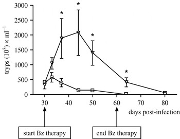
As seen in Fig. 2, all T. cruzi-infected mice showed proven parasitaemias prior to Bz therapy, at 30 days post-infection (p.i.) (Fig. 2). Then, the level of parasitaemia among infected mice that received Bz began to decrease at 37 days p.i. (7 days after the onset of the treatment) (n=10) and dropped to negative results even by the microhaematocrit method at the end of the Bz therapy (60 days p.i.). This group was designated ‘Mice with Low Parasitaemia’ (LP). In contrast, the infected untreated mice (n=10) showed an increase in the number of circulating parasites reaching a peak between 37 and 45 days p.i.; parasites remained detectable throughout the observation period (80 days p.i.). This group was designated ‘Mice with High Parasitaemia’ (HP).
Kinetics of parasitaemia in Trypanosoma cruzi-infected untreated females (triangles) and T. cruzi-infected Bz-treated females (squares) during the observation period. The arrows show the beginning and the end of Bz treatment. Values are the mean of 10 individual data±S.E. * P<0·01.

Screening of pregnancy outcome
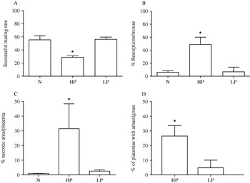
To evaluate whether the decrease in the systemic parasite load exerted an effect on the reproductive capacity of T. cruzi-infected females and on the progression of the fetus, both the rate of successful mating and the fetal resorption rate were evaluated. Regarding pregnancy outcome, LP mice showed similar behaviour to that of N mice. In contrast, HP mice displayed a worse mating performance since vaginal plugs were observed only in 29·68±2·69% of females during the observation period (P<0·02) (Fig. 3A). The number of resorptions also diminished among the LP females when compared with HP mice (P<0·02), showing similar values to controls (Fig. 3B).
Effect of parasiticide treatment on mating rate, pregnancy outcome, tissue injury and placental parasite load. (A) Rate of successful mating among Mice with High Parasitaemia (HP), Mice with Low Parasitaemia (LP) and untreated uninfected controls (N). The presence of a vaginal plug was evaluated daily up to day 50 post-mating (10–20 animals/group) and the rate of successful mating was calculated as described in the Materials and Methods section. (B) Fetal resorptions were quantified among HP, LP and N females on day 14 of gestation (4–7 animals/group) as described in the Materials and Methods section. (C) Percentage of necrotic areas was calculated by using Image Pro Plus software after microscopic examination of each placenta from Mice with High Parasitaemia (HP) (n=3), Mice with Low Parasitaemia (LP) (n=3) and untreated uninfected control mice (N) (n=4). * P<0·05, HP vs LP. Values are the mean±S.D. (D) The percentage of placental samples with amastigote nests detected by microscopic examination was calculated from each animal of each experimental group as described in the Materials and Methods section. * P<0·05. Values are the mean±S.E. Results of 1 representative experiment out of 3 are shown.

Histopathology of the placental samples from T. cruzi-infected mice
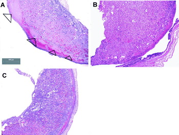
The improvement of the reproductive performance in LP females might be associated with lower parasite load and milder inflammatory changes in the fetomaternal interface. Therefore, the presence of amastigote nests and the area of necrotic damage in placental samples obtained from T. cruzi-infected females (both HP and LP) were studied. The low number of fetal resorptions recorded among LP females correlates with a reduction in the percentage of necrotic areas in placental samples obtained from mice of this group (Fig. 3C and Fig. 4B). Necrosis was identified as fibrin deposits and nuclear debris surrounded by few inflammatory cells, mostly polymorphonuclear leukocytes and scarce lymphocytes. In contrast, HP females showed increased tissue damage at the placental level (Fig. 3C and Fig. 4A) (P<0·05). These results are in agreement with a decrease in the persistence of the parasite in placental samples where amastigotes were microscopically searched for (P<0·05) (Fig. 3D). Only minimal areas of necrosis were detected in placental samples obtained from mice of the N group (Fig. 4C).
Histological analysis of HP, LP and N mice. (A) Tissue section from HP mouse showing extensive necrotic compromise of the placenta (arrow heads). (B) Tissue section from LP mouse showing reduction of necrotic areas in the placenta. (C) Placental section of N mouse. Results of a representative fetoplacental unit from 1 animal out of 3 in each group are shown. The marker shown in (A) is the same for all sections.

Analysis of IgG isotypes against live trypomastigotes in sera from T. cruzi-infected mice
In order to determine whether the improvement of reproductive performance observed among LP mice correlates with changes in the immune response at mating time, the immune status before mating was assessed.
It is known that the control of parasitaemia level is exerted in part by opsonizing/lytic antibodies. For this reason, the presence of IgG1 and IgG2a directed against live trypomastigotes was studied in sera from LP and HP mice at the end of the Bz treatment and before mating.
Titration of individual sera from HP, LP mice and controls using an indirect ELISA with epimastigote whole homogenate as antigen, showed that the appropriate serum dilution to be used in FC-ALTA was 1/50 for LP or controls and 1/1800 for HP mice (data not shown).
Individual sera were subjected to analysis by flow cytometry to determine the binding of specific antibodies bearing particular isotypes to live trypomastigotes. Parasites were identified on the basis of their forward (FSC) and side (SSC) light-scattering properties. The median fluorescence intensity and the mean percentage of positive fluorescent parasites (PPFP) for each IgG isotype were calculated for each serum sample. The analysis revealed that sera from LP females reduced both the number of parasites that bind specific IgG1 (PPFP) as well as the density of specific IgG1 molecules bound to the surface of live parasites (MFI) when compared to HP mice (P<0·05, Table 1). In contrast, analysis of IgG2a of sera from HP and LP revealed no differences in MFI and PPFP values (Table 1).
Detection of anti-live trypomastigote membrane-bound IgG1 and IgG2a present in sera from HP, LP and N females using flow cytometry

a Results are expressed as the Median Fluorescence Intensity (MFI) and the percentage of positive fluorescent parasites (PPFP) for each IgG isotype, calculated by subtracting the corresponding value for irrelevant matched isotype controls Serum dilutions: 1/50 for LP or N and 1/1800 for HP mice. Data are given as means of 3 independent experiments. Three to four animals per group were used. * P<0·05, HP vs LP, Mann-Whitney U test. HP: ‘Mice with High Parasitaemia’, LP: ‘Mice with Low Parasitaemia’ and N: Uninfected Untreated Mice.
Sera from both N and N-Bz mice showed similar MFI and PPFP values (data not shown). These results show that the Bz treatment was able to modulate the specific IgG isotype profile towards the predominance of IgG2a over IgG1 in LP mice.
Analysis of lymphocyte populations in spleens from T. cruzi-infected mice prior to mating
Previous reports have demonstrated that Bz is able to modulate activation/memory T cell response and this modulation is directly related to the persistence of parasite antigen (Bustamante et al. Reference Bustamante, Bixby and Tarleton2008; Sathler-Avelar et al. Reference Sathler-Avelar, Vitelli-Avelar, Lima Massara, de Lana, Pinto Dias, Teixeira-Carvalho, Eloi-Santos and Martins-Filho2008). In this work we studied the effect of Bz therapy on the activation/memory pattern of T cells previous to mating and its potential impact on placental injury.
As expected, T. cruzi-infected mice showed enlargement of the spleen in comparison with control mice. However, no differences in cellularity between LP and N mice were observed. On the contrary, HP mice showed higher numbers of splenocytes than LP mice (P=0·05, Table 2). The frequency of CD4+ splenocytes failed to exhibit changes between experimental groups. Nevertheless, the absolute cell numbers of CD4+ T cells were reduced in the LP group in comparison with the HP group (P<0·02, Table 2). On the other hand, the CD8+ compartment displayed no difference either in the frequency or in the cellularity between HP and LP groups (Table 2).
Effect of benznidazole therapy on the frequency and total number of CD4+ and CD8+ splenocytes of Trypanosoma cruzi-infected mice and controls

§ Data are expressed as 106 cells, mean±S.D.
# Data are expressed as percentage, mean±S.D.
N: normal untreated mice; N-Bz: normal Bz-treated mice; HP: Mice with High Parasitaemia; LP: Mice with Low Parasitaemia.
* P=0·05, ** P<0·02, HP vs LP. Five to 8 animals per group were used.
Although both control groups failed to show statistical differences in the measured parameters, an effect of the drug on the splenic leukopoiesis cannot be ruled out since the number of splenocytes in the Bz-N group showed a trend to be lower than in N group (Table 2).
IFN-γ production by spleen cells (SC) from LP and HP mice
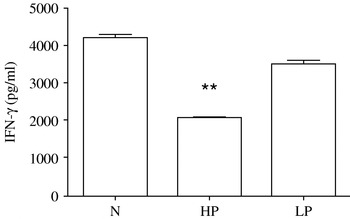
To establish whether the reduction in parasitaemia in LP mice correlated with an effector immune response driven by cytokines, the production of IFN-γ by SC from LP and HP mice was compared with the control group. Spontaneous production of IFN-γ in cultures from mice of all experimental groups was below detection levels (62·5 pg/ml). However, SC from HP mice produced lower levels of this cytokine than those from LP mice after polyclonal stimulation with Con A (P<0·001). Mitogen-stimulation of SC from N mice exhibited the highest level of IFN-γ production (Fig. 5).
IFN-γ production in ConA-stimulated cultures of splenocytes from HP, LP and N mice. Mice were infected with Trypanosoma cruzi (K98 clone) and treated with benznidazole at day 30 post-infection. After 30 days of therapy, spleen cell suspensions were cultured with 5 μg of ConA and the 24-h supernatants were tested by capture ELISA. Values are means±S.D. from duplicate cultures; n=3–5 animals/group. ** P<0·001, HP vs LP mice. Data are representative of 2 independent experiments.

Expression of activation/memory markers in the splenocyte T compartment
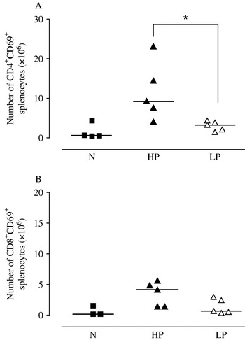
In order to study whether there is a role of pre-mating Bz treatment in the modulation of the spleen T cell compartment that could contribute to reduce placental inflammation in our experimental model, phenotypic analysis of the experienced-memory/activation cell surface markers CD44 and CD69 of the CD4+ and CD8+ subsets was performed at the end of the Bz therapy. Flow cytometry analysis showed that HP mice had increased both CD4+CD69+ and CD8+CD69+ T lymphocyte numbers (Fig. 6A and B), exhibiting a status of recent activation. This effect was almost abolished among LP mice which displayed marked differences for CD4+CD69+ (P<0·02) (Fig. 6A) and exhibited a tendency to decrease values for CD8+CD69+ in relation to those shown by HP mice (Fig. 5B).
Scatter plots of the number of CD4+CD69+ and CD8+CD69+ splenocytes. Data are from uninfected mice (N, filled squares), Mice with High Parasitaemia (HP, filled triangles) and Mice with Low Parasitaemia (LP, open triangles). Horizontal bars represent median values of a representative experiment of 3 independent assays. Each symbol represents 1 animal. * P<0·05, HP vs LP mice.

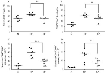
When CD44 expression was studied in both T cell subsets, expansion of this experienced/memory subpopulation was observed among both CD4+ and CD8+ cells of HP group (P<0·005). When absolute cell numbers were considered, T cells from LP mice showed significantly smaller numbers of CD44+ cells, both in CD4+ and CD8+ T cell subsets (P<0·0005 and P<0·05, respectively) when compared with the HP group (Fig. 7A and B).
Scatter plots of the frequency and absolute number of CD4+CD44+ and CD8+CD44+ splenocytes. Data are from non-infected mice (N, filled squares), Mice with High Parasitaemia (HP, filled triangles) and Mice with Low Parasitaemia (LP, open triangles). Frequency of CD4+ CD44+ and CD8+CD44+ cells (A and B) and absolute numbers of CD4+CD44+ and CD8+CD44+ cells (C and D). Horizontal bars represent median values of a representative experiment of 3 independent assays. Each symbol represents 1 animal.* P<0·05, ** P<0·005 and *** P<0·0005; HP vs LP mice.

DISCUSSION
Previous reports have shown that Bz interrupts in vivo parasite replication (Maya et al. Reference Maya, Cassels, Iturriaga-Vásquez, Ferreira, Faúndez, Galanti, Ferreira and Morello2007). In the present study, we used Bz as a way to decrease the systemic parasite load before mating and to study its impact in a murine model of T. cruzi infection that induces alterations in pregnancy outcome but fails to achieve vertical transmission. For this purpose, the Bz scheme began at 30 days p.i. when parasite multiplication was already established. Nonetheless, a significant decrease in the parasitaemia levels at mating time was observed. Therefore, the supply of bloodstream trypomastigotes to the placenta would be clearly diminished during its development. This assumption is confirmed, in the present work, by the reduction in the placenta injury observed in the LP group that is associated with low tissue parasitism. Since previous studies indicate that the parasite load at the acute phase of T. cruzi infection influences the development of Chagas' disease pathology at the chronic phase of the disease (Marinho et al. Reference Marinho, D'Imperio Lima, Grisotto and Alvarez1999), we can assume that the reduction of parasitaemia achieved by Bz therapy in LP mice results in amelioration of pregnancy outcome.
During the last few years, substantial evidence from experimental and clinical studies supports the conclusion that parasite persistence plays a crucial role in the pathogenesis of Chagas disease (Tarleton, Reference Tarleton2001). This is supported by the presence of T. cruzi at or near the inflammatory foci and by the evidence that parasiticide treatment results in a decrease of the disease severity (Añez et al. Reference Añez, Carrasco, Parada, Crisante, Rojas, Fuenmayor, Gonzalez, Percoco, Borges, Guevara and Ramirez1999; Garcia et al. Reference Garcia, Ramos, Senra, Vilas-Boas, Rodrigues, Campos-de-Carvalho, Ribeiro-dos-Santos and Soares2005; Zhang and Tarleton, Reference Zhang and Tarleton1999). In the present experimental model, the resorptions triggered by the K98 T. cruzi clone are strongly associated with inflammatory infiltrates and with the persistence of the parasite in the murine genital tract as well as with high parasitaemia levels in these females (Solana et al. Reference Solana, Celentano, Tekiel, Jones and González Cappa2002). Indeed, using a strain of T. cruzi with a biological behaviour similar to that of the K98 clone, Mjihdi et al. (Reference Mjihdi, Lambot, Stewart, Detournay, Noël, Carlier and Truyens2002) stated that infertility and early fetal losses were related to high maternal parasitaemia.
There is little information on the influence of T. cruzi strains from different lineages on the course of pregnancy in experimental models and/or infected pregnant women. Previous reports performed with Bolivian and Argentine human samples have demonstrated that sublineage IId of T. cruzi (T. cruzi IId) is present in the blood of congenitally infected newborns (Virreira et al. Reference Virreira, Truyens, Alonso-Vega, Brutus, Jijena, Torrico, Carlier and Svoboda2007; Burgos et al. Reference Burgos, Altcheh, Bisio, Duffy, Valadares, Seidenstein, Piccinali, Freitas, Levin, Macchi, Macedo, Freilij and Schijman2007). This is not surprising, since T. cruzi II is the lineage that usually circulates in the domestic cycle in these areas of South America (Marcet et al. Reference Marcet, Duffy, Cardinal, Burgos, Lauricella, Levin, Kitron, Gürtler and Schijman2006; Virreira et al. Reference Virreira, Serrano, Maldonado and Svoboda2006; Rozas et al. Reference Rozas, Botto-Mahan, Coronado, Ortiz, Cattan and Solari2007; Steindel et al. Reference Steindel, Kramer Pacheco, Scholl, Soares, de Moraes, Eger, Kosmann, Sincero, Stoco, Murta, de Carvalho-Pinto and Grisard2008). In this regard, previous results from our laboratory have also reported that congenital transmission is achieved in mice born to mothers infected with the RA strain (Solana et al. Reference Solana, Celentano, Tekiel, Jones and González Cappa2002) which has been classified as T. cruzi II (Risso et al. Reference Risso, Garbarino, Mocetti, Campetella, Gonzalez Cappa, Buscaglia and Leguizamon2004) but not in those born to mothers infected with K98. The clone used here (K98) belongs to the T. cruzi I (Risso et al. Reference Risso, Garbarino, Mocetti, Campetella, Gonzalez Cappa, Buscaglia and Leguizamon2004) and displays a high myotropism, showing a persistent parasitic load in the uterus and placenta of female infected mice (Solana et al. Reference Solana, Celentano, Tekiel, Jones and González Cappa2002). T. cruzi I genotype circulates mainly among infected people in Mexico, Colombia and Venezuela (Higo et al. Reference Higo, Miura, Horio, Mimori, Hamano, Agatsuma, Yanagi, Cruz-Reyes, Uyema, Rojas de Arias, Matta, Akahane, Hirayama, Takeuchi, Tada and Himeno2004) and there are still no studies related to pregnancy and vertical transmission. Hence, the effect of parasite populations belonging to T. cruzi I on fetal progression and/or on congenital transmission may be underestimated.
The effect of the immune response on the control of T. cruzi infection results from the combined action of T and B cells (Gazzinelli et al. Reference Gazzinelli, Pereira, Romanha, Gazzinelli and Brener1991; Dos Reis, Reference Dos Reis1997). B cells are known to contribute to immune resistance against the parasite through the production of specific antibodies, the maintenance of experienced/memory CD8+ T cells and the determination of an adequate T cell cytokine pattern associated with parasite control (Rodriguez et al. Reference Rodriguez, Santero, Afchain, Bazin and Capron1981; Cardillo et al. Reference Cardillo, Postol, Nihei, Aroeira, Nomizo and Mengel2007). IFN-γ elicits the immunoglobulin switch to IgG2a, the IgG subclass with higher opsonizing and complement-activating potential in the mouse model. In IFN-γ−KO mice, the humoral response is completely shifted to IgG1, with no production of specific IgG2a isotype, and the animals succumb to the acute infection due to high parasitaemias and intense inflammatory lesions with numerous amastigote nests in the heart (Marinho et al. Reference Marinho, Nuñez-Apaza, Martins-Santos, Bastos, Bombeiro, Bucci, Sardinha, Lima and Álvarez2007). On the other hand, disease-associated immunosuppression during the acute phase is believed to facilitate the multiplication and dissemination of T. cruzi parasites in mammalian hosts (Sztein and Kierszenbaum, Reference Sztein and Kierszenbaum1992). Therefore, the reduction of systemic parasite load by parasiticide treatment could help to avoid these alterations, allowing the host to mount an effective immune response.
The duration and/or strength of antigenic stimulation due to parasite persistence regulates the isotypic immunoglobulin profile and effector T cell function (Kalia et al. Reference Kalia, Sarkar, Gourley, Rouse and Ahmed2006). Recently, the absence of anti-live trypomastigote IgG antibodies (ALTA) that include the previously described lytic antibodies, has been described as a very reliable cure marker (Krautz et al. Reference Krautz, Kissinger and Krettli2000; Martins-Filho et al. Reference Martins-Filho, Eloi-Santos, Teixeira Carvalho, Oliveira, Rassi, Luquetti, Rassi and Brener2002). A flow cytometric method to detect these antibodies (FC-ALTA) has been described as a new tool for monitoring the efficacy of Bz treatment in human Chagas' disease (Martins-Filho et al. Reference Martins-Filho, Eloi-Santos, Teixeira Carvalho, Oliveira, Rassi, Luquetti, Rassi and Brener2002; Vitelli-Avelar et al. Reference Vitelli-Avelar, Sathler-Avelar, Wendling, Rocha, Teixeira-Carvalho, Martins, Dias, Rassi, Luquetti, Elói-Santos and Martins-Filho2007). In the present work, we used this test to evaluate changes in IgG homeostasis through the detection of IgG1 or IgG2a by FC-ALTA. LP mice showed a decline in both IgG isotypes being particularly evident for IgG1. These changes in humoral immune response towards a predominant effector IgG2a isotype should play a role in trypomastigote blood clearance. This is also supported by the increased IFN-γ production by mitogen stimulated splenocytes from mice of the LP group. In this sense, a previous report has described levels of IFN-γ production higher than those found by us in this work, even in the absence of polyclonal stimulus. The observed discrepancies may be related to differences in therapy schedule, mouse and parasite strains used (Murta et al. Reference Murta, Ropert, Alves, Gazzinelli and Romanha1999). Our results suggest that Bz treatment positively modulates IFN-γ production contributing to parasite clearance and diminishing parasite supply to placental tissues.
Several mechanisms have been postulated to trigger tissue inflammation in experimental Chagas disease, including self-antigen exposure secondary to the tissue damage that elicits cross-reactive autoimmune responses. Autoreactive and T. cruzi-specific responses derived from parasite persistence are not incompatible or mutually exclusive in Chagas disease, and a combination of both may be involved in the establishment of myocardial damage (Marin-Neto et al. Reference Marin-Neto, Cunha-Neto, Maciel and Simões2007). Accordingly, previous reports from our laboratory have demonstrated that CD4+ but not CD8+ T lymphocytes from mice infected with CA-I, the parental strain of the K98 clone, are responsible for muscle injury when transferred to normal recipients. Besides, only CD4+ lymphocytes from these mice are able to proliferate against muscle antigens (Mirkin et al. Reference Mirkin, Celentano, Malchiodi, Jones and González Cappa1997). A noticeable reduction was detected in the CD4+ splenocyte subset among LP females. This finding correlates with a diminution of the necrotic area in placental samples obtained from LP mice. To establish whether this T cell subset is involved in placental damage, further studies need to be carried out.
In addition to its known direct trypanocidal activity, Bz also modulates the immune response during T. cruzi experimental infection (Michailowsky et al. Reference Michailowsky, Murta, Carvalho-Oliveira, Pereira, Ferreira, Brener, Romanha and Gazzinelli1998; Piaggio et al. Reference Piaggio, Roggero, Pitashny, Wietzerbin, Bottasso and Revelli2001; Romanha et al. Reference Romanha, Alves, Murta, Silva, Ropert and Gazzinelli2002). Studies addressing the activation of the immune system by effect of Bz have shown contradictory results. Sathler-Avelar et al. (Reference Sathler-Avelar, Vitelli-Avelar, Lima Massara, de Lana, Pinto Dias, Teixeira-Carvalho, Eloi-Santos and Martins-Filho2008) suggest that Bz-treatment induces substantial T and B-cell activation status, while others have demonstrated that Bz-therapy reduces T-cell activation but sustains expansion of effector and experienced/memory CD8+ T cells (Olivieri et al. Reference Olivieri, Cotta-De-Almeida and Araújo-Jorge2002). Together, these data support the hypothesis that the impact of Bz on the immunological features in T. cruzi infection could be related to the time when Bz is administered, the drug resistance of the parasite population and the intrinsic characteristics of the parasite strain. The hypothesis of ‘decreasing-potential’ establishes that down-modulation of antigen stimulation may lead to a decrease in the number of effector and memory T cells (Ahmed et al. Reference Ahmed and Gray1996). In this sense, patients at the chronic phase of the disease exhibit decreases in experienced/memory T cells due to the effect of Bz treatment (Bertocchi et al. Reference Bertocchi, Álvarez, Pérez, Armenti, Viotti, Lococo, Petti, Postan, Albareda, Laucella and Tarleton2008). Our data indicate that treatment reduces the activation as well as the experienced/memory status of spleen T cells in mice. This correlates with a reduction in parasitaemia levels at mating time by the effect of the parasiticide treatment. If it is considered that activated/memory-experienced CD4+ T cells are involved in placental injury, it can be assumed that a decreased number of activated (CD69) and experienced/memory (CD44) CD4+ could be beneficial for pregnancy outcome in LP mice. However, further studies are needed to confirm the role of these T cells in placental injury.
On the whole, these findings indicate that a reduction in parasitaemia before mating achieved by the effect of parasiticide therapy fosters the emergence of immune mechanisms that result in the reduction of tissue injury with a favourable effect on pregnancy outcome. These changes may account for the benefits of the aetiological treatment of Chagas disease prior to pregnancy.
This work was partially supported by grants of Fondo Nacional de Ciencia y Tecnología (FONCyT, PICT 05-14241) and Universidad de Buenos Aires (UBA, ME036). We thank Dr Gerardo Mirkin for critical reading of the manuscript. M. P. and S. M. G. C. are members of the Research Career of the Consejo Nacional de Ciencia y Tecnología (CONICET), Argentina. C. D. A. S. and C. V. P. are fellows from CONICET.