Introduction
Ti6Al4V has been used for biomedical applications because it exhibits elevated mechanical and corrosion resistance, and good biocompatibility [Reference Huang, Hsu and Yu1]. Superficial treatments for texturing, such as electropolishing, have been proposed to control the roughness and wettability, aiming at increasing the biomaterial–implant contact.
In the process of architecture and formation of the bone, nanostructured biomaterials have been used [Reference Landis and Silver2]. Many studies have revealed that nanostructured biomaterials promote bone regeneration, thus favoring adhesion, spreading, proliferation, and differentiation of the cells [Reference Martinez, Engel, Planell and Samitier3, Reference Lord, Foss and Besenbacher4]. Nanostructured biomaterials have strong interaction with cells; hence, they help the stimulation of osseointegration in these cells [Reference Wang, Zhao, Liu, Weir, Zhou and Xu5].
Zhao et al. (2012) showed that nanostructured titanium surfaces have significative effect on cell differentiation [Reference Zhao, Liu, Wu, Zhang and Chu6]. Moreover, Liao et al. (2014) reported that nanotopography can stimulate the differentiation process of stem cells [Reference Liao, Nguyen, Ngiam, Wang, Cheng, Chan and Ramakrishna7] and that a nanostructured surface can create binding sites in cells, which can facilitate the process of osseointegration [Reference Fricain, Schlaubitz, Le Visage, Arnault, Derkaoui, Siadous, Catros, Lalande, Bareille, Renard, Fabre, Cornet, Durand, Léonard, Sahraoui, Letourneur and Amédéd8].
Based on the knowledge that nanostructured surfaces favor cell growth, it is important to consider the changes caused by the sterilization processes on the surface of these biomaterials. Gossla et al. (2016) [Reference Gossla, Tonndorf, Bernhardt, Kirsten, Hund, Aibibu, Cherif and Gelinsky9] evaluated the cytocompatibility of chitosan samples obtained by electrostatic agglomeration after autoclave sterilization (121 °C, 20 min), 70% ethanol, γ radiation (greater than 25 KGy, 60Co), and supercritical carbon dioxide (0.25% water, 0.15% hydrogen peroxide, and 0.5% acetic anhydride, at 38 °C, 8.5 MPa, for 5, 15, 30, and 45 min). For all sterilization methods, a decrease in cell viability was observed over time. In addition, the authors showed that the 70% ethanol and the supercritical CO2 sterilization methods were the ones in which cell viability presented greater variation, probably because of the morphological changes caused by these methods. This study pointed out that those sterilization methods (autoclave, 70% ethanol, γ radiation, and supercritical carbon dioxide) can affect the materials’ biological, mechanical, chemical, and physical properties. Moreover, the authors argue that choosing the appropriate sterilization method should be based on the type of material to be worked on. Furthermore, they have also observed that all sterilization methods decreased Young’s modulus of the chitosan samples, and that autoclave sterilization changed the staining of the samples.
Wang et al. (2014) [Reference Wang, Wen, Hodgson and Li10] observed that during cell growth, the cells had few and no elongated cytoplasmic extensions (filopodia) on those titanium surfaces that were autoclaved, thus indicating a low interaction force of the cells with the surface.
Giai et al. (2016) [Reference Giai, Ortiz, Kappes, Senko and Iannizzi11] conducted a study involving the efficiency of dry heat sterilization methods (170 °C for 3 h), ethanol (75 vol% ethanol solution for 15 or 18 h), and 2% vol. glutaraldehyde for 15 h in carbon steel samples. The authors emphasized that metal sterilization should neither alter the surface nor affect the susceptibility of the metal to corrosion, even though, at the same time, it must continue to act to deactivate the biological activity.
A study developed by Baldin et al. (2018) [Reference Baldin, Garcia, Henriques, Ely, Birriel, Brandalise and Malfatti12] evaluated the effect of three sterilization methods (steam autoclave, ethylene oxide, and hydrogen peroxide plasma) on hybrid coatings applied on Ti6Al4V. The authors found that the hybrid coating protection was compromised regardless of the sterilization method. Moreover, they showed that the steam autoclave sterilization caused more morphological changes on the silane hybrid coating and on the Ti6Al4V substrate than the ethylene oxide and the hydrogen peroxide plasma methods. Altogether the sterilization methods changed the morphological and physicochemical properties of the Ti6Al4V substrate, as well as the hybrid silane film applied on Ti6Al4V.
However, Duske et al. (2012) [Reference Duske, Koban, Kindel, Schröder, Nebe, Holtfreter, Jablonowski, Weltmann and Kocher13] showed that plasma technology has been used for cleaning titanium surfaces, and, as a consequence, it improves the adhesion of biocompatible materials to the tissues. The oxygen plasma treatment (OPT) modifies the physicochemical properties of the materials, including hydrophobicity/hydrophilic to improve surface properties. Hsu et al. (2013) [Reference Hsu, Chang, Tu, Tsai and Su14] have highlighted that OPT is very effective in long-range oxidation, going centimeters deep into the material and showing good uniformity on the surface.
Matuska et al. (2014) [Reference Matuska and McFetridge15] reported that sterilization methods by gamma irradiation and ethylene oxide damaged the structure of stem cells, thus contributing to a decrease in cell adhesion. Conway et al. (1999) [Reference Conway and Kiernan16] demonstrated that a 70% ethanol sterilization treatment induces protein denaturation and tissue dehydration, which leads to changes in cellular interactions. Schmidt et al. (2000) [Reference Schmidt and Baier17] showed that glutaraldehyde, as a sterilizing agent, causes poor cellular infiltration, which leads to failure in the calcification process.
Seunghan et al. (2011) [Reference Oh, Brammer, Moon, Bae and Jin18] established that autoclave sterilization drastically altered the adhesion and proliferation of osteoblasts on TiO2 nanotubes. In addition, the authors showed that this sterilization method negatively altered the nanofeatures of protein adhesion as well.
In this research, the effects of the following sterilization methods were studied: ethanol + PBS, autoclave, and air plasma. These methods were conducted on a nanostructured surface obtained by electrochemical treatment at 25 V and 7 °C for 4 min. These processes, especially the first two, have been widely used in studies mentioned in the literature; however, few studies highlight the changes on the surfaces of the samples when they are subjected to these sterilization methods.
This article presents a study to evaluate the changes the sterilization processes cause on the surface of the materials, considering practical parameters usually used in sterilization processes. The autoclave and ethanol + PBS sterilization processes are widely used in medical procedures, and many studies have been conducted on these methods. The air plasma sterilization was used as an alternative to solve the changes on the surface of the biomaterials, normally reported after the sterilization processes.
Results and discussion
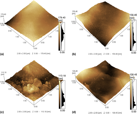
Figure 1(a) shows an atomic force microscopy (AFM) image of the electrochemically treated sample before sterilization. The formation of a well-defined nanostructure in the entire analyzed area can be seen.
3D AFM images of the electrochemically treated Ti6Al4V samples for 4 min after the sterilization methods. (a) Before sterilization methods; (b) air plasma sterilization; (c) ethanol + PBS sterilization; and (d) autoclave sterilization.

After sterilization, a significant change on surface morphology was observed on samples sterilized with ethanol + PBS [Fig. 1(c)] and autoclave [Fig. 1(d)]. However, the air plasma–sterilized sample [Fig. 1(b)] showed no changes in the morphology before and after the sterilization process, as indicated in Fig. 1(a).
A study developed by Junkar et al. (2016) [Reference Junkar, Kulkarni, Drasler, Rugelj, Mazare, Flasker, Drobne, Humpolícek, Resnik, Schmuki, Mozetic and Iglic19] had already observed the destruction of TiO2 nanotubes and morphological changes after autoclave sterilization. The authors also observed that after sterilization by UV light (253.7 nm length, 40 cm light source distance, 36W Philip lamp), there was a slight disruption of the titanium nanotubes, thus causing a disordering of the nanotubular structure. Nevertheless, an intact nanotubular structure was observed after oxygen plasma sterilization (RF generating source, frequency of 13.56 MHz, output power of 200 W, pressure of 75 Pascal for 60 s).
To strengthen the importance of using plasma sterilization, Wen (2015) [Reference Wen20] showed in her study that the oxygen plasma sterilization method changes the morphology of the nanotubes, because during the sterilization, the temperature is low on the surface, and there are no molecules of water (vapor) involved. This decreases the possibilities for the evaporation of the fluoride, which induces the crystallization or modification of the nanotubes.
In relation to autoclave sterilization process, Lamberti et al. (2015) [Reference Lamberti, Chiodoni, Shahzad, Bianco, Quaglio and Pirri21] suggest that the destruction of TiO2 nanotubes in a wet autoclave can be related to the combined effect of humidity and high temperature (127–134 °C) from the sterilization vapor, which induces the crystallization of the TiO2 nanotubes, thus resulting in a morphological alteration. According to Lamberti et al. (2015) [Reference Lamberti, Chiodoni, Shahzad, Bianco, Quaglio and Pirri21], the changes observed on the surface morphology of the nanotube may be associated with the combination of water vapor effects and the temperature used during the sterilization process. Wang et al. (2011) [Reference Wang, Liu, Zhang, Tao, Pippel and Domen22] discussed the proposed mechanisms for water vapor and crystallization causing changes in the morphology of the nanotubes.
As shown by Yu et al. (2010) [Reference Yu, Dai and Cheng23], water vapor molecules from the sterilization process interact with the walls of the amorphous TiO2 nanotubes and condense, as hot water, acting as a catalyst and favoring the rearrangement of TiO2 in TiO62− octahedron. For Wang et al. (1998) [Reference Wang, Hashimoto, Fujishima, Chikuni, Kojima, Kitamura, Shimohigoshi and Watanable24], the destruction of TiO2 nanotubes can also be related to the presence of F− ions that evaporated from the nanotubes surface because of the high temperature used in the sterilization process. Kulmarni et al. (2015) [Reference Kulkarni, Patil-Sen, Junkar, Kulkarni, Lorenzetti and Iglic25] and Kulkarni et al. (2015) [Reference Kulkarni, Flasker, Lokar, Mrak-Poljsak, Mazare, Artenjak, Cucnuk, Kralj, Velikonja, Schmuki, Kralj-Iglic, Sodin-Semrl and Iglic26] demonstrated that these F− ions come from the anodization of the electrolytes used for the production of the nanotubes, being, usually, in low concentration on the surface of the nanotubes.
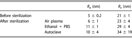
The sterilization methods that altered the nanometric morphology of the samples treated electrochemically were the ethanol + PBS and the autoclave. The effect on the values of the nanometric roughness (R a) (Table I) was seen to be higher for the samples sterilized by ethanol + PBS. The increase in the surface roughness can be related to the deposition of a salt layer from the evaporating water from the PBS during the ethanol + PBS sterilization. The autoclaved sample showed few changes in the nanometric roughness. For the air plasma–sterilized samples, no difference or change in the roughness values at the nanometer scale after sterilization was identified. Considering the operational parameters used in the present work, no changes on the sample surface morphology were observed after air plasma sterilization.
Nanometric roughness measurements for the electrochemically treated Ti6Al4V samples at 25 V, and 7 °C for 4 min.

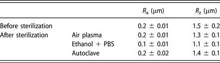
Regarding micrometer roughness (Table II), no significant difference was observed in the samples analyzed before and after the sterilization process, regardless of the sterilization process used. This demonstrates that the changes caused by the sterilization processes studied were only evidenced on a nanometric scale.
Micrometric roughness measurements for the electrochemically treated Ti6Al4V samples at 25 V, and 7 °C for 4 min.

According to Junkar et al. (2016) [Reference Junkar, Kulkarni, Drasler, Rugelj, Mazare, Flasker, Drobne, Humpolícek, Resnik, Schmuki, Mozetic and Iglic19], the air plasma sterilization process can eventually cause a slight increase in micrometric roughness, thus modifying the surface activity due to the action of the oxygen from the plasma. However, in the present work, under the conditions used for the air plasma sterilization process, no modification on the nanostructured surface of Ti6Al4V was observed, even at a nanoscale.
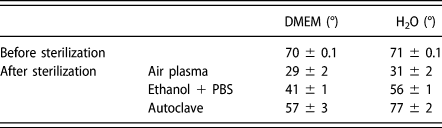
The contact angle values used as DMEM solution are shown in Table III. The sterilization methods by ethanol + PBS and autoclave (Table III) presented higher values of contact angle than the air plasma sterilized surface. This more hydrophobic behavior is probably associated to the morphological changes that occurred on the surfaces, as shown in Figs. 1(c) and 1(d), as a consequence of adsorption and/or deposition of material.
Contact angle measurements for the electrochemically treated samples in DMEM, and H2O for 4 min before and after sterilization methods.

In addition, the air plasma sterilization method tends to make the surface more hydrophilic, because it can be observed even on the sample that did not undergo a sterilization process, which presented a higher contact angle than the air plasma–sterilized sample.
This effect of the air plasma sterilization method is probably associated with the removal of apolar molecules present in the air, which are inevitably adsorbed by the metal surface of the Ti6Al4V alloy. Moreover, this method does not cause morphological changes on the surfaces after sterilization, as seen in Fig. 1(b).
A study developed by MacDonald et al. (2002) [Reference Macdonald, Deo, Markovic, Stranick and Somasundaran27] using cleaning processes with butyl alcohol for 30 min on titanium surfaces showed an increase in hydrophobicity of the surface. Similarly, more hydrophobic behavior was also found by Vezeau et al. (2000) [Reference Vezeau, Keller and Wightman28]. The contact angle for the nonsterilized surface was lower for the ethanol-sterilized sample; nevertheless, this contact angle value is still higher than that found on the air plasma sterilized surface.
Four sterilization methods (autoclave, UV light, hydrogen peroxide plasma, and oxygen plasma) on TiO2 nanotubes were evaluated in the research developed by Junkar et al. (2016) [Reference Junkar, Kulkarni, Drasler, Rugelj, Mazare, Flasker, Drobne, Humpolícek, Resnik, Schmuki, Mozetic and Iglic19], yet no influence on the wettability of these nanotubes was observed. According to Lamberti et al. (2015) [Reference Lamberti, Chiodoni, Shahzad, Bianco, Quaglio and Pirri21] the decrease in the contact angle of TiO2 nanotubes after autoclaving (90 °C, 240 min) can be related to the formation of surface defects that act as −OH adsorption sites. There was a decrease in the contact angle values for the autoclaved sample when these were compared to the nonsterilized sample; however, these values are still higher than those found on the air plasma–sterilized sample.
For Junkar et al. (2016) [Reference Junkar, Kulkarni, Drasler, Rugelj, Mazare, Flasker, Drobne, Humpolícek, Resnik, Schmuki, Mozetic and Iglic19], the oxygen plasma sterilization on TiO2 nanotubes causes a surface exposure by reactive oxygen species, which interacts with the surface reducing the contamination by hydrocarbons and forming a dense layer of TiO2 oxide. However, titanium is known to have a natural TiO2 layer on the surface, which is altered after its exposure to oxygen plasma. According to these authors, many of the changes on the surface properties of the TiO2 layer can be attributed to changes on the surface chemistry and wettability, properties which strongly influence the biocompatibility results. This study [Reference Junkar, Kulkarni, Drasler, Rugelj, Mazare, Flasker, Drobne, Humpolícek, Resnik, Schmuki, Mozetic and Iglic19] also points out that surface treatments influence the oxide layer, making it denser, less contaminated with hydrocarbons, and more hydrophilic.
The increasing surface hydrophilicity after air plasma sterilization evidenced by the decrease in the contact angle can contribute to the increase in the interaction between the surface of the implant and the biological environment. Schrader (1982) [Reference Schrader29], and Baier et al. (1984) [Reference Baier, Meyer, Natiella, Natiella and Carter30] demonstrated that a layer of cells appears more easily on a surface with hydrophilic character, presenting, at the same time, a strong cellular adhesion.
For Hurbett et al. (1996) [Reference Hurbett, Ratner, Schakenraad and Schoen31], the surface hydrophilicity is a determinant factor in the biocompatibility of biomaterials. Eriksson et al. (2004) [Reference Eriksson, Nygren and Ohlson32] stressed that when implants with high hydrophilicity are implanted in the bone, the rate of bone formation is increased, contributing to the maturation and differentiation of bone cells. Buser et al. (2004) [Reference Buser, Broggini, Wieland, Schenk, Denzer, Cochran, Hoffmann, Lussi and Steinemann33] emphasized that a hydrophilic surface promotes bone mineralization, which, potentially, contributes to rapid osseointegration and early bone loading.
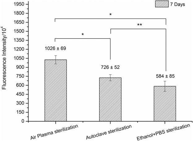
The results of the cell viability after 7 days of contact with human embryonic stem cell-derived mesenchymal progenitors (hESC-MPs) on the electrochemically treated samples and sterilized by different sterilization methods are presented in Fig. 2.
Cell viability of hESC-MPs after 7 days of contact with biomaterials. *P < 0.05, **P < 0.1.

The electropolished and sterilized with ethanol + PBS samples showed lower cell growth (less cell viability) when compared to the other samples that were sterilized by autoclaving and air plasma, as seen in Fig. 2. The lower cell viability of stem cells on the sample sterilized by ethanol + PBS is believed to be related to larger modifications caused on the surface of the sample, as seen in Fig. 1(c). Nonetheless, the electropolished and sterilized by air plasma sample, seen in Fig. 2, presents higher cell growth (cell viability). This may be related to the fact that no changes were observed on the surface of the sample after being sterilized by air plasma, which contributed to the permanence of the integrity of the nanostructured surface. This nanostructured surface [Fig. 1(b)], which is associated with lower nanometric roughness values (Table I), with greater wettability, and lower values of contact angle surface (Table III), contributed to the increase in cell growth, resulting in higher cell viability (Fig. 2). As reported in many studies in the literature [Reference Antonini, Kothe, Reilly, Owen, Marcuzzo and Malfatti34, Reference Antonini, Junior, Reilly and Malfatti35], a surface with higher wettability (lower contact angle) can improve cell growth. It can also be related to the removal of apolar functional groups that can be adsorbed on the surface, which can facilitate and improve the interactions of the cellular structure with the surface.
In relation to the surface that was electropolished and sterilized by autoclave, a decrease in cellular viability, when compared to the air plasma–sterilized sample, was evidenced. Such decrease can be related to the changes that also occurred on the nanostructured surface after the autoclave sterilization method [Fig. 1(d)]. In addition to the changes in the nanometric morphology, higher R z value (Table I) and lower wettability, which is evidenced by higher contact angle values (Table III), may have contributed to a reduction in the cellular viability of the nanostructured sample that was sterilized by autoclave.
Conclusions
The sterilization methods by autoclave and ethanol + PBS altered the nanometric morphology of the samples. Considering the operational parameters used in the present work, no changes in the morphology of the sample after sterilization by air plasma was verified, which is evidenced by the similar nanometric roughness values before and after the sterilization process. The sterilizations by ethanol + PBS and autoclave increased the nanometric average roughness, whereas no changes occurred in the roughness values of the samples sterilized by air plasma.
The samples sterilized by air plasma had their hydrophilicity increased (reduction of the contact angle) when compared to the nonsterilized samples. This can be related to the removal of apolar molecules coming from the atmosphere, which are unavoidably adsorbed by the surface.
The ethanol + PBS and the autoclave sterilization processes altered the nanometric morphology of the nanostructured samples, which resulted in lower cell growth and viability. Even though the air plasma sterilization process did not alter the nanostructured surface, the integrity, and permanence of the nanostructures associated with lower nanometric roughness, and greater wettability (lower values of contact angle) may have contributed to higher growth of hESC-MPs and higher cell viability.
The results shown in this article pointed out the surface modifications due to the sterilization processes in metallic materials and evidenced no changes in the morphology of the sample after sterilization by air plasma, considering the operational parameters used in the present work. This indicated that this process can be used as an alternative to avoid modifications on the surface of metallic biomaterials. In addition, the surface characteristics after sterilization by air plasma contributed to hESC-MPs growth and increase in cell viability.
The scope of this investigation aimed to evaluate the changes the sterilization processes (air plasma, ethanol + PBS, and autoclave) caused on the surface of the materials, considering practical parameters usually used in sterilization processes. Although the research has reached its aims, there were some unavoidable limitations, such as the study of the time, temperature, and humidity of these sterilization processes. Another limitation was the increase in the surface roughness; it can be related to the deposition of a salt layer from the evaporating water from the PBS because during the ethanol + PBS sterilization, but this measurement was not realized in this study, once that this paper aimed to alert the changes that the sterilization processes cause in the surface of the materials, considering practical parameters usually employed in sterilization processes.
Material and methods
Materials
The Ti6Al4V samples were obtained as billets (provided by TiBrasil Titânio Ltda), having approximately 14 mm (diameter) and 2 mm (thickness). The samples were sliced in a CNC machine (Romi) of maximum revolution of 4000 rpm and maximum potency of 10 KW.
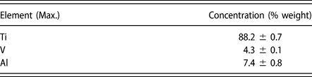
The chemical composition analyses of the samples were performed by X-ray fluorescence (Niton® XL3t GOLDD+ Series X-ray fluorescence; Thermo Fisher Scientific, Billerica, MA). The alloy used in this study is predominantly composed of Ti, Al, and V, as shown in Table IV. According to the ASTM B348-13 standard, the concentration indicated for this alloy is Al (5.5–6.5 wt%) and V (3.5–4.5 wt%), thus being the material used in this study in accordance with the standard.
Chemical composition of the Ti6Al4V used in this research.

Electropolishing procedure
All samples were mechanically polished with silicon carbide sandpaper of up to 4000 grit, neutral soap, and water. Before the electropolishing process, samples were washed in deionized water using an ultrasonic bath and dried with cold air, with subsequent cleaning by means of RF plasma glow discharge. Ti6Al4V disks with an exposed area of approximately 1.3 cm2 and 2 mm thickness were used for electropolishing. They were electropolished in an acidic solution consisting of sulfuric acid, hydrofluoric acid, and glycerine in a 6:3:1 (v/v) ratio, using a power source (MPC-303DI, Minipa, São Paulo, Brazil) with platinum as the cathode (area of approximately 2.0 cm2) and the sample as the anode. Electropolishing was performed at 25 V at 7 °C ± 0.5 °C for 4 min. After the electrochemical process and before the sterilization procedures, all samples were washed in deionized water using an ultrasonic bath for 10 min and dried with cold air. However, after the sterilization processes, aiming to follow clinical protocol, the samples were not submitted to any other procedure before tests.
Roughness and wettability analyses
Surface morphology and nanometric roughness were characterized by atomic force microscopy (AFM), using a SHIMADZU SPM 9500J3 microscope (Shimadzu, Barueri, São Paulo, Brazil). The micrometric roughness was determined using a MITUTOYO SJ-400 profilometer (Mitutoyo America Corporation, Aurora, Illinois).
Surface roughness (in micrometric scale) was determined by measuring the R a (arithmetical mean of the absolute values within a sampling length), and the R z (the vertical distance between the highest peak and the deepest valley within a sampling length). Nanometric roughness was obtained by measuring the R a (arithmetical mean of the absolute values of the profile deviations from the mean line of the roughness profile) and the R z (mean value of the sum of the height of the highest profile of five peaks, and the depth of the deepest profiles valley, relative to the mean line, within a sampling length).
The surface wettability was determined by the sessile drop method using an equipment developed by the Corrosion Research Laboratory (LAPEC) at the Universidade Federal do Rio Grande do Sul (UFRGS, Porto Alegre, Brazil). With this equipment, it was possible to determine the contact angle between a drop of DMEM (Dulbecco’s Modified Eagle’s Medium from Sigma-Aldrich Co., LLC., Merck, Darmstadt, Germany) and the analyzed substrate. The contact angle was determined by image analyses using a SURFTENS 4.5 software (OEG, Frankfurt, Germany).
Three samples were used in each analysis and all results showed in this work are presented as mean ± standard deviation.
Sterilization methods
The air plasma sterilization was conducted with an RF plasma glow discharge, with maximum power of 65 W and sterilizing time of 5 min in atmosphere of N2 and O2 in the same proportion of the atmospheric air (78.1% N2, 20.9% O2, 1% argon and other gases) [Reference Ettwein and Maslin36], at 250 µatm of pressure.
The ethanol + PBS sterilization was conducted from immersion of the samples in ethanol 70% v/v for 3 h in ultrasonic bath. Subsequently, the samples were immersed in PBS in an ultrasonic bath for 3 times of 20 min each.
In steam autoclave sterilization, the samples were exposed to 121 °C for 30 min under 1.2 bar of pressure.
Cell culture
Human embryonic stem cell-derived mesodermal progenitors (hES-MPs) (Cellartis, Gothenburg, Sweden) and mesenchymal progenitor cells were used for all cell culture experiments. Cells were cultured at 37 °C, 5% CO2 in basal media (BM), containing minimum essential alpha medium, 10% fetal bovine serum, 2 mM L-glutamine, and 100 mg/mL penicillin/streptomycin in gelatin-coated T75 flasks. During passage, BM was supplemented with 4 ng/mL human fibroblastic growth factor (Life Technologies, Warrington, U.K.).
Samples were seeded at 10,000 cells per sample. They were allowed to attach onto the surface for 45 min before the disc was submerged in 2 mL of BM and incubated overnight. On day 1, samples were transferred to a new well plate, and 2 mL of OIM was added to selected samples. The medium was changed every 2–3 days.
To assess the suitability of the Ti6Al4V samples for cell culture, resazurin reduction (RR) assays were performed. Fluorescence was measured using a microplate reader, and it was correlated with cell viability [Reference O’Brien, Wilson, Orton and Pognan37]. One mM resazurin sodium salt in dH2O was diluted in BM (10 vol%) to create the RR solution. The medium was removed from the samples and replaced with 2 mL of the RR solution. Well plates were wrapped in foil and incubated at 37 °C for 4 h. And, 200 µL of the reduced solution was added to a 96-well plate in triplicate. They were measured using a spectrofluorometer at λex: 540 nm and λem: 590 nm in an Infinite F200 Pro spectrophotometer (Tecan Trading AG, Männedorf, Switzerland).
Statistical analysis
All statistical analyses were conducted in a GraphPad Prism (version 7.00). Three samples were used per analysis. Depending on whether a response was affected by one or two factors, either one- or two-way ANOVA with a Tukey post-test was used to evaluate significant differences. Differences were considered significant when P < 0.05. All graphs are presented as mean ± standard deviation, and notable significant differences are indicated either in the graphs or in the captions.
Acknowledgments
The present work was conducted with the support of CAPES and CNPq, Brazilian government agencies focused on the development of human resources.